Article Text

Abstract
Background The concept of the synovio-entheseal complex (SEC) anatomical functional unit has been developed in the context of arthritic disease (1). Previous studies have demonstrated the involvement of SECs in the progression of ligament derived hand osteoarthritis (OA) and in erosion formation in OA (2).Thus far the role of SECs as potential factors in large joint OA has not been reported.
Objectives First, to investigate whether the posterior cruciate ligament (PCL) had a SEC structure and to determine whether this could contribute to degenerative changes in the knee joints in OA. Second, to look at morphometric measurements of the PCL-SEC to determine whether there was an association with MRI observable lesions of the PCL in early OA. Third, to provide histological validation for MRI findings.
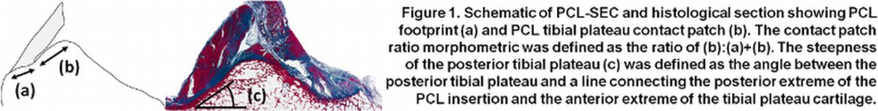
Methods 49 subjects with bilateral MRI images available were chosen from the progression cohort of the National Institute of Health Osteoarthritis Initiative (OAI) study. 3T MRI images taken at enrolment were read by an experienced radiologist using a novel scoring system. The PCL and the area surrounding its tibial insertion were assessed for the presence and severity of bone marrow oedema, osteophyte formation, cortical defects, intra-osseous and synovial cysts and posterior capsular synovitis. Sagittal plane MR images running through the mediolateral centre of the PCL were used to define morphometrics including: PCL-posterior tibial plateau contact patch (Fig. 1b) and posterior tibial plateau steepness (Fig 1c). Spearman’s rank correlation coefficients were calculated to test for associations between the morphometric measurements and ligament pathology scores. Histological sections of normal cadaveric tissue were taken and stained with Masson’s trichrome to assess the histology and variation of the SEC.
Results The PCL contact patch ratio (mean=0.605, SD=0.07) and posterior tibial plateau steepness (36.6°, 4.0) were used to describe anatomical variation in the PCL-SEC. Significant correlation was observed between posterior tibial plateau steepness and the dimension of osteophytes lateral to the PCL insertion (ρ=0.258, P=0.011). The correlation was greater in the 61 knees with less severe OA (Kellgren-Lawrence (K-L) score = 0, 1 or 2), (ρ=0.414, P=0.001). In the same mild OA sub-cohort, a negative correlation was seen between the PCL contact patch morphometric and the presence of oedema at the PCL insertion site (ρ= -0.297, P=0.020). When all 98 knees were considered (K-L score = 0, 1, 2, 3 or 4) no such correlation was seen. Histological findings confirmed the SEC structure of the PCL tibial enthesis and anatomical variation.
Conclusions Anatomical variations in the morphology of the PCL-SEC may influence the way in which mechanical stresses are dissipated throughout the SEC functional unit. In this study we observed a smaller PCL contact patch being associated with a higher risk of MRI determined disease. Thus, the morphology of the PCL-SEC plays an important role in the phenotypic expression of early knee OA.
McGonagle D et al Arthritis & Rheumatism 2007;56(8):2482-91.
Tan AL et al Arthritis & Rheumatism 2005;52(8):2355-65.
Disclosure of Interest None Declared