

Abstract
Objective. To investigate whether patients with a very early diagnosis of systemic sclerosis (VEDOSS) may already present circulating markers and in vitro signs of microvascular dysfunction.
Methods. Serum samples were obtained from 55 patients with systemic sclerosis (SSc), 25 patients with VEDOSS, and 55 matched healthy controls (HC). Serum levels of pan-vascular endothelial growth factor (VEGF) and soluble neuropilin-1 (sNRP-1) were measured by ELISA. Human dermal microvascular endothelial cells (H-MVEC) were cultured and stimulated with SSc, VEDOSS, and HC sera. Protein expression of NRP-1 was analyzed by Western blotting, cell proliferation by 5′-bromodeoxyuridine assay, migration capacity by wound-healing assay, and capillary-like tube formation by Matrigel assay.
Results. Serum levels of pan-VEGF were increased in patients with VEDOSS and SSc versus HC (p = 0.05 and p = 0.003, respectively). Serum levels of sNRP-1 were significantly reduced in patients with VEDOSS and SSc compared with controls (p = 0.012 and p = 0.027, respectively). NRP-1 expression was decreased in H-MVEC stimulated with VEDOSS sera (p < 0.001 vs HC). Proliferation was reduced in H-MVEC stimulated either with VEDOSS or SSc sera in comparison with HC sera (p = 0.015 and p = 0.043, respectively). Wound healing was compromised in H-MVEC stimulated with VEDOSS and SSc sera versus HC sera (p < 0.01 for both). Capillarogenesis was decreased in H-MVEC stimulated with VEDOSS sera (p < 0.01) and SSc sera (p < 0.001) compared with cells stimulated with HC sera.
Conclusion. Similar to patients with SSc, patients with VEDOSS already present biological signs of endothelial dysfunction. Our data demonstrate that VEDOSS sera significantly modify endothelial cell behavior and impair the angiogenic potential of the microvascular system.
Systemic sclerosis (SSc) is characterized by widespread vascular injury and dysfunction, impaired angiogenesis, immunological abnormalities, and progressive fibrosis of the skin and internal organs1,2,3,4. In the pathogenetic cascade of SSc, the vascular system seems primarily affected, together with the derangement of the immune system. In this scenario, endothelial cell activation, damage, and apoptosis are the main features that favor vascular tone dysfunction and ischemia-reperfusion injury, vessel wall remodeling, and reduced capillary blood flow. These events progressively lead to extracellular matrix accumulation and fibrosis4,5,6,7,8. The involvement of the microvascular system is also characterized by capillary loss, mainly from an aberrant regeneration of capillaries and a defective angiogenesis5,9. In SSc, it has been shown that vascular endothelial growth factor (VEGF)-A/VEGF receptor (VEGFR) system is profoundly disturbed. Moreover, a deficiency in the coreceptor neuropilin (NRP)-1 (initially described as an axonally expressed receptor for secreted class-3 semaphorins) may be an additional factor contributing, with the perturbed VEGF-A/VEGFR-2 system, to peripheral microvasculopathy and defective angiogenesis in SSc10.
The deregulation of vascular tone control, clinically evident as Raynaud phenomenon (RP), and microcirculatory abnormalities are the earliest clinical manifestations of SSc and may precede skin and visceral involvement by months or years6,7,11,12. As evident in nailfold videocapillaroscopy (NVC), vascular damage and angiogenic disturbances are present since the “early” NVC pattern of SSc, and further aggravate in the “active” and “late” patterns, culminating in the loss of capillaries with formation of avascular areas6,7. Many vascular biomarkers, including proangiogenic and angiostatic factors, have been linked to peripheral vasculopathy in SSc13.
VEGF is strongly overexpressed in the skin and serum of patients with SSc, together with the VEGF receptors (VEGFR-1 and -2), although no effective angiogenesis is observed. VEGF levels are mainly increased in the earliest stages of the disease, which may be related to compensatory mechanisms and may have deleterious effects on the vascular network. Although elevated levels of VEGF are consistent with active angiogenesis, an uncontrolled chronic overexpression throughout various disease stages, as seen in patients with SSc, might contribute to disturbed vessel morphology rather than promote new vessel formation13.
It has been shown that RP and puffy fingers, together with abnormal capillaroscopy and positive SSc-specific antinuclear antibodies, may allow identification of patients with the preliminary criteria for very early diagnosis of SSc (VEDOSS)14,15,16,17. These patients already present modifications of the microvasculature and complications such as digital ulcers (DU)18. For this reason, the aim of our work was to investigate whether the sera of patients with VEDOSS present modifications of factors involved in angiogenesis and may elicit a reduction of the angiogenic potential of microvascular endothelial cells in vitro.
MATERIALS AND METHODS
Patients, controls, and serum samples
Patients were included who were followed regularly at the Department of Experimental and Clinical Medicine, Division of Rheumatology, Azienda Ospedaliero-Universitaria Careggi (AOUC), Florence, Italy, or at the Autoimmunity Outpatient Clinic of the Department of Internal Medicine, Centro Hospitalar de São João (CHSJ), Porto, Portugal. Inclusion criteria consisted of being classified as SSc19 or VEDOSS15, having clinical information available for chart review (demographic, clinical manifestations, imaging, and immunology), and being able to give written informed consent for chart review and for performing blood tests. Patients with a concomitant autoimmune disease were excluded. The presence of primary RP was an exclusion criterion for healthy controls (HC).
Serum samples were obtained from 55 patients with SSc (49 women; median age 64 yrs, range 37–81 yrs; mean disease duration 10 years, range 1–31 yrs) classified as limited cutaneous SSc (n = 40) or diffuse cutaneous SSc (n = 15)20, from 25 patients with VEDOSS (21 women; median age 50 yrs, range 19–77 yrs; median disease duration 1 yr, range 0–8 yrs), and from 55 age-matched and sex-matched healthy individuals (51 women; median age 52 yrs, range 29–70 yrs).
All patients reported the occurrence of RP. At the time blood was withdrawn, the presence of DU was recorded. NVC was performed on 8 fingers by 2 operators (IC and SG). Recorded images were then saved and scored blindly afterward by both doctors to divide patients into 3 capillaroscopic patterns (i.e., “early,” “active,” and “late”)21.
The clinical and demographic characteristics of the patients with SSc and VEDOSS are shown in Table 1. Patients were not receiving immunosuppressive medications, corticosteroids, or other disease-modifying drugs. Before blood sampling, patients were washed out for 10 days from oral vasodilating drugs and for 2 months from intravenous prostanoids. Fresh venous blood samples were drawn, left to clot for 30 min before centrifugation at 1500 g for 10 min, and serum was collected and stored in aliquots at −80°C until used.
Demographic and clinical characteristics of the patients with SSc and VEDOSS included for collection of serum samples. Values are n (%) unless otherwise specified.
The study was approved by the local institutional review board of AOUC (AOUC 69/13), as well as by the Health Ethical Committee of CHSJ (CHSJ 84/13). All subjects provided written informed consent.
ELISA for serum pan-VEGF, soluble NRP-1 (sNRP-1), and Semaphorin 3A (Sema3A)
Serum levels of pan-VEGF (catalogue number DVE00; R&D Systems), sNRP-1, and Sema3A (catalogue number ABIN415191 and ABIN481720, respectively; Antibodies-on line) were measured by commercial quantitative colorimetric sandwich ELISA, according to the manufacturer’s protocol. Each sample was measured in duplicate. For all ELISA assays, the interassay and intraassay variances were < 10%.
Culture and stimulation of human dermal microvascular endothelial cells (H-MVEC)
H-MVEC purchased from ATCC were cultured in RPMI 1640 medium (Invitrogen Life Technologies) supplemented with 10% fetal bovine serum (FBS; Invitrogen Life Technologies), 1% penicillin/streptomycin (Invitrogen Life Technologies), 1.176 g/l of sodium bicarbonate, 4.76 g/l of HEPES, 1 ml/l of EGF, and 1 mg/l of hydrocortisone > 98% (Sigma-Aldrich), and maintained at 37ºC in a humidified 5% CO2 atmosphere. H-MVEC were used between the third and seventh passages in culture. In stimulation experiments, H-MVEC were serum-starved overnight before treatment with sera from patients with VEDOSS, patients with SSc, or HC.
Western blotting analysis
Proteins were isolated from H-MVEC lysates using RIPA buffer (Chemicon International) and 20 µg of protein were separated by 8% sodium dodecyl sulfate-polyacrylamide gel electrophoresis and transferred to Hybond nitrocellulose membrane (Amersham Life Science). Membranes were then incubated with primary antibodies against NRP-1 (1:1000; Abcam) and α-tubulin (1:1000; Sigma-Aldrich). After overnight incubation at 4°C, membranes were washed with Tris-buffered saline containing 0.1% Tween 20 and incubated with secondary antibodies at room temperature for 1 h. Immunoreactive bands were then visualized by the enhanced chemiluminescence detection system (ECL kit, Amersham Life Science) as previously described22. The expression of NRP-1 in H-MVEC was measured at basal condition and after stimulation with 10% VEDOSS sera (n = 8) and HC sera (n = 8) for 24 h.
H-MVEC proliferation assay
H-MVEC (6 × 10⁴ cells/ml) were grown for 24 h and then incubated with 10% sera from patients with SSc (n = 10), patients with VEDOSS (n = 10), and HC (n = 10) for 24 h. Basal or stimulated H-MVEC were then incubated with 5′-bromodeoxyuridine (BrdU) solution at a final concentration of 0.01 mM during the treatment period. Optical density of proliferating cells (positive for BrdU) after ELISA assay using anti-BrdU–specific antibodies (Roche Diagnostics) was evaluated at the microplate reader according to the manufacturer’s instructions and as previously reported22.
H-MVEC migration assay: wound healing
H-MVEC were seeded in 24-well plates precoated with 0.1% gelatin and allowed to grow to 100% confluence. Cell monolayer was injured by a 10-µl tip and cells were washed twice with phosphate buffered saline and then incubated in basal medium or medium containing 10% sera of patients with SSc (n = 10), patients with VEDOSS (n = 10), and HC (n = 10). Cell migration into the wounded area was then visualized and photographed on a phase contrast microscope (Nikon) at a magnification of 40×, after 24, 29, 42, and 46 h of incubation. Wound healing capacity was assessed by comparing the images of the wounded area at the beginning and at 46 h to quantify the migration rate of the cells after wounding.
H-MVEC capillary-like tube formation assay
In vitro capillary morphogenesis assay was performed in 96-well plates covered with Matrigel (BD Biosciences). Matrigel (50 µl; 10–12 mg/ml) was pipetted into culture wells and polymerized for 30 min to 1 h at 37°C, as described elsewhere23. H-MVEC (30 × 103 cells/well) were incubated in basal RPMI 1640 medium with 2% FBS or 10% sera from patients with SSc (n = 10), patients with VEDOSS (n = 10), and HC (n = 10). Plates were photographed at 6 h and 24 h. Results were quantified at 24 h by measuring the percent field occupancy of capillary projections, as determined by image analysis.
Statistical analysis
Statistical analyses were performed using the Statistical Package for Social Sciences software for Windows, version 20.0 (SPSS). Categorical variables are presented as frequencies and percentages and continuous variables as mean and SD or median and interquartile range (IQR) for variables with skewed distribution. Normal distribution was checked using Shapiro–Wilk test or skewness and kurtosis. The Student t test and nonparametric Mann–Whitney U test were used where appropriate for statistical evaluation of the differences between 2 independent groups, while ANOVA or nonparametric Kruskal-Wallis tests were used for statistical evaluation of the differences between 3 independent groups. Posthoc analyses were performed with Mann–Whitney U test, considering Bonferroni correction (α/number of comparisons). All reported p values are 2-tailed, with a p value of < 0.05 indicating statistical significance.
RESULTS
Serum levels of pan-VEGF, sNRP-1, and Sema3A in patients with VEDOSS
Serum levels of pan-VEGF were increased either in patients with VEDOSS (median 283.96, IQR 191.01–360.29 pg/ml) or in patients with SSc (median 323.13, IQR 151.28–507.51 pg/ml) compared with HC (median 227.81, IQR 114.94–300.64 pg/ml; p = 0.05 and p = 0.003, respectively; Figure 1A). There were no significant differences in levels of pan-VEGF between the VEDOSS and SSc groups (Figure 1A). Higher levels of pan-VEGF were found in patients with VEDOSS with both “early” and “active” NVC patterns (median 313.47 pg/ml and 296.62 pg/ml, respectively) versus HC, although these differences did not reach statistical significance (Figure 1B).

(A) Serum levels of pan-VEGF determined by colorimetric sandwich ELISA in healthy controls and patients with VEDOSS and SSc. (B) Serum levels of pan-VEGF in patients with VEDOSS according to NVC pattern and in healthy controls. Each box represents the 25th to 75th percentiles. Lines inside the boxes represent the median. Lines outside the boxes represent the 10th and the 90th percentiles. Circles indicate outliers, and asterisks indicate the extreme values. Kruskal-Wallis test was used for statistical analysis. Pan-VEGF: pan-vascular endothelial growth factor; SSc: systemic sclerosis; VEDOSS: very early diagnosis of SSc; NVC: nailfold videocapillaroscopy; NS: not significant.
We next addressed whether circulating levels of sNRP-1 were affected in patients with VEDOSS. Serum levels of sNRP-1 were significantly reduced in patients with VEDOSS (median 0.12, IQR 0.03–0.24 ng/ml) and SSc (median 0.23, IQR 0.0–0.45 ng/ml) compared with HC (median 0.38, IQR 0.05–1.56 ng/ml; p = 0.012 and p = 0.027, respectively; Figure 2A). There were no significant differences in levels of sNRP-1 between the VEDOSS and SSc groups (Figure 2A). Regarding NVC changes in VEDOSS, as an additional measure of peripheral microvascular involvement, we also found no differences in sNRP-1 between the patients with “early” and “active” NVC patterns and those with normal NVC (Figure 2B).

(A) Serum levels of sNRP-1 determined by colorimetric sandwich ELISA in healthy controls and patients with VEDOSS and SSc. (B) Serum levels of sNRP-1 in patients with VEDOSS according to NVC pattern and in healthy controls. Each box represents the 25th to 75th percentiles. Lines inside the boxes represent the median. Lines outside the boxes represent the 10th and the 90th percentiles. Circles indicate outliers, and asterisks indicate the extreme values. Kruskal-Wallis test was used for statistical analysis. sNRP-1: soluble neuropilin-1; SSc: systemic sclerosis; VEDOSS: very early diagnosis of SSc; NVC: nailfold videocapillaroscopy; NS: not significant.
Further, in accordance with data from SSc10, no significant differences in serum levels of Sema3A were detected between patients with VEDOSS (median 2.44, IQR 0.99–4.31 ng/ml) and HC (median 4.04, IQR 1.94–4.80 ng/ml; p = 0.29; data not shown).
Expression of NRP-1 in H-MVEC stimulated with VEDOSS sera
The effect of patients’ sera on endothelial cell NRP-1 expression was investigated. In agreement with what was previously reported for SSc10, the expression of NRP-1 was decreased in H-MVEC after stimulation with VEDOSS sera compared with HC sera (p < 0.001; Figure 3).
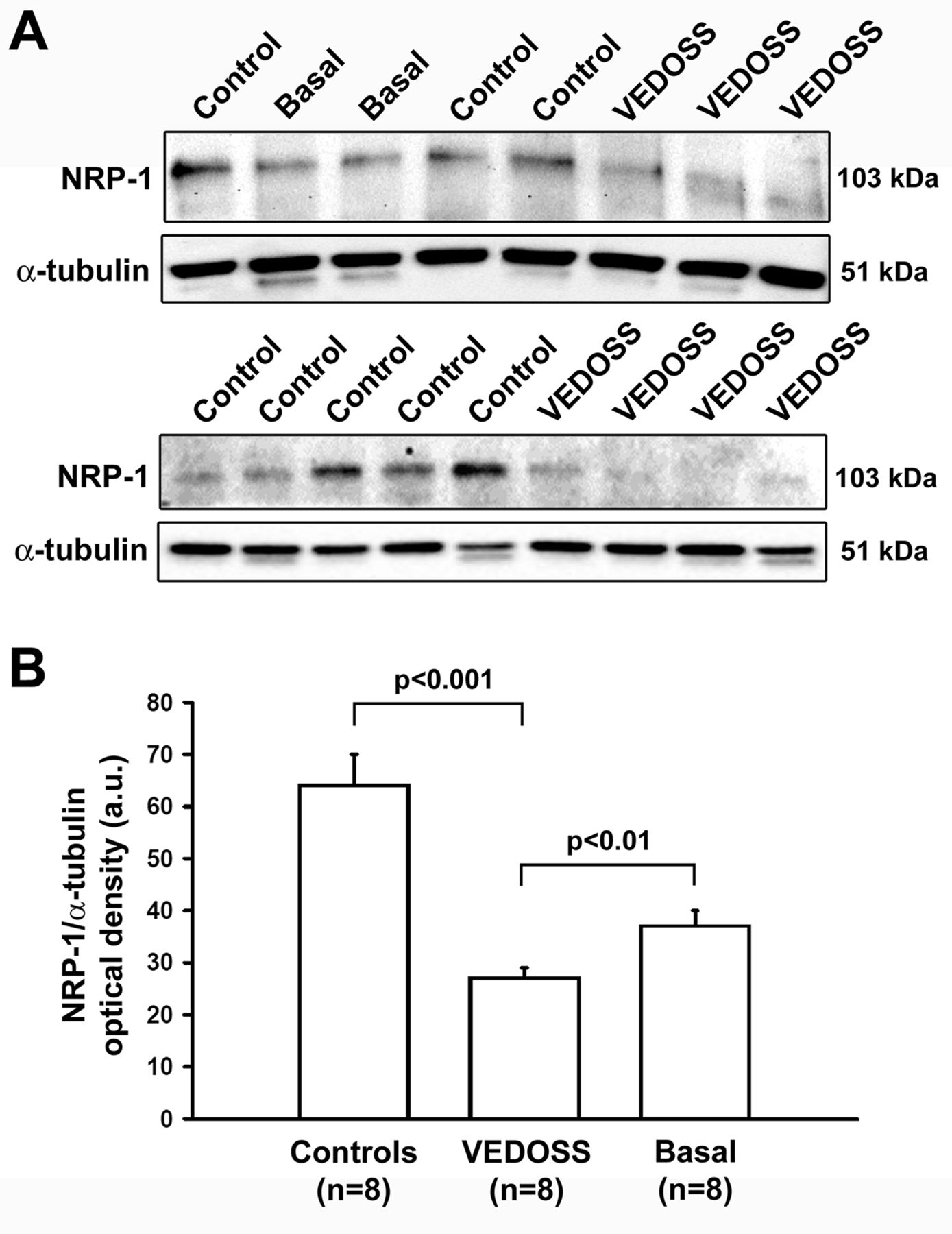
Expression of NRP-1 in H-MVEC. (A) Representative immunoblots. Western blotting was carried out on total protein extracts from H-MVEC at basal condition (n = 8) or treated with 10% sera from healthy control subjects (n = 8) and patients with VEDOSS (n = 8) for 24 h. (B) The densitometric analysis of the bands normalized to α-tubulin is reported in the histograms. Data are means ± SD of optical density in arbitrary units (a.u.). Mann–Whitney U test was used for statistical analysis. NRP-1: neuropilin-1; H-MVEC: human dermal microvascular endothelial cells; VEDOSS: very early diagnosis of systemic sclerosis.
Proliferation, migration, and capillary-like tube formation of H-MVEC stimulated with VEDOSS sera
Proliferation of H-MVEC was decreased after stimulation either with VEDOSS sera or SSc sera in comparison with HC sera (p = 0.015 and p = 0.043, respectively; Figure 4A). Further, cell migration was compromised in H-MVEC stimulated with VEDOSS and SSc sera versus HC (p < 0.01 for both; Figure 4B and Figure 4C) as determined by wound-healing assay after 46 h of stimulation (the time by which almost complete healing was observed in cultures treated with HC sera).

(A) Effect of sera from patients with VEDOSS and SSc on H-MVEC proliferation. Cell proliferation was measured by BrdU assay in H-MVEC at basal condition (n = 10) or treated with 10% sera from healthy controls (n = 10), patients with VEDOSS (n = 10), and patients with SSc (n = 10) for 24 h. Each box represents the 25th to 75th percentiles. Lines inside the boxes represent the median. Lines outside the boxes represent the 10th and the 90th percentiles. (B and C) Effect of sera from patients with VEDOSS and patients with SSc on H-MVEC migration. Wound-healing capacity was assessed after 46 h in H-MVEC at basal condition (n = 10) or treated with 10% sera from healthy controls (n = 10), patients with VEDOSS (n = 10), and patients with SSc (n = 10). (B) Four representative images of the wounded area at 46 h are shown for each experimental group; dotted lines represent wound margins. (C) Quantitative analysis of the percentage of wound repair. Data are means ± SD. * p < 0.01 vs basal H-MVEC and H-MVEC treated with healthy control sera. SSc: systemic sclerosis; VEDOSS: very early diagnosis of SSc; H-MVEC: human dermal microvascular endothelial cells; BrdU: 5′-bromodeoxyuridine; NS: not significant.
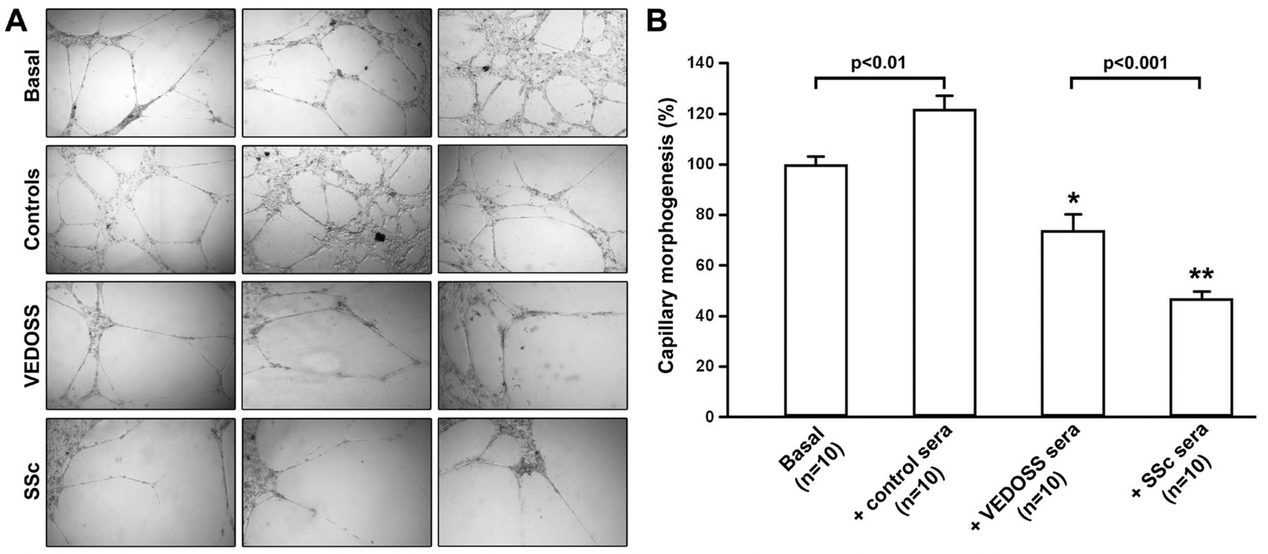
The assembly of H-MVEC into capillary-like structures was also addressed. Capillarogenesis was decreased in H-MVEC stimulated for 24 h with VEDOSS (p < 0.01) and SSc sera (p < 0.001) versus HC sera (Figure 5A and Figure 5B). Interestingly, exposure of H-MVEC to VEDOSS sera resulted in a significantly higher angiogenic capacity as compared with cells treated with SSc sera (p < 0.001; Figure 5A and Figure 5B).

Effect of sera from patients with VEDOSS and SSc on H-MVEC capillary-like tube formation. (A) Representative images of capillary-like tube formation assay on Matrigel after 24 h in H-MVEC at basal condition (n = 10) and after stimulation with 10% sera from healthy controls (n = 10), patients with VEDOSS (n = 10), and patients with SSc (n = 10). Three representative images are shown for each experimental group. (B) Capillary-like tube formation quantified as percentage field occupancy of capillary projections. Statistical analysis was carried out with Mann–Whitney U test. * p < 0.01 vs basal H-MVEC and H-MVEC treated with healthy sera. ** p < 0.001 vs basal H-MVEC and H-MVEC treated with healthy sera. SSc: systemic sclerosis; VEDOSS: very early diagnosis of SSc; H-MVEC: human dermal microvascular endothelial cells.
DISCUSSION
SSc is primarily a vascular disease5,6. It is generally accepted that initial vascular injury because of autoimmunity and/or environmental factors causes structural and functional abnormalities of microvasculature, resulting in the constitutive activation of fibroblasts in various organs24. Because peripheral vasculopathy appears to be present since the very early onset of SSc pathogenesis, our study analyzed the microvascular derangement in patients clinically classified as VEDOSS.
VEGF-A/VEGF receptor signaling pathway, including its coreceptor NRP-1, is involved in the disturbance of angiogenesis in SSc10. VEGF has been shown to be strongly overexpressed in the skin and serum of patients with SSc, although without effective angiogenesis, and VEGF levels are mainly increased in the earliest stages of the disease13. In our study, both patients with VEDOSS and SSc presented higher serum levels of VEGF when compared with HC, with a tendency to higher levels in SSc versus VEDOSS. In a recent study comparing HC and different stages of SSc25, VEGF serum levels did not show a highly significant linear trend across the different study groups, while other vascular biomarkers did.
Interestingly, this increase in serum VEGF levels was accompanied by lower circulating levels of sNRP-1 both in patients with VEDOSS and SSc. Moreover, levels of sNRP-1 tended to be lower in VEDOSS than in SSc, though not statistically significant. Corroborating ELISA findings, NRP-1 expression was significantly decreased in H-MVEC treated with VEDOSS sera when compared with HC sera. Thus, the higher VEGF serum levels observed in VEDOSS might be a way of compensating for the lack of NRP-1 response in endothelial cells. In contrast to our findings in SSc10, we found no correlations between serum levels of sNRP-1 and NVC patterns in VEDOSS, perhaps due to either the smaller number of patients in the VEDOSS group or to a less severe peripheral vasculopathy in those patients. Further studies are needed to elucidate this topic. However, regarding the comparison with HC, the findings of circulating sNRP-1 previously reported for SSc were similar to those found in the VEDOSS group of patients in our present study. Of note, there were no significant differences between VEDOSS and SSc results, suggesting that the VEDOSS “environment” already presents characteristics of the established disease, rather than being a “pre-disease”. This hypothesis seems further corroborated by the data obtained in vitro. In fact, to further analyze vascular derangement in VEDOSS, we performed assays of cell proliferation, migration, and capillarogenesis in vitro. Interestingly, stimulation with VEDOSS sera compromised the ability of H-MVEC to proliferate and to perform wound healing. Moreover, Matrigel assay clearly showed a gradual decrease in capillary-like tube formation from VEDOSS to SSc, supporting the progressive antiangiogenic features of the disease.
Of note, our present observations are also in agreement with a recent study showing that distinct SSc subsets have different degrees of vasculopathy and that markers of abnormal endothelial function are increased in the earliest stages of the disease, in which clinical and laboratory findings of advanced disease cannot yet be observed25.
Patients with VEDOSS already present circulating biomarkers of defective angiogenesis and their sera significantly alter the normal behavior of endothelial cells. This evidence suggests that the involvement of the microvascular system and endothelial cell injury do in fact occur in very early SSc, even when only a few clinical signs and symptoms are present. Further studies, with larger samples of patients with VEDOSS, will be required to identify other potential circulating biomarkers of vascular dysfunction in VEDOSS.
Research into the cellular and molecular basis of SSc has provided new insights into its pathophysiology and potential targets for intervention. Emerging therapies based on immunomodulation, antifibrotic agents, and vasoprotection hold the possibility of preventing end-organ damage and improving longterm outcomes in patients with SSc, who can benefit from an early and accurate diagnosis18. There must be a window of opportunity for effective therapy for SSc, and this appears to be confined to the very early phase of the disease26. In the near future, widening our knowledge about VEDOSS pathophysiology and its pathogenic mechanisms may help to identify new candidate therapeutic agents for this very early phase of SSc.
Footnotes
IC was supported by a research grant from the Foundation for the Development of Internal Medicine in Europe. The study was partially funded by the Portuguese National Funding Agency (FCT: UID/BIM/04293/2013).
- Accepted for publication April 11, 2017.


