Article Text

Abstract
Background Interleukin 17A (IL-17A) exerts pivotal proinflammatory functions in chronic inflammatory and autoimmune diseases.
Objective To investigate IL-17A expression in temporal artery lesions from patients with giant-cell arteritis (GCA), and its relationship with disease outcome.
Methods Fifty-seven patients with biopsy-proven GCA were prospectively evaluated, treated and followed for 4.5 years (52–464 weeks). Relapses, time (weeks) required to achieve a maintenance prednisone dose <10 mg/day, and time (weeks) to complete prednisone withdrawal were prospectively recorded. IL-17A mRNA was measured by real-time quantitative RT-PCR in temporal arteries from all patients and 19 controls. IL-17 protein expression was assessed by immunohistochemistry/immunofluorescence.
Results IL-17A expression was significantly increased in temporal artery samples from GCA patients compared with controls (6.22±8.61 vs 2.50±3.9 relative units, p=0.016). Surprisingly, patients with strong IL-17A expression tended to experience less relapses, and required significantly shorter treatment periods (median 25 vs 44 weeks to achieve <10 mg prednisone/day, p=0.0079). There was no correlation between IL-17A and RORc or RORα expression suggesting that these transcription factors may not exclusively reflect Th17 differentiation, and that cells other than Th17 cells might contribute to IL-17 expression in active patients. Accordingly, FoxP3+IL-17A+ cells were identified in lesions by confocal microscopy and were dramatically reduced in specimens from treated patients.
Conclusions IL-17A expression is increased in GCA lesions, and is a predictor of response to glucocorticoid treatment. The contribution of FoxP3+ cells to IL-17A production in untreated patients suggests that induced-Tregs may facilitate disease remission when proinflammatory cytokine production is downregulated by glucocorticosteroids.
- Systemic vasculitis
- Giant Cell Arteritis
- Cytokines
- Disease Activity
- Inflammation
Statistics from Altmetric.com
Introduction
Giant-cell arteritis (GCA) is a large and medium-sized vessel vasculitis considered to be a Th1-mediated disease on the basis of its granulomatous appearance and the strong expression of IFNγ and IFNγ-induced products in lesions.1–5
Patients with GCA experience a dramatic improvement with high-dose glucocorticoids (GC). However, response is not sustained, and about 40%–50% of patients relapse when GC are tapered.6 ,7 Persistent activity results in prolonged GC treatment, and more than 80% of patients experience GC-related complications.8 Mechanisms involved in disease persistence are unknown. Increased expression of TNFα and CCL2 in vascular lesions and elevated serum concentrations TNFα and IL-6 are associated with relapsing disease and prolonged GC-requirements.5 ,9 ,10 However, blocking TNFα with infliximab did not result in reduced relapse rate or cumulated GC doses when compared with placebo in a randomised clinical trial, indicating that blocking TNFα is not sufficient to abrogate disease activity.11 In the context of this trial, comparison of gene expression in second temporal artery biopsies performed in four patients after 1 year of treatment disclosed that IFNγ and IL12/23p40, but not IL12p35, were upregulated in relapsing patients.3 Discrepancy between expression of the two IL-12 subunits, p35 and p40, suggested that upregulated IL-12/23p40 might be part of IL-23. This led to the search for IL-23 and its related cytokine IL-17 in GCA lesions.3 ,12 ,13
IL-17A production characterises a distinct T cell phenotype, Th17, with pivotal proinflammatory functions in several autoimmune and chronic inflammatory disorders previously thought to be Th1-mediated.14 ,15 Several cytokines participate in Th17 differentiation in humans, including IL-1β, IL-6 and TGFβ. IL-21 also contributes, particularly in the absence or paucity of IL-6, and IL-23 participates in the expansion/maintenance of the Th17 phenotype.15 All these cytokines are abundantly expressed in GCA lesions.3 ,9 ,16 ,17
The aim of this study was to expand these initial observations by investigating the expression of IL-17A in temporal artery biopsies from a sizable series of patients with GCA to investigate the relationship between IL-17A expression and clinically relevant findings, such as the intensity of the systemic inflammatory response, relapses and response to therapy.
Patients and methods
Patients
The study group consisted of 57 patients with biopsy-proven GCA diagnosed between 1997 and 2006 at our institution (Hospital Clinic, Barcelona) (see online supplementary data S1 and figure S1 for selection criteria). All patients were prospectively evaluated and treated by the authors (GEF, JHR, SPG and MCC) with a predefined homogeneous glucocorticoid-tapering schedule.6 ,10 Clinical data recorded at the time of diagnosis included disease symptoms, number of relapses, time to achieve a prednisone dose <10 mg/day, and time to complete prednisone discontinuation with no relapse within the following 6 months. The following baseline blood tests were recorded: erythrocyte sedimentation rate (ESR), haemoglobin, C-reactive protein (CRP) and haptoglobin concentrations, and platelet counts. Relapse was defined as reappearance of cranial symptoms, polymyalgia rheumatica, or systemic symptoms that could not be attributed to other conditions.3 ,11 Isolated fluctuations on ESR or CRP were not considered relapses. Symptoms of relapse had to resolve by an increase of 10 mg above the previous effective dose.
Clinical data of the patients are displayed in online supplementary table S1. In 38 patients, the temporal artery was removed before starting treatment, and the remaining 19 had been treated with prednisone (60 mg/day) for a median of 7 days (range 2–12). In four patients, a contralateral temporal artery biopsy was performed after 1 year of treatment.3
Uninvolved temporal arteries from 19 patients (13 women and 6 men) with a median of 77 years (range 64–91) in whom GCA was considered but not confirmed, served as controls. All of them were subsequently diagnosed with other diseases (see online supplementary methods).
The study was approved by the Ethics Committee of Hospital Clínic (Barcelona), and patients signed an informed consent.
RNA Isolation and cDNA synthesis
Temporal artery biopsies were embedded in optimal cutting temperature (OCT, Sakura, The Netherlands), snap-frozen in liquid nitrogen and stored at −80°C until used. Sections consecutive to those that provided the histopathologic diagnosis were processed for RNA isolation using TRIzol Reagent (Invitrogen, Carlsbad, California, USA).
Total RNA (1 μg) was reverse-transcribed to cDNA using the Archive kit (Applied Biosystems, Foster City, California, USA), employing random hexamer priming.
Real-time quantitative PCR
cDNA was measured by quantitative real-time PCR using specific Pre-Developed TaqMan gene expression assays (see online supplementary methods) from Applied Biosystems as previously described.9 ,16 All samples were normalised to the expression of the housekeeping gene, GUSb. The comparative CT method was used to assess relative gene expression.
Immunohistochemistry
To determine the topography of IL-17 expression, serial 4–6 µm sections were obtained from frozen temporal arteries of two of the patients and two controls. Sections were air-dried, fixed with cold acetone and permeabilised with 0.1% saponin. Endogenous peroxidase was blocked with H2O2, and the slides were incubated with the primary polyclonal antibody goat antihuman IL-17A (R&D Systems, Minneapolis, Minnesota, USA). Optimal dilutions were tested on frozen sections of human tonsils (positive control). Immunoglobulins obtained from the same species served as negative controls. Immunodetection was performed with a HRP-labeled polymer conjugated to a secondary antibody (EnVision Visualisation method, Dako, Glostrup, Denmark) using 3,3′-diaminobenzidine as a chromogen.
Enzyme-linked immunosorbent assay (ELISA) detection of IL-17A in tissue
Temporal artery protein extracts could be obtained from seven of the patients (five responders and two relapsers) from the phenolic phase during RNA extraction. IL-17A concentration was measured by immunoassay using Quantikine Human IL-17A from R&D Systems, according to the manufacturer's protocol.
Immunofluorescence staining and confocal microscopy
For qualitative assessment of cytokine distribution at the cellular level, immunofluorescence staining was performed in three additional temporal artery biopsies obtained from an active patient and from two patients who had received treatment with prednisone at 60 mg/day for 8 days at the time of the artery excision. A fragment of these biopsies was fixed in 4% paraformaldehyde with increasing concentrations of sucrose, frozen with OCT and stored at −80°C. Cryostat 10 μm sections were fixed with 4% paraformaldehyde, permeabilised with Triton 0.1% and immunostained with the primary and secondary antibodies detailed in online supplementary methods. Nuclei were stained with Hoechst dye (Molecular Probes, Life Technologies Ltd, Paisley, UK) at 1 : 1000. Slides were mounted in Mowiol 4-88 Reagent (Merck4Biosciences, Nottingham, UK) and examined using a laser scanning confocal Leica TCS SP5 microscope (Leica Microsystems, Heildelberg, Germany). Images were processed with Leica Confocal software and Image J software (Wayne Rasband, Bethesda, Massachusetts, USA). The number of IL-17 positive and IL-17/FoxP3 double positive cells per field was counted in 10 fields per specimen at 200×magnification.
Statistical analysis
Mann–Whitney test, Spearman's rho correlation coefficient, and Kaplan–Meier survival curves analysed with log-rank test were used for statistical analysis. With the exception of comparison between untreated (38) and treated (19) patients, statistical analysis was restricted to the cohort of 38 treatment-naïve patients.
Results
IL-17A is upregulated in GCA lesions
IL-17A mRNA was significantly more abundant in temporal arteries from untreated patients than in control arteries (6.22±8.61 vs 2.50±3.90 relative units; p=0.016) (figure 1A). By contrast, plasma IL-17A was not detectable, or was slightly above the detection threshold in the majority of patients and controls (figure 1B).

IL-17A concentrations in temporal artery biopsies and serum from patients with giant-cell arteritis (GCA). (A) IL-17A mRNA expression (relative units) in temporal arteries from 38 treatment-naïve patients with GCA and 19 controls. (B) IL-17A concentrations in sera from 33 GCA treatment-naïve patients and seven controls. Dotted line indicates detection threshold. NA, not applicable.
As shown in figure 2, immunostaining revealed intense IL-17A protein expression by inflammatory cells in GCA lesions. Giant-cells immunostained negative for IL-17A. Interestingly, although control arteries had some constitutive IL-17A mRNA expression, control arteries immunostained negative for IL-17A.

Immunohistochemical detection of IL-17A expression in temporal artery lesions. (A) IL-17 expression in a giant-cell arteritis (GCA)-involved temporal artery section. (B) Lack of IL-17A immunostaining in a temporal artery from a control individual. (C) Closeup view of a GCA-involved artery where distinct IL-17A+cells can be observed in all arterial layers: adventitia (adv), media (med) and intima (int). (D) Giant cells (arrow) do not express IL-17A. (E) IL-17A expression in the temporal artery from a treatment-naive patient compared with IL-17A expression in the contralateral biopsy obtained after being treated with prednisone for 1 year (F).
Lack of correlation between IL-17A expression and systemic inflammatory findings
Given the known proinflammatory functions of IL-17A, we explored whether IL-17A mRNA expression correlated with the intensity of the systemic inflammatory response. No differences in IL-17A mRNA expression were observed between patients with strong versus weak systemic inflammatory response at diagnosis (see online supplementary table S2). No significant correlation was found between IL-17A mRNA expression and acute-phase reactants including ESR (r=0.0886, p=0.60), haemoglobin (r=−0.03563, p=0.82) or CRP concentrations (r=0.1495, p=0.45).
Correlation between IL-17A and other proinflammatory cytokines in GCA lesions
We next tested the potential correlation between IL-17A expression in treatment-naïve samples and the expression of cytokines known to participate in Th17 differentiation (IL-6, TGFβ) or in the maintenance and expansion of the Th17 phenotype (IL23p19).15 IL-17A mRNA concentrations in untreated GCA arteries significantly correlated with IL-6 (r=0.363, p=0.025) and with IL23p19 mRNA (r=0.397, p=0.008). These findings suggest coordinated regulation in accordance with the role for IL-6 and IL-23 in inducing and maintaining the Th17 phenotype, respectively. No correlation was found between IL-17A and TGFβ (r=−0.037, p=0.848).
IL-17A concentration is decreased in temporal arteries from treated GCA patients
IL-17A mRNA concentrations in temporal artery biopsies from the 19 treated patients were significantly lower than those found in the 38 treatment-naïve patients (1.74±2.48 relative units vs 6.22±8.61; p=0.017) (figure 3A). In four patients who underwent a second temporal artery biopsy after being treated for 1 year, immunohistochemical detection of IL-17 protein, which was intense at the time of diagnosis (figure 2E), was limited to scattered remaining inflammatory cells in the second specimen (figure 2F).

Relationship between IL-17A and response to therapy. (A) IL-17A mRNA expression (relative units) in temporal arteries from 38 treatment-naïve and 19 prednisone treated giant-cell arteritis (GCA) patients. (B) IL-17A mRNA content in temporal arteries from 38 treatment-naïve GCA patients according to relapses. (C) IL-17 protein content in temporal arteries from seven GCA patients according to relapses (patient numbers are too small for reliable statistics). (D) IL-17A mRNA concentration in initial temporal artery biopsies from patients still requiring prednisone compared with that obtained from patients in sustained treatment-free remission, 3 years after diagnosis. NA: not applicable
IL-17A expression and long-term response to glucocorticoid treatment
Surprisingly, IL-17A mRNA concentrations in lesions tended to be higher in patients who achieved sustained remission or experienced just one disease flare compared with those who experienced multiple relapses (7.46±9.73 vs 3.19±3.70 relative units; p=0.058) (figure 3B). The same trend was observed in IL-17A protein concentration which tended to be higher in non-relapsing patients (19.30±29.96 vs 0.68±0.63 pg/ml) (figure 3C). Accordingly, IL-17A mRNA levels tended to be higher in patients able to completely discontinue prednisone at 3 years (5.17±8.11 vs 0.29±0.46, p=0.06) (figure 3D).
Similarly, untreated patients with high IL-17A mRNA content (above 75% percentile) in their arteries achieved a maintenance prednisone dose <10 mg/day significantly sooner than patients with lower IL-17A mRNA values (median 25 vs 44 weeks, p=0.0079) (figure 4A). Likewise, patients with elevated IL-17A mRNA concentrations were able to completely withdraw prednisone earlier than patients with lower IL-17 mRNA levels (p=0.028) (figure 4B).
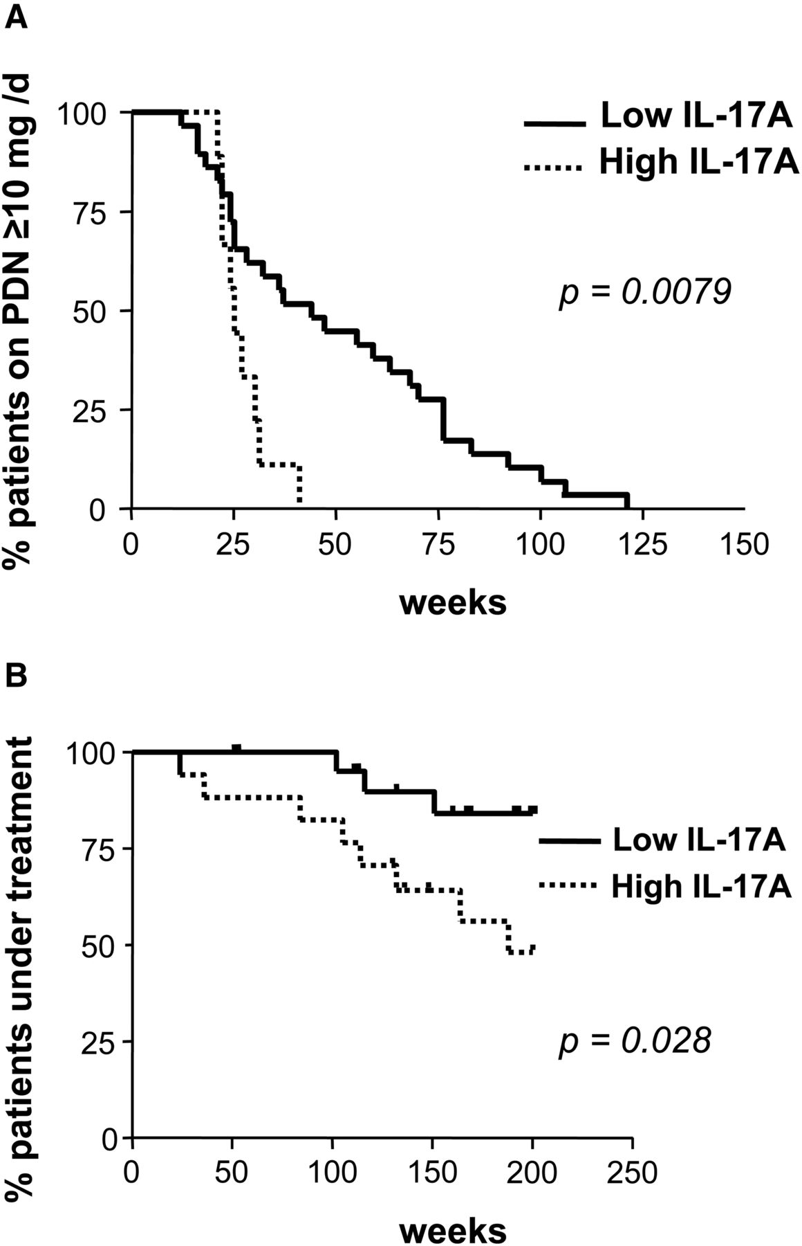
Association between IL-17A expression in giant-cell arteritis (GCA) lesions and treatment requirements. (A) Percentage of patients requiring >10 mg prednisone/day to maintain remission, according to the intensity of IL-17A expression in vascular lesions. High IL-17A refers to mRNA concentration within the 75th percentile, and low IL-17A below the 75th percentile. (B) Percentage of patients requiring prednisone treatment over time, according to the intensity of IL-17A expression in lesions as in A.
Plasticity between regulatory T lymphocytes (Tregs) and Th17 lineages may contribute to the association between increased IL-17A expression and response to therapy
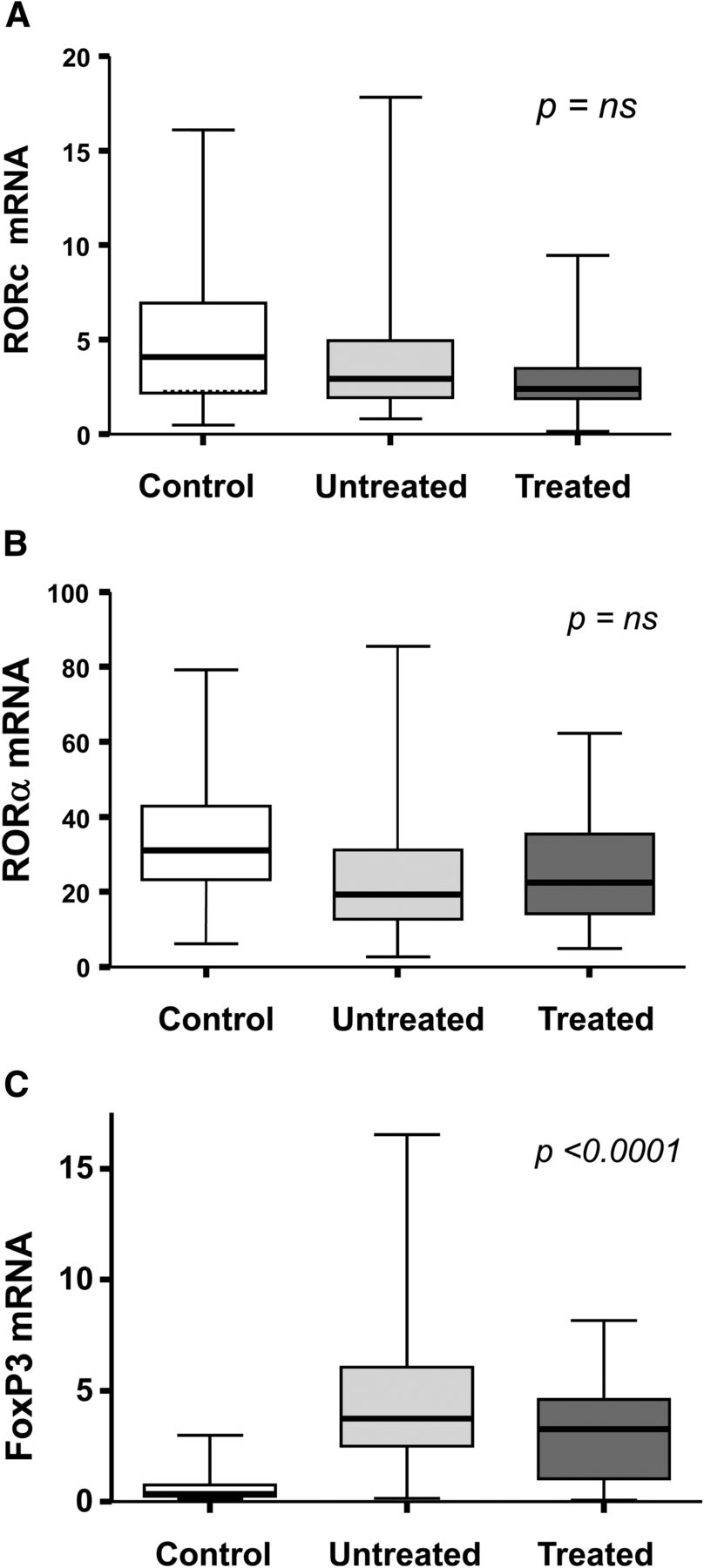
We next investigated the expression of transcription factors RORα and RORc, master regulators of Th17 lineage. RORα and RORc were expressed at similar levels in GCA and control arteries; their expression tended to correlate (r=0.33, p=0.053), and were not influenced by treatment (figure 5). No correlation was found between IL-17A and RORα (r=−0.20, p=0.36) or RORc expression (r=−0.11, p=0.596). Taken together, these findings suggest that RORα and RORc may be expressed by cells other than Th17 lymphocytes, and that Th17 cells may not be the only producers of IL-17A in GCA. By contrast, FoxP3 was significantly upregulated in GCA lesions and significantly decreased with treatment (figure 5).

Expression of master regulators of Th17 (RORc and RORα) and Treg differentiation (FoxP3) in temporal artery lesions. (A) Expression of RORc (relative units) in 19 control arteries, 38 treatment-naïve patients and 19 prednisone-treated patients. Expression of RORα (B) and FoxP3 (C) in the same patient subsets.
It has been recently shown that induced regulatory Tregs may transiently lose their suppressive capacity and produce IL-17A in an inflammatory microenvironment.18–21 Confocal microscopy of temporal artery sections showed that a subset of Tregs (identified as FoxP3-positive cells) contributed, indeed, to IL-17A production in active GCA lesions, and that these cells may be reduced in specimens from treated patients (figure 6). Initial IL-17A expression may then reflect the functional activity of Th17 cells and also the contribution of IL-17A-producing FoxP3 cells with their potential to recover suppressive activity when inflammatory stimuli decrease in the microenvironment.

Expression of IL-17A and Foxp3 cells in temporal artery lesions from patients with giant-cell arteritis. (A) IL-17A expression (red colour) in a patient with active giant-cell arteritis (GCA). (B) IL-17A expression in a patient treated with 60 mg prednisone/day for 1 week. (C) Higher magnification of an inflammatory cell expressing only IL-17A (red, merged). (D) A magnified inflammatory cell intensively expressing both IL-17A and FoxP3 (orange, merged). (E) A magnified inflammatory cell from a treated patient with lower coexpression (orange, merged). (F) A magnified cell from a treated patient expressing primarily Foxp3 (green, merged). (G) IL-17A+ and IL-17A+FoxP3+ double positive cells (mean±SD) in 10 different fields (200×) per specimen containing similar number of nuclei in lesions from an active patient versus two treated patients. p<0.05 (due to the small number of cases, statistics are only indicative).
Discussion
We found that IL-17A expression is prominent in GCA lesions. Consistently, IL-17A expression correlated with expression of IL-6, a cytokine involved in Th17 differentiation, and particularly, with expression of the IL-23 subunit IL-23p19, a cytokine involved in the maintenance and expansion of the Th17 phenotype.15 ,21 The lack of correlation with TGFβ, also required for Th17 differentiation, as well as the significant correlation with IL-23, may be related to the fact that temporal artery biopsies are rarely performed during early events and are usually obtained when the inflammatory process is fully developed. At the stages when temporal arteries are obtained, TGFβ may play additional roles in vascular remodelling.3 ,22 Our findings, obtained in a sizable series of patients with prospectively recorded clinical and follow-up data confirm preliminary observations performed in small series of patients, and support the participation of Th17 mechanisms in the pathogenesis of GCA.3 ,12 ,13
IL-17A, typically, but not exclusively, produced by Th17 cells, plays a central role in the development of tissue inflammation in a variety of experimental models and human diseases.15 ,21 IL-17A exerts strong proinflammatory functions on a variety of cells, including endothelial cells, by inducing expression of classical proinflammatory cytokines IL-1β, IL-6 and TNFα, endothelial adhesion molecules, chemokines and metalloproteinases.14 ,15 ,23 All these molecules are strongly expressed in GCA, and are thought to participate in proinflammatory amplification cascades.2 ,9 ,16 ,23–,25 The remarkable expression of IL-17A found in GCA supports a prominent role in vascular inflammation. Supporting this concept, deletion of IRF-4-binding protein leading to sustained activation of IRF-4, a transcription factor involved in Th17 differentiation, results in large-vessel vasculitis in mice.26
While IL-17A was remarkably upregulated in lesions, it was barely detectable in serum. Consistent with this finding, we could not find a relationship between IL-17A expression and systemic symptoms or acute-phase proteins, suggesting that IL-17A functions are predominantly exerted locally at the vascular lesions. IL-17A strongly amplifies the expression of other proinflammatory cytokines (IL-6, TNFα) which, in turn, are effectively released into the bloodstream and do correlate with the intensity of the systemic inflammatory response in GCA.6 ,10
As others and we have previously shown in small series,3 ,12 ,13 IL-17A mRNA and IL-17A-expressing cells were dramatically reduced in specimens obtained from treated patients. This finding supports the prominent proinflammatory role of IL-17A in GCA, and suggests that downregulation of IL-17A may at least partially account for the dramatic effect of high-dose glucocorticoids in ameliorating disease-related symptoms, and in substantially decreasing vascular inflammation after long-term treatment.3
The most intriguing finding of this study was the association between strong initial expression of IL-17A and response to therapy. Patients with strong IL-17A expression tended to experience less relapses and were able to reduce and completely discontinue prednisone earlier than patients with weaker IL-17A expression. This may indicate that patients who develop a predominantly Th17 response are more sensitive to glucocorticoid treatment.
Recent studies have shown that, on one hand, Th17 cells are not the only producers of IL-17A15 ,27 and that, on the other hand, there is remarkable plasticity among T cell lineages depending on the conditions of the microenvironment.18–20 Particularly interesting is the fact that induced Tregs, which role was initially identified as immunosuppressive, are able to produce IL-17A when exposed to an inflammatory milieu.
We show that both RORα and RORc, markers and master regulators of Th17 are equally expressed in GCA and control arteries. Constitutive expression of RORα and RORc by non-inflamed arteries may indicate an important, previously unknown role of these factors in vascular biology. By contrast, FoxP3 was upregulated in GCA and decreased with treatment, consistent with our early findings demonstrating expression of CD25+ lymphocytes in lesions and their reduction upon treatment.4 The lack of correlation between RORα/RORc and IL-17A expression also suggests that other cells, in addition to Th17 cells, may contribute to IL-17A production in GCA. FoxP3-positive cells contributed, indeed, to IL-17A production. These preliminary findings suggest that induced Tregs may functionally evolve over time, and may have a role in limiting disease activity in GCA. Expanding Tregs by administration of low-dose IL-2 has been recently shown to ameliorate cryoglobulinemic-related vascular inflammation in humans.28 Moreover, it has recently become apparent that not all Th17 cells are equally pathogenic, and that abundance of TGFβ expression may switch Th17cells to the alternative non-pathogenic phenotype.29 We have recently shown that prednisone treatment increases TGFβ expression,3 which may also limit the pathogenicity of Th17 cells.
In conclusion, strong IL-17A expression in involved arteries is a biomarker and predictor of response to therapy. Our preliminary data suggests that different cell lineages may contribute to IL-17A expression in GCA. However, the observational nature of this and other existing studies addressing IL-17A expression in GCA does not allow to draw strong mechanistic conclusions about the specific role of IL-17A and Th17 cells in GCA. Our findings suggest complex interplay among T cell lineages in delineating disease fate and response to therapy that deserves further investigation.
Acknowledgments
We thank Mrs Montse Sánchez and Mrs Ester Tobías for excellent technical support with immunohistochemistry and Dr María Calvo for her invaluable advice with confocal microscopy.
References
Supplementary materials
Supplementary Data
This web only file has been produced by the BMJ Publishing Group from an electronic file supplied by the author(s) and has not been edited for content.
Files in this Data Supplement:
- Data supplement 1 - Online supplement
Footnotes
Handling editor Tore K Kvien
-
Contributors GEF, MCC: designed the study and wrote the paper; GEF, MCB, EPR, EL, MS: performed experimental work; JHR, SPG, AGM, JMG, MCC: collected and analysed clinical data; MUR: designed the study, analysed data, provided important inputs to the writing of the manuscript and approved the final version. All the authors repeatedly discussed the results of the paper, made suggestions for improvement and contributed to generate further data. All the authors read and reviewed the drafts and approved the final version of the manuscript.
-
Funding Supported by Ministerio de Economía y Competitividad (SAF 08/04328 and SAF 11/30073).
-
Competing interests None.
-
Patient consent Obtained.
-
Ethics approval Internal Review Board, Hospital Clínic.
-
Provenance and peer review Not commissioned; externally peer reviewed.