Article Text

Abstract
Background Fibrosis and vascular disease are cardinal features of systemic sclerosis (SSc). Stimulators of soluble guanylate cyclase (sGC) are vasoactive drugs that are currently being evaluated in phase III clinical trials for pulmonary arterial hypertension.
Objective To study the antifibrotic potency of sGC stimulators.
Methods The effect of the sGC stimulator BAY 41-2272 on the release of collagen from dermal fibroblasts was examined. The antifibrotic effects of BAY 41-2272 on prevention and regression of fibrosis in bleomycin-induced dermal fibrosis and in Tsk-1 mice were also studied. Telemetric blood pressure studies in conscious mice were used to study potential hypotensive effects of sGC stimulation.
Results sGC stimulation with BAY 41-2272 dose-dependently inhibited collagen release in dermal fibroblasts from patients with SSc and healthy individuals. Furthermore, BAY 41-2272 stopped the development of bleomycin-induced dermal fibrosis and skin fibrosis in Tsk-1 mice, preventing dermal and hypodermal thickening, reducing the numbers of myofibroblasts and reducing the hydroxyproline content. In addition, BAY 41-2272 was highly effective in the treatment of established fibrosis in the modified models of bleomycin-induced skin fibrosis and Tsk-1 mice. Treatment with sGC stimulators was well tolerated. Relevant antifibrotic doses of BAY 41-2272 did not affect systemic blood pressure and heart rate in mice.
Conclusions These findings demonstrate potent antifibrotic effects and good tolerability of sGC stimulators in various experimental models of SSc. Given their potential vasoactive properties, sGC stimulators may be promising candidates for the dual treatment of fibrosis and vascular disease in SSc.
Statistics from Altmetric.com
Introduction
Fibrosis of the skin and internal organs is a hallmark of systemic sclerosis (SSc). During the course of the disease, progression of fibrosis disrupts physiological tissue architecture and causes organ failure, resulting in high morbidity and mortality among patients with SSc. Fibroblasts are the key players in the development of fibrosis in SSc. They show a pathological and persistent activation with enhanced expression of contractile proteins and excessive release of extracellular matrix components.1 2 The mechanisms of the pathological fibroblast activation are only partially known, and targeted antifibrotic treatments are not available for clinical use.3
During development of antifibrotic drugs, attention needs to be paid to other disease manifestations of SSc—in particular, vascular disease. Since vascular disease does not always share common disease mechanisms with fibrosis, targeting of specific signalling pathways may result in opposite effects (ie, improving fibrosis but worsening vascular disease).
The soluble guanylate cyclase (sGC) catalyses the production of cyclic guanosine monophosphate (cGMP) upon binding of nitric oxide (NO). NO-binding causes conformational changes of the sGC to enable cGMP formation from guanosine triphosphate. Once released by the sGC, cGMP can act as a second messenger to activate further downstream targets, such as cGMP-regulated ion channels, protein kinases (G-kinases) and phosphodiesterases (PDEs). Through those effectors, cGMP regulates a variety of physiological processes, including cell growth and proliferation, vascular tone and remodelling, immune responses and neuronal transmission.4
For more than 100 years, the sGC has been targeted by NO donor drugs (eg, glyceryl trinitrate) or inhaled NO to treat cardiovascular disease. Nevertheless, insufficient biometabolism, the development of tolerance and non-specific interactions of NO with other biological molecules hamper the usefulness of these compounds. Side effects not mediated by sGC occur, especially, upon prolonged application and high concentrations of NO. Under these conditions, the free radical NO reacts with reactive oxygen species to cause DNA damage, prompt lipid peroxidation, and alteration of protein function, which includes oxidation of the sGC rendering the enzyme unresponsive to NO.4 5 Of note, the toxic effects of NO may be particularly harmful in patients with SSc, because oxidative stress has been implicated in fibroblast activation.1 Therefore, sGC stimulators that cause an NO-independent, direct stimulation of the sGC can overcome these shortcomings of NO donor drugs and inhaled NO.6 Thus, sGC stimulators induce a strong and prolonged increase of cGMP levels without causing oxidative stress.5 7 Different sGC stimulators have been identified,5 and, even more importantly, a recent phase II clinical trial has demonstrated that sGC activation may be safe and efficient in patients with pulmonary arterial hypertension (PAH), including those with SSc-related PAH.8
In view of these first promising findings on efficacy and tolerability in PAH, which is a common disease manifestation in SSc, we examined the antifibrotic potential of specific sGC stimulation in different experimental models of SSc.
Materials and methods
Patients and fibroblast cultures
Fibroblast cultures were obtained from skin biopsy samples of four patients with SSc and five healthy individuals. All patients with SSc presented with diffuse-cutaneous SSc, and 3 mm punches were taken from lesional skin at the volar side of the forearm; punches from healthy controls were taken similarly. Fibroblast isolation and culture were performed as described previously.9 Fibroblasts from passages 4–8 were used for the experiments. All patients with SSc and healthy volunteers provided written informed consent as approved by the institutional ethics committee.
sGC stimulation with BAY 41-2272
SSc and healthy dermal fibroblasts were cultured in Dulbecco's modified Eagle's medium containing 1% fetal bovine serum for 48 h. Two hours after BAY 41-2272 was added, fibroblasts were stimulated with recombinant transforming growth factor β (TGFβ) (10 ng/ml; R&D Systems, Abingdon, UK). Forty-eight hours after TGFβ stimulation, supernatants were collected (to measure collagen content) and cells lysed in RAI buffer (for RNA analysis). BAY 41-2272 (kindly provided by Bayer Pharma, Wuppertal, Germany) was dissolved in dimethyl sulphoxide (DMSO). The final concentration of DMSO in the experiments did not exceed 0.1%.
Collagen measurements
Total soluble collagen in cell culture supernatants was quantified by Sircol collagen assay, according to the manufacturer's instructions (Biocolor, Belfast, UK)
Quantitative real-time PCR
RNA isolation and transcription as well as quantitative real-time PCR were performed as described previously (supplementary file 1).9,–,14
Preventing progressive bleomycin-induced dermal fibrosis
Skin fibrosis was induced in 6-week-old DBA/2 mice (JANVIER SAS, Le Genest Saint Isle, France) by subcutaneous injections of bleomycin for 3 weeks, as described previously.11 15 Controls were injected with 0.9% NaCl. BAY 41-2272 was suspended in 0.5% tylose and administered orally in concentrations of 1 mg/kg and 3 mg/kg twice daily. Sham treatment was performed with 0.5% tylose. In total, the experiments consisted of four different groups with 10 mice each: (1) NaCl subcutaneously (SC) and sham treatment; (2) bleomycin SC and sham treatment; (3) bleomycin SC and BAY 41-2272 1.0 mg/kg twice a day and (4) bleomycin SC and BAY 41-2272 3.0 mg/kg twice a day. After the treatment period, mice were killed and the injected skin processed for further analysis.
Treating established bleomycin-induced dermal fibrosis
A modified bleomycin model was used to study the effectiveness of sGC stimulation in the reduction of established fibrosis.15 16 The treatment protocol was similar to the bleomycin-prevention model as described above, but treatment groups and time periods differed. Each group consisted of five mice: (1) NaCl SC for 6 weeks and sham treatment for 6 weeks; (2) bleomycin SC for 3 weeks followed by NaCl for 3 weeks, sham treatment for 6 weeks; (3) bleomycin SC for 6 weeks and sham treatment for 6 weeks and (4) bleomycin SC for 6 weeks and BAY 41-2272 3.0 mg/kg twice a day for 6 weeks. After 6 weeks of treatment, mice were killed by cervical dislocation, and the skin was analysed.
Preventing progressive fibrosis in Tsk-1 mice
Tsk-1 mice develop spontaneous fibrosis of dermal and hypodermal layers.15 Treatment of Tsk-1 and pa/pa control mice started at the age of 5 weeks and was performed for 5 weeks.10 The following groups were examined: (1) pa/pa and sham treatment; (2) Tsk-1 and sham treatment; (3) Tsk-1 and BAY 41-2272 1.0 mg/kg twice a day and (4) Tsk-1 and BAY 41-2272 3.0 mg/kg twice a day. After 5 weeks of treatment (corresponding to 10 weeks of age), mice were killed by cervical dislocation, and the skin was analysed.
Treating established fibrosis in TSK-1 mice
To examine efficacy in reducing established fibrosis in Tsk-1 mice, the following groups were evaluated: (1) pa/pa and sham treatment for 5 weeks, killed at 10 weeks of age; (2) Tsk-1 killed at the age of 5 weeks; (3) Tsk-1 killed at 10 weeks of age after 5 weeks of sham treatment and (4) Tsk-1 killed at age of 10 weeks after 5 weeks of BAY 41-2272 3.0 mg/kg twice a day.
Analysis of murine skin
Dermal thickness in the bleomycin models and hypodermal thickness in Tsk-1 mice, α-smooth muscle actin counts and hydroxyproline content were analysed as described previously (supplementary file 1).9,–,14
Assessment of blood pressure and heart rate in conscious CD1 mice with telemetric implants
After implantation of telemetric transmitters, CD1 mice received either BAY 41-2272 or sham treatment (tylose 0.5%) in a single application, and haemodynamics were monitored for 24 h (details in supplementary file 1).
Statistical analysis
All data are presented as median with IQR, and differences between the groups were tested for their statistical significance by Mann–Whitney U non-parametric test. p Values are expressed as follows: *0.05>p>0.01; **0.01>p>0.001; ***p<0.001. p Values <0.05 were considered significant.
Results
sGC stimulation reduces the collagen production in vitro
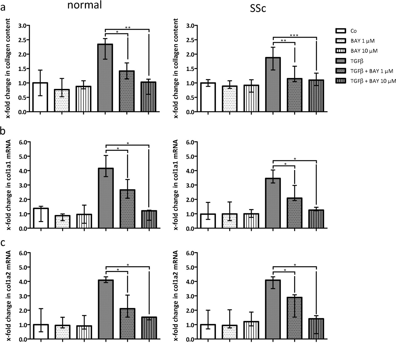
We found that sGC stimulation with BAY 41-2272 effectively reduced collagen release in fibroblasts from both healthy individuals and patients with SSc dose-dependently (figure 1A). In detail, BAY 41-2272 at doses of 1 µmol/l and 10 µmol/l inhibited the increase of the collagen content by 69.1% (p=0.033) and 98.1% (p=0.006) in dermal fibroblasts from healthy individuals. Similarly in SSc dermal fibroblasts, collagen content was diminished by 86.9% (p=0.012) and 87.5% (p<0.001) at doses of 1 µmol/l and 10 µmol/l. In accordance with collagen release, mRNA levels for procollagens col1a1 and col1a2 were reduced upon sGC stimulation (figure 1B).

Stimulation of soluble guanylate cyclase (sGC) with BAY 41-2272 reduces transforming growth factor (TGF)β-dependent collagen release by fibroblasts from healthy individuals and patients with systemic sclerosis (SSc). BAY 41-2272 was applied in doses of 1 µmol/l and 10 µmol/l to either unstimulated or TGFβ-stimulated fibroblasts (final TGFβ concentration: 10 ng/ml). All experiments are expressed as x-fold of the unstimulated control. (A) Collagen content in the supernatant as measured by Sircol assay. (B) Col1a1 mRNA levels with β-actin as internal control. (C) Col1a2 mRNA levels with β-actin as internal control. n=8 for all experiments.
sGC stimulation inhibits progressive bleomycin-induced skin fibrosis
After we had demonstrated that sGC stimulation inhibited the release of collagen in vitro, we examined its effects on the development and progression of fibrosis in the mouse model of bleomycin-induced dermal fibrosis. When challenged with bleomycin, mice developed skin thickening by 71.5% (p<0.001) compared with NaCl-injected controls. Treatment with the sGC stimulator prevented skin thickening by 51.7% (p<0.001) at 1 mg/kg twice a day and 79.7% (p<0.001) at 3 mg/kg twice a day (figure 2A,B). Stimulation of the sGC also prevented the increase of dermal hydroxyproline content and myofibroblast counts in a dose-dependent manner (figure 2C,D). Thus, sGC stimulation potently inhibited fibroblast activation and progressive fibrosis in bleomycin-induced fibrosis as a model of inflammation-driven fibrosis.

Stimulation of soluble guanylate cyclase (sGC) with BAY 41-2272 prevents the development of bleomycin-induced dermal fibrosis in a dose-dependent manner. (A) Trichrome staining with blue staining for collagens. Pictures are shown at 100-fold magnification. (B) Skin thickening as determined by trichrome staining. Values are expressed in relation to skin thickness of NaCl-treated and sham-treated mice. (C) Hydroxyproline (HP) content expressed in relation to HP content of NaCl-treated and sham-treated mice. (D) α-Smooth muscle actin (α-SMA)-positive myofibroblasts expressed in relation to α-SMA counts in NaCl- treated and sham-treated mice. All groups consisted of 10 mice each.
sGC stimulation induces regression of bleomycin-induced skin fibrosis
Having shown that sGC stimulation prevents the development of fibrosis, we next investigated whether it is also effective in the treatment of established fibrosis. We therefore used a modified bleomycin model, in which the onset of treatment with the sGC stimulator started after dermal fibrosis was already established. In this model, we observed a progression of fibrosis upon prolonged bleomycin treatment (figure 3A). Treatment with BAY 41-2272 at a dose of 3 mg/kg twice a day during the last 3 weeks of bleomycin treatment inhibited this progression despite continuing bleomycin injections (p=0.006; compared with sham-treated mice challenged with bleomycin for 6 weeks). The dermal thickness even decreased below pretreatment levels represented by mice treated with bleomycin for 3 weeks followed by NaCl for 3 weeks (reduction by 34.1%; p=0.006; figure 3A). Stimulation of the sGC was also effective in reducing the hydroxyproline content and the myofibroblast counts below the results of the 3-week bleomycin/3-week NaCl group (p=0.006 and p=0.07; figure 3B,C), demonstrating that sGC stimulation prevents progression of fibrosis and also induces regression of established inflammation-dependent fibrosis.

Stimulation of soluble guanylate cyclase (sGC) induces the regression of established dermal fibrosis. (A) Skin thickening as determined by trichrome staining. Values are expressed in relation to skin thickness of NaCl- treated and sham-treated mice. (B) Hydroxyproline (HP) content expressed in relation to hydroxyproline content of NaCl- treated and sham-treated mice. (C) α-Smooth muscle actin (α-SMA)-positive myofibroblasts expressed in relation to α-SMA counts in NaCl-treated and sham-treated mice. All groups consisted of five mice each.
sGC stimulation inhibits progressive skin fibrosis in Tsk-1 mice
To evaluate the antifibrotic potential of sGC stimulation in an inflammation-independent model of fibrosis, we used Tsk-1 mice. Stimulation of sGC by BAY 41-2272 was highly effective in preventing progressive hypodermal thickening in a dose-dependent manner. BAY 41-2272 at 1 mg/kg twice a day reduced hypodermal thickening by 37.0% (p<0.001) and, at a dose of 3 mg/kg twice a day, by 56.8% (p<0.001; figure 4A,B). These results were confirmed by reduction of the hydroxyproline content and myofibroblast counts upon treatment with low and high doses of the sGC stimulator (figure 4C,D).
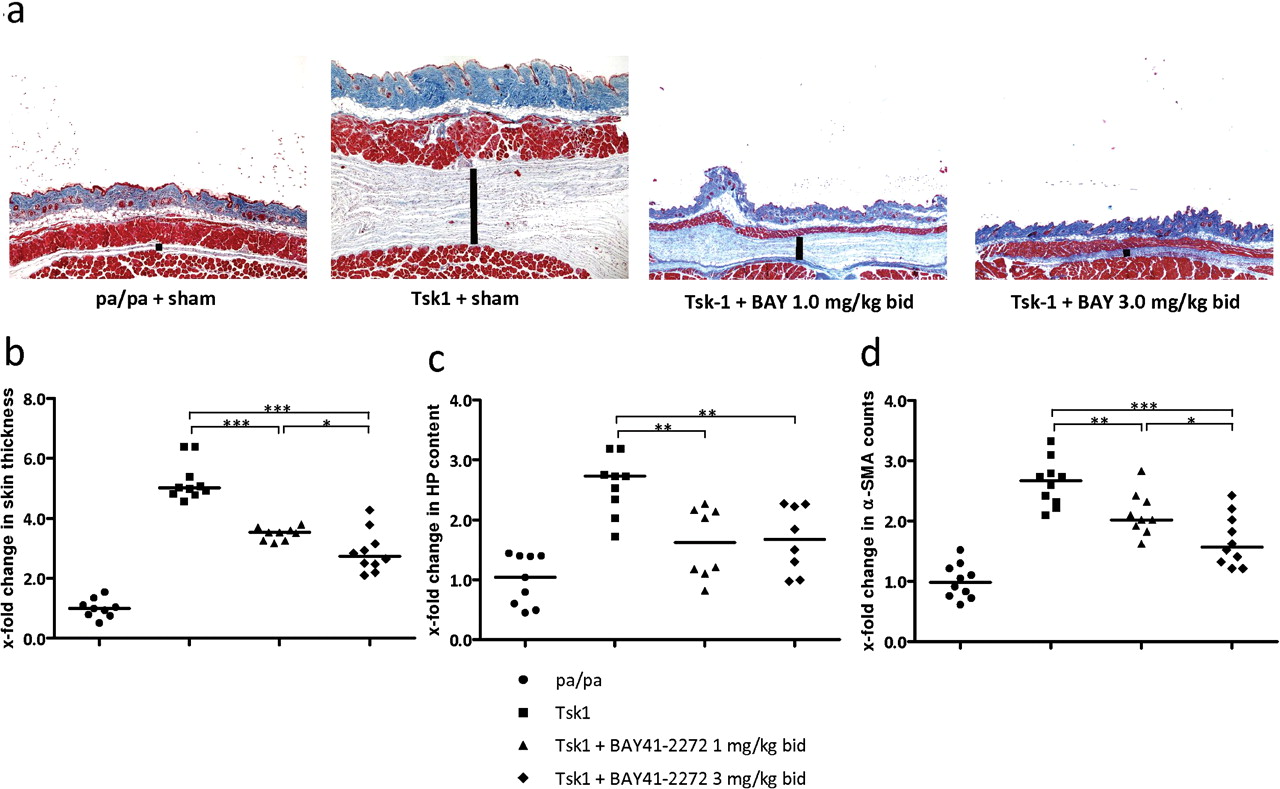
Stimulation of soluble guanylate cyclase (sGC) with BAY 41-2272 prevents the development of spontaneous fibrosis in Tsk-1 mice. (A) Trichrome staining with blue staining for collagens. Figures are shown at 100-fold magnification. (B) Skin thickening as determined by trichrome staining. Values are expressed in relation to skin thickness of sham-treated pa/pa mice. (C) Hydroxyproline (HP) content expressed in relation to HP content of sham-treated pa/pa mice. (D) α-Smooth muscle actin (α-SMA) positive myofibroblasts expressed in relation to α-SMA counts in sham-treated pa/pa mice. All groups consisted of 10 mice each.
sGC stimulation is effective in the treatment of established, inflammation-independent fibrosis
We also evaluated whether sGC stimulators induce regression of fibrosis in the Tsk-1 model. In sham-treated Tsk-1 mice, hypodermal thickening progressed from 5 weeks to 10 weeks of age (p=0.006; figure 5A). Treatment with the sGC stimulator BAY 41-2272, however, prevented the progression of fibrosis, and even reduced hypodermal thickness below that of 5-week-old Tsk-1 mice (p=0.004) with reductions of 68.1% (compared with skin thickness at 10 weeks of age) and 47.7% (compared with 5 weeks of age). Consistently, hydroxyproline content and myofibroblast counts were also lower in 10-week-old Tsk-1 mice treated with BAY 41-2272 than in sham-treated 5-week-old Tsk-1 mice (p=0.016 and p=0.004, figure 5B,C) demonstrating that sGC stimulators are effective in preventing and treating fibrosis in both inflammatory and inflammation-independent models of dermal fibrosis.

Stimulation of soluble guanylate cyclase (sGC) with BAY 41-2272 induces regression of skin fibrosis in Tsk-1 mice. (A) Hypodermal thickening as determined by trichrome staining. Values are expressed in relation to skin thickness of sham-treated pa/pa mice. (B) Hydroxyproline (HP) content expressed in relation to HP content of sham-treated pa/pa mice. (C) α-Smooth muscle actin (α-SMA)-positive myofibroblasts expressed in relation to α-SMA counts in sham-treated pa/pa mice. All groups consisted of five mice each.
sGC stimulation is well tolerated and does not cause clinically relevant changes of systemic blood pressure in relevant antifibrotic doses
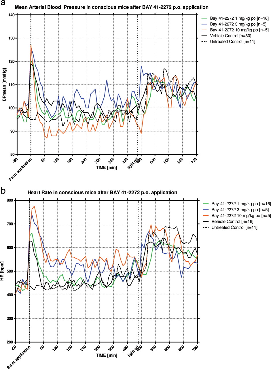
Mice treated with the sGC stimulator BAY 41-2272 appeared healthy with normal activity, behaviour and texture of the fur. To exclude the possibility that antifibrotic doses of sGC stimulators are accompanied by hypotensive effects, and to determine whether antifibrotic effects are driven by systemic haemodynamic effects, we performed telemetric blood pressure and heart rate measurements. The sGC stimulator BAY 41-2272 caused a dose-dependent decrease in blood pressure accompanied by compensatory heart rate increase at 3 and 10 mg/kg (figure 6A,B). However, no relevant alterations in blood pressure and heart rate occurred during treatment with 1 mg/kg BAY 41-2272, a dose that showed antifibrotic efficacy in SSc models. At a dose of 10 mg/kg, which exceeded the doses we used in the fibrosis models, blood pressure levels dropped and the heart rates rose, indicating significant systemic haemodynamic effects. Hence, while very high doses of the sGC stimulator caused the expected systemic decrease in blood pressure with reflex tachycardia, sGC stimulation at antifibrotic doses was well tolerated and did not exhibit relevant blood pressure and heart rate alterations.

Telemetry studies assessed blood pressure and heart rate over 24 h after treatment with BAY 41-2272 in conscious mice. Non-treated and sham-treated CD1 mice served as negative controls. BAY 41-2272 was applied at 09:00 in doses of 1 mg/kg, 3 mg/kg and 10 mg/kg by mouth in tylose suspension. (A) Mean arterial blood pressure levels in mm Hg after treatment with placebo (tylose suspension), 1, 3 and 10 mg/kg BAY 41-2272 given as tylose suspension. (B) Heart rates in beats per minute after treatment with placebo (tylose suspension), 1, 3 and 10 mg/kg BAY 41-2272 given as tylose suspension.
Discussion
We demonstrated that sGC stimulation is highly effective and well tolerated in various models of fibrosis. Stimulation of sGC with BAY 41-2272 has potent antifibrotic effects in bleomycin-induced dermal fibrosis and Tsk-1 mice, representing inflammatory and inflammation-independent dermal fibrosis. In both models, sGC stimulation prevents progressive dermal fibrosis, and also induces the regression of established fibrosis. Therefore, sGC stimulators may be effective in various stages of fibrosis. These include inflammatory stages, which often occur early in the disease course, as well as late stages of the disease when fibrosis has already become manifest. Given its efficacy in a variety of different SSc models, which implies a broad antifibrotic potential of sGC stimulators and a pivotal antifibrotic role of cGMP increase, the antifibrotic activity of these compounds may be transferred to other organ systems, including the liver and kidneys.17 18 The broad antifibrotic effects of sGC stimulators in different models and settings might be explained by their mode of action: we demonstrated that sGC stimulation directly and potently targets the release of extracellular matrix from fibroblasts, the key cellular players of fibrosis.
In view of the well-known vasoactive properties of sGC in higher doses, we directly compared antifibrotic effects and systemic haemodynamic activity. Our telemetry experiments showed that BAY 41-2272 does not cause a systemic decrease of blood pressure at doses that have significant antifibrotic effects. Only at very high doses, which we did not use in the fibrosis experiments, did a decrease in blood pressure and reflex tachycardia become evident. In line with our findings, a recent phase II clinical trial in patients with PAH demonstrated that sGC stimulation with riociguat, a chemically related compound, is well tolerated. While riociguat effectively improved haemodynamics in the pulmonary arteries, no serious adverse events occurred, and mild side effects had resolved by study completion.8 Of note, PAH is a common and serious disease manifestation in SSc, and sGC stimulation may be beneficial in these patients as well. In view of its vasodilatory activity, sGC stimulation might be effective on large vessels including the pulmonary arteries and also on peripheral vessels and capillaries. Thus, sGC stimulators might also be beneficial for small-vessel vasculopathy in SSc. Since our models, however, are not representative for SSc vascular disease, this hypothesis still needs to be proved with additional studies.
NO-independent, direct sGC stimulation may have several advantages over other vasoactive treatments that target sGC-cGMP signalling at other levels, such as PDE5 inhibitors and inhaled NO. Because PDE5 inhibition only prevents degradation of cGMP but does not increase its formation, PDE5 inhibition requires endogenous NO formation with a baseline production of cGMP. Indeed, failure of PDE5 inhibitor treatment in a subset of patients with PAH has been suggested to result from insufficient NO-induced cGMP production at baseline.19 In addition, antifibrotic efficacy in dermal fibrosis, as we observed for sGC stimulation in experimental fibrosis, is unlikely, because PDE5 expression is low in dermal fibroblasts.20 This is of paramount importance since high PDE5 activity might limit the effect of NO donors and sGC stimulators via effective cGMP degradation. Inhaled NO, which is clinically used in pulmonary hypertension after surgery and in persistent pulmonary hypertension in the newborn, has only short-term efficacy owing to rapid adaptation, and harbours the risk of rebound hypertension after withdrawal.21 Moreover, NO in high concentration reacts with oxygen to form reactive oxygen species, which can activate fibroblasts and may contribute to the progression of the disease.1 Bypassing NO via direct sGC stimulation, however, overcomes these risks, since application of additional NO beyond the endogenous levels is not necessary.
Tolerability is a major concern in the development of antifibrotic treatments in SSc. Experience suggests that new antifibrotic treatments can interfere with other disease manifestations, particularly small-vessel vasculopathy associated with Raynaud's phenomenon and digital ulcers. For example, the clinical use of tyrosine kinase inhibitors in SSc seems to be limited by vascular side effects—in particular, massive peripheral oedema, which are far less prominent in the treatment of other diseases such as chronic myeloid leukaemia.22 Thus, optimal antifibrotic treatments for patients with SSc should effectively treat both, fibrosis and vascular disease or, at least, not cause worsening of the patients' vascular manifestations. Accumulating evidence from clinical trials indicates that sGC stimulators might soon extend our arsenal of treatments for PAH. Based on its mode of action, sGC stimulation might also have beneficial effects on peripheral vascular disease, but this needs further confirmation. If this concept holds true, sGC stimulators will be promising candidates for a simultaneous targeted treatment of fibrosis and vascular disease in SSc.
Taken together, we showed that sGC stimulation is effective in various experimental models of dermal fibrosis. These antifibrotic effects occur in well tolerated doses without haemodynamic effects. In view of its mode of action and first clinical studies in the treatment of PAH, sGC stimulators might be used to treat fibrosis and also improve vascular manifestations in SSc. Since sGC modulators, such as riociguat, are already in phase III clinical trials, stimulation of sGC may be a promising new approach for targeted treatments in SSc.
Acknowledgments
The authors thank Maria Halter and Anna-Maria Herrmann for excellent technical assistance.
References
Supplementary materials
Supplementary Data
This web only file has been produced by the BMJ Publishing Group from an electronic file supplied by the author(s) and has not been edited for content.
Files in this Data Supplement:
- Web Only Data - This web only file has been produced by the BMJ Publishing Group from an electronic file supplied by the author(s) and has not been edited for content.
Footnotes
-
Funding CB: Research scholar at the Interdisciplinary Center of Clinical Research (IZKF) in Erlangen; grant from the Erlanger Leistungsbezogene Anschubfinanzierung und Nachwuchsförderung (ELAN). MT: CMH research projects No 00000023728. JHWD: grant A40 of the Interdisciplinary Center of Clinical Research (IZKF) in Erlangen; grants from the Deutsche Forschungsgesellschaft (grants DI 1537/1-1, DI 1537/2-1, DI 1537/4-1, AK 144/1-1, SCHE 1583/7-1, and DI 1537/5-1); and the Career Support Award of Medicine of the Ernst Jung Foundation.
-
Competing interests JHWD is member of the advisory board of Bayer HealthCare and has received speaker fees from Bayer HealthCare; and is also stock owner of 4D Science GmbH, which cooperates with Bayer HealthCare. CH-D, GvD, PS are employees of Bayer HealthCare.
-
Ethics approval Ethical committee of the Universit of Erlangen-Nuremberg.
-
Provenance and peer review Not commissioned; externally peer reviewed.
-
Correction notice This article has been corrected since it was published Online First. Under funding an additional grant was included for JHWD.