Article Text

Abstract
Objective The clinical features of rheumatic patients with coronavirus disease 2019 (COVID-19) have not been reported. This study aimed to describe the clinical features of COVID-19 in rheumatic patients and provide information for handling this situation in clinical practice.
Methods This is a retrospective case series study. Deidentified data, including gender, age, laboratory and radiological results, symptoms, signs, and medication history, were collected from 2326 patients diagnosed with COVID-19, including 21 cases in combination with rheumatic disease, in Tongji Hospital between 13 January and 15 March 2020.
Results Length of hospital stay and mortality rate were similar between rheumatic and non-rheumatic groups, while the presence of respiratory failure was more common in rheumatic cases (38% vs 10%, p<0.001). Symptoms of fever, fatigue and diarrhoea were seen in 76%, 43% and 23% of patients, respectively. There were four rheumatic patients who experienced a flare of rheumatic disease during hospital stay, with symptoms of muscle aches, back pain, joint pain or rash. While lymphocytopaenia was seen in 57% of rheumatic patients, only one patient (5%) presented with leucopenia in rheumatic cases. Rheumatic patients presented with similar radiological features of ground-glass opacity and consolidation. Patients with pre-existing interstitial lung disease showed massive fibrous stripes and crazy-paving signs at an early stage. Five rheumatic cases used hydroxychloroquine before the diagnosis of COVID-19 and none progressed to critically ill stage.
Conclusions Respiratory failure was more common in rheumatic patients infected with COVID-19. Differential diagnosis between COVID-19 and a flare of rheumatic disease should be considered.
Trial registration number ChiCTR2000030795.
- autoimmune diseases
- autoimmunity
- inflammation
This article is made freely available for use in accordance with BMJ’s website terms and conditions for the duration of the covid-19 pandemic or until otherwise determined by BMJ. You may use, download and print the article for any lawful, non-commercial purpose (including text and data mining) provided that all copyright notices and trade marks are retained.
https://bmj.com/coronavirus/usageStatistics from Altmetric.com
Key messages
What is already known about this subject?
COVID-19 has been characterised as a global pandemic, affecting over 200 countries and territories.
Most rheumatic patients have immune dysregulation and rely on immunosuppressive agents to control disease progression.
Some antirheumatic agents have been proposed to possess beneficial effects in COVID-19.
What does this study add?
We describe, for the first time, the clinical features of COVID-19 in rheumatic patients.
Respiratory failure was more common in rheumatic patients infected with COVID-19.
CT, pathogen test and disease-specific symptoms provide important information for differential diagnosis between COVID-19 and a flare of rheumatic disease.
How might this impact on clinical practice or future developments?
This study describes the clinical features of rheumatic patients infected with COVID-19.
Differential diagnosis between COVID-19 and a flare of rheumatic disease should be considered during this pandemic.
Introduction
Coronavirus disease 2019 (COVID-19) caused by severe acute respiratory syndrome coronavirus 2 (SARS-CoV-2) has progressed rapidly into a worldwide pandemic. The estimated mortality ranges from about 1% to as high as 20% in different regions.1
Functional and effective antivirus immune response is critical for the defence against SARS-CoV-2. Subsets of immune effector cells responsible for antiviral immunity, such as antibody-secreting cells (CD3−CD19+CD27hiCD38hi) and activated follicular helper T cells (CD4+CXCR5+ICOS+PD-1+), were increased preceding the clearance of SARS-CoV-2 and symptomatic recovery, suggesting that antiviral immune response is crucial for SARS-CoV-2 clearance.2 The progression to acute respiratory distress syndrome in COVID-19 was associated with the manifestation of excessive release of inflammatory cytokines, especially the upregulation of interleukin (IL)-2, IL-6, IL-10, IL-12 and tumour necrosis factor-α (TNF-α).3 SARS-CoV-2-infected patients, especially critically ill patients, had increased plasma levels of inflammatory cytokines, in parallel with an increase in a variety of tissue damage markers such as aspartate aminotransferase, alanine aminotransferase, creatine kinase-MB, hypersensitive troponin I and lactate dehydrogenase.3–5
As of 15 March 2020, a total of 21 patients with rheumatic diseases were diagnosed with COVID-19 at Tongji Hospital (Wuhan, China), a hospital reconstructed and designated to care for moderately to critically ill patients with COVID-19. In this retrospective observational study, we describe the clinical characteristics and medication history prior to hospitalisation for COVID-19 in these patients, aiming to delineate the clinical, laboratory and radiological features, outcome, and medical history of rheumatic patients with laboratory-confirmed infection of SARS-CoV-2.
Methods
Study design and patients enrolled
This is a retrospective case series study. A total of 21 rheumatic patients out of the 2326 patients with COVID-19 admitted to Tongji Hospital between 13 January and 15 March 2020 were enrolled in this study. All these patients were diagnosed with COVID-19 according to the seventh edition of the Guidance for Corona Virus Disease 2019 released by the National Health Commission of the People’s Republic of China (www.nhc.gov.cn/yzygj/s7653p/202003/46c9294a7dfe4cef80dc7f5912eb1989.shtml). Clinical classification (mild, moderate, severe and critically ill) was defined as described earlier.6 Written informed consent was waived due to the rapid spread of this emerging infection.
Data collection
Deidentified data on demographics (gender, age), clinical (medical history, admission/discharge time, classification of disease severity, symptoms and signs, respiratory failure status, disease outcome and so on), radiological (characteristics of chest CT scans) and laboratory information (leucocyte/lymphocyte counts, haemoglobin, serum cytokines and so on), and treatments (before and after COVID-19 diagnosis) were obtained from the electronic hospital information system of Tongji Hospital. CT images of the 21 rheumatic patients were obtained from the Department of Radiology in Tongji Hospital. Presence of radiological features (including ground-glass opacity (GGO), pleural lesions and air bronchogram sign) in each case was independently evaluated by at least two radiologists.
Laboratory measurements of SARS-CoV-2
RNA detection
A real-time reverse transcription PCR assay was applied to detect the presence of SARS-CoV-2 RNA in throat swab samples. Extraction of SARS-CoV-2 RNA from throat swab samples was done with respiratory sample RNA isolation kit (Biogerm, Shanghai, China) within 2 hours. Two target genes, including open reading frame 1ab (forward primer: CCCTGTGGGTTTTACACTTAA; reverse primer: ACGATTGTGCATCAGCTGA) and nucleocapsid protein N (forward primer: GGGGAACTTCTCCTGCTAGAAT; reverse primer: CAGACATTTTGCTCTCAAGCTG), were simultaneously amplified and tested. SARS-CoV-2 nucleic acid detection kit was used according to the manufacturer’s protocol (Shanghai Biogerm Medical Technology). A cycle threshold value less than 37 was defined as a positive test result, and a cycle threshold value of 40 or more was defined as a negative test. A further confirmation by retesting should be made when a cycle threshold value located in the range between 37 and 40. The detection limit for the SARS-CoV-2 nucleic acid detection kit is 1000 copies/mL, and there were no cross-reactions with human genome DNA or nucleic acids from other common human respiratory viruses (including HCoV-OC43, HCoV-HKU1, HCoV-229E, MERS-CoV, SARS-CoV, HCoVNL63, adenovirus, respiratory syncytial virus A and B, H1N1 influenza virus, H3N2 influenza virus, H5N1 influenza virus, H7N9 influenza virus, H9N2 influenza virus, Epstein-Barr virus, Mycoplasma pneumoniae, influenza B virus and so on).
Antibody detection
Detection of IgM/IgG anti-SARS-CoV-2 antibody in sera samples was performed using a chemiluminescence immunoassay kit for 2019-nCov detection from Xiamen Innodx Biotech, according to the manufacturer’s instruction.
Statistical analysis
Categorical variables were presented as numbers and percentages, and continuous variables as median±IQR. Comparison of ordered categorical variables between two different groups was done by Mann-Whitney U test, while comparison of proportions for unordered categorical variables was realised using χ2 test. Statistical analyses were conducted using SPSS software (V.21) or R software (V.3.6.2). Pie plots, upset plot and stacked bar plots were drawn with R package ggplot2 and UpSetR. Grouped bar plots and heatmap were plotted with GraphPad Prism software (V.8). A p value less than 0.05 was regarded as statistically significant.
Results
Basic information and disease severity of rheumatic patients in this study
Between 13 January and 15 March 2020, 2326 patients with COVID-19 were admitted to Tongji Hospital, 21 of whom were patients suffering from rheumatic diseases. In these 21 rheumatic patients, 20 were SARS-CoV-2 RNA-positive, while the only RNA-negative case showed positive IgM and IgG antibody against SARS-CoV-2. There were eight rheumatoid arthritis (RA), four systemic lupus erythematosus (SLE), three primary Sjögren’s syndrome (pSS), two undifferentiated connective tissue disease, one polymyalgia rheumatica (PMR), one juvenile idiopathic arthritis (JIA) and two ankylosing spondylitis (AS) cases (figure 1A). Compared with non-rheumatic patients, the ratio of female patients was significantly higher in the rheumatic disease group (rheumatic group vs non-rheumatic group: 81% vs 50%, χ2=6.73, p=0.009; figure 1B). The age structures of rheumatic group and non-rheumatic group were similar (|Z|=0.79, p=0.43); most patients in both groups were more than 40 years old (rheumatic group: 90%; non-rheumatic group: 89%) at the time of diagnosis with COVID-19 (figure 1C).

Basic information on rheumatic cases enrolled in this study. Ratio of rheumatic cases to the total number of patients with COVID-19 admitted to Wuhan Tongji Hospital (China), from 13 January 2020 to 15 March 2020 (left). Our series in this study consisted of eight RA cases, four SLE, three pSS, two UCTD, two AS, one JIA and one PMR (A). Gender distribution (B), age distribution (C), ratio of respiratory failure (D), hospitalisation time distribution (E) and mortality rate (F) of patients with COVID-19 with and without rheumatic diseases. Comparison of ordered categorical variables between two different groups was done using Mann-Whitney U test, while comparison of proportions for unordered categorical variables was realised using χ2 test. P<0.05 was regarded as statistically significant. AS, ankylosing spondylitis; JIA, juvenile idiopathic arthritis; PMR, polymyalgia rheumatica; pSS, primary Sjögren’s syndrome; RA, rheumatoid arthritis; SLE, systemic lupus erythematosus; UCTD, undifferentiated connective tissue disease.
More patients in the rheumatic group presented with respiratory failure, compared with those in the non-rheumatic group (rheumatic group vs non-rheumatic group: 38% vs 10%, χ2=13, p<0.001; figure 1D). Despite no statistical difference observed in the distribution of hospitalisation time between the two groups (|Z|=0.74, p=0.46), there was a tendency for the rheumatic group to have more patients with a length of hospital stay of 20 days or more (rheumatic vs non-rheumatic: 57% vs 47%; figure 1E). However, no difference in mortality rates was observed between rheumatic and non-rheumatic groups (9.52% vs 9.54%, χ2 <0.001, p>0.99; figure 1F).
Clinical characteristics and medication of rheumatic patients suffering from COVID-19
Symptoms
Fever was observed in 18 out of the 21 rheumatic patients (figure 2A). Of the cases with fever, the highest temperatures ranged between 38.1℃ and 39℃ in 61%, while temperatures higher than 39℃ were observed only in 11.1% of cases (figure 2B). Most of the rheumatic cases presented with at least one respiratory symptom (90%), but the pattern differed in different patients: cough, expectoration and dyspnoea were present in 57%, 43% and 67% of rheumatic patients, respectively. Besides respiratory symptoms, it is worth mentioning that a large portion of rheumatic cases in our study presented with fatigue and/or diarrhoea, which could be seen in both the active status of rheumatic diseases and COVID-19 (figure 2A,C). Fatigue and diarrhoea have been noted as initial symptoms in many COVID-19 cases. Fatigue is also one of the most common complaints in autoimmune rheumatic disease,7 while diarrhoea could be caused either by certain antirheumatic medications such as leflunomide8 or a rheumatic disease itself such as Behçet disease and Ehlers-Danlos syndrome.9

Symptoms and signs of rheumatic patients with COVID-19. Ratio of symptoms and signs presented by rheumatic patients with COVID-19: the lower panel shows the composition structure, while the upper panel shows the concrete percentage of each component of the corresponding parameters (A). Structure of the highest temperature of patients with fever (B) and ratio of cases who presented with at least one respiratory symptom (C).
Laboratory indices
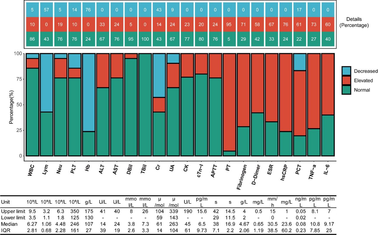
Information on laboratory indices is shown in figure 3. Unlike the lymphocyte count in peripheral blood which was downregulated in 57% of rheumatic cases in our study, leucopenia was rarely seen (5%). Elevated count of neutrophils was observed in nearly one-fifth of patients, while 16 (76%) of 21 patients showed anaemia (haemoglobin, median±IQR: 107±27 g/L). Liver dysfunction was less common. Alanine aminotransferase and aspartate aminotransferase were increased, respectively, in 7 (33%) and 5 (24%) patients, while elevation of serum bilirubin level was nearly absent (only 1 (5%) patient showed elevated serum direct bilirubin (DBil) level (9.1 mmol/L)). Elevated creatine kinase (CK) and troponin I (Tn-I) were observed in 3 (23%) of 13 and 4 (20%) of 20 patients. The highest level of CK and cardiac Tn-I presented, respectively, in patient 4 (CK: 1248 U/L) and patient 5 (124.8 pg/mL), who eventually passed away.

Laboratory indices of rheumatic patients with COVID-19. The middle panel shows the composition structure, the upper panel shows the concrete percentage of each component, while the lower panel shows the unit, upper/lower limit, median value and IQR of the corresponding parameters. ALT, alanine aminotransferase; APTT, activated partial thromboplastin time; AST, aspartate aminotransferase; CK, creatine kinase; Cr, creatinine; cTn-I, Cardiac troponin- I; DBil, direct bilirubin; ESR, erythrocyte sedimentation rate; Hb, hemoglobin; hsCRP, high-sensitivity C-reactive protein; IL-6, interleukin-6; Lym, lymphocyte; Neu, neutrophil; PCT, procalcitonin; PLT, platelet; PT, prothrombin time; TBil, total bilirubin; TNF-α, tumour necrosis factor-α; UA, uric acid; WBC, white blood cell.
System of coagulation and anticoagulation seemed to be activated in most rheumatic cases: increased prothrombin time and elevated fibrinogen level were, respectively, observed in 95% and 71%, while elevated D-dimer level was observed in 58% of rheumatic cases. The internal milieu of rheumatic cases was relatively proinflammatory: 11 (73%) and 9 (60%) of 15 cases, whose cytokine levels had been detected, showed elevated serum TNF-α (median±IQR: 10.8±7.85 pg/mL) and IL-6 (9.17±25 pg/mL) levels, which corresponded to a similar portion of cases with increased erythrocyte sedimentation rate (ESR) and C reactive protein (CRP) levels. Procalcitonin, which may indicate potential bacterial infection, was also elevated in 11 (61%) of 18 cases.
Radiological features
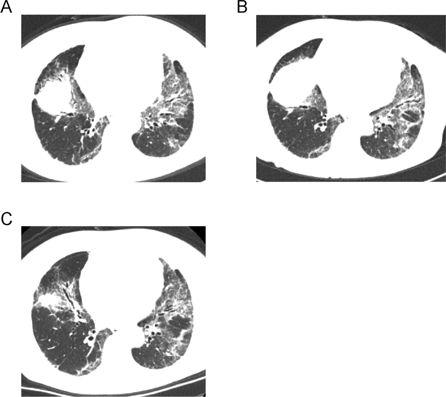
The most common sign in rheumatic patients with COVID-19 was GGO, which was observed in 86% of rheumatic cases. The presence of patchy shadow (57%) and pleural lesions (29%) was also noticed. Pleural effusion, which is rarely seen in non-rheumatic patients with COVID-19, was observed in 2 (10%) of 21 cases. Signs of GGO, consolidation and patchy shadow, in combination with massive fibrous stripes and crazy-paving signs, were observed at early stage in both cases with pre-existing RA-associated interstitial lung disease (ILD) (patients 6 and 7). The crazy-paving signs and fibrous stripes remained after the absorption of GGO and consolidation on CT images, suggesting a manifestation of ILD (figure 4).

Chest CT of a rheumatoid arthritis patient with COVID-19 and interstitial lung disease. (A) CT image on the day of diagnosis showing ground-glass opacity (GGO), consolidation and fibrous stripes in both lungs. There was crazy-paving sign in the left upper lobe. (B) CT scan image 20 days after diagnosis showed that the GGO was partially absorbed and the fibrous stripes/crazy-paving sign had no significant changes. (C) CT scan image 36 days after diagnosis showing a significant absorption of GGO, while the fibrous stripes and crazy-paving sign remained unchanged.
Medication
Before the diagnosis of COVID-19, 6 (29%) of 21 patients were administered glucocorticoids, while 5 (24%) patients were administered hydroxychloroquine (HCQ). Non-steroidal anti-inflammatory drugs (NSAIDs), methotrexate, leflunomide, tripterygium and glycosides of Tripterygium wilfordii Hook were also administered according to the type and status of their underlying rheumatic diseases. During hospital stay, 4 (19%) of 21 cases had undergone flare of rheumatic disease, and therapeutic agents including HCQ, NSAID, mycophenolate mofetil and high-dose prednisone (40 mg/day) were added to help attenuate the disease activity. Among these four cases with rheumatic flare, patient 6 (RA) presented with pain in involved joints, patient 12 (SLE) presented with rash and haemolytic anaemia, patient 19 (AS) presented with back pain and ankle pain, and patient 21 (PMR) presented with muscle aches. During the hospital stay, 10 cases received glucocorticoids with an average dosage of 37 mg/day (converted to the corresponding dosage of prednisone), for the treatment of COVID-19. Three doses of tocilizumab (TCZ) were used in patient 1 with RA in order to prevent the possibility of cytokine storm (80 mg, 160 mg and 80 mg, respectively). After three doses of TCZ treatment, high-sensitivity C-reactive protein (hsCRP) level reduced from 253 to 5 mg/dL and serum IL-6 reduced from 483.8 to 251 pg/mL, suggesting that TCZ treatment is effective in controlling the RA activity and preventing potential cytokine storm. None of the five cases with long-term HCQ treatment for antirheumatic therapy progressed to critically ill, and three of them (60%) remained moderately ill until discharge. Besides these five cases, two patients (patients 12 and 21) received HCQ treatment after hospitalisation due to a flare of rheumatic disease or as anti-COVID-19 therapy. All these patients achieved full recovery (figure 5). Thirteen out of the 21 patients had comorbid conditions, such as hypertension, coronary heart disease, diabetes and chronic obstructive pulmonary disease. Hypertension was the most common comorbidity, affecting 7 out of the 21 patients (figure 5).

Medication of rheumatic patients with COVID-19 before and during the hospitalisation. Heatmap in the upper panel records the medication of each case before the diagnosis of COVID-19. Heatmap in the lower panel records the information during hospitalisation, including the flare condition of rheumatic disease, medication for flare, glucocorticoids usage (converted to the corresponding dosage of prednisone) during hospitalisation, and some other parameters recording the process of hospitalisation and outcome. Mean of different colours illustrated by legends located on the right side of each heatmap. Numbers shown in different units are concrete values of the corresponding indices of specific patients. ACI, atherosclerotic cerebral infarction; AS, ankylosing spondylitis; CEG, chronic erosive gastritis; CHD, coronary heart disease; COPD, chronic obstructive pulmonary disease; DM, diabetes mellitus; EP, epilepsy; HBP, high blood pressure; HCQ, hydroxychloroquine; HCV, hepatitis C virus; JIA, juvenile idiopathic arthritis; MMF, mycophenolate mofetil; NSAIDs, non-steroidal anti-inflammatory drugs; PMR, polymyalgia rheumatica; pSS, primary Sjögren’s syndrome; RA, rheumatoid arthritis; SLE, systemic lupus erythematosus; UCTD, undifferentiated connective tissue disease.
Discussion
The condition of rheumatic patients under the COVID-19 pandemic is worrisome. Our data suggest that rheumatic patients may present with more severe symptoms when infected with COVID-19, with a higher risk of respiratory failure compared with non-rheumatic group. Several recent studies reported that proinflammatory cytokines including IL-6, TNF-α and IL-1, which have been shown as the pathogenic factors in many rheumatic diseases, may also be responsible for the tissue damage in multiple organs in patients with COVID-19.10–13 The long-term treatment of glucocorticoids and/or immunosuppressive agents also increases the risk of infection of microbes.14 It is possible that COVID-19 and rheumatic condition can affect each other at the level of pathogenic mechanism, and finally lead to the catastrophic deterioration of patients’ health. Thus, identification of (active) rheumatic condition in COVID-19 should be taken seriously, and vice versa.
There are many common clinical characteristics between COVID-19 and a flare of rheumatic diseases, such as fatigue, fever, and abnormalities in laboratory tests (ESR, CRP, lymphocytopaenia and so on) and CT (GGO is also commonly presented in rheumatic patients with lung involvement). The presence of these symptoms may lead to a misinterpretation of rheumatic disease flare and further delay the recognition of COVID-19. The use of glucocorticoid may also cover up the symptom of fever, one of the most well-known symptoms of COVID-19 recognised by the public. In addition, several laboratory indices commonly used in the assessment of COVID-19, such as lymphocytopaenia and inflammatory biomarkers, are also very similar in both conditions. In our series, lymphocytopaenia was observed in 57% of cases, while elevation of CRP, ESR, IL-6 and TNF-α was observed in 76%, 67%, 60% and 73% of cases, respectively. Although it is difficult to distinguish the precise origin of these clinical features, in combination with other parameters (like immune-related parameters), these may provide some easily ignored but useful information indicating the presence of non-COVID-19 conditions.
Chest CT of rheumatic patients with COVID-19 should also be carefully evaluated. Generally, the radiological features on chest CT of rheumatic patients with COVID-19 were similar to those in non-rheumatic COVID-19 patients, including the presence of GGOs, patchy shadows and consolidation. However, lungs are also the target of many systemic diseases including rheumatic diseases. Connective tissue disease-associated interstitial lung disease (CTD-ILD) is commonly seen in RA, pSS, scleroderma and dermatomyositis.15 Thus, differential diagnosis should also be made to identify the origin of interstitial lesions. In our cases, we noticed an early presence of massive fibrous stripes and crazy-paving signs on CT images of patients with CTD-ILD, which were suggested to present at later stages.16 17 More importantly, these signs remained unchanged after absorption of GGO/consolidation and symptomatic recovery. Therefore, these features can be considered during the differential diagnosis between ILD and COVID-19.
How to balance the medication of antirheumatic and antivirus therapy is another important task in clinical practice. In this study, none of the five cases using long-term HCQ progressed to critically ill stage. In vitro studies and a recent human trial support the antiviral activity of chloroquine and HCQ.18–20 Their use in patients with COVID-19 has been shown to improve pneumonia symptoms and lab tests and decrease progression to severe/critical conditions in randomised clinical trials in China.21 22 Nevertheless, there were also reports showing no benefits,23 even hazardous effects,24 of chloroquine/HCQ in COVID-19. Some studies indicated that there was a high frequency of side effects (eg, cardiotoxicity) in chloroquine-treated or HCQ-treated patients with COVID-19, especially when used at a high dose.25 26 Therefore, the use of HCQ/chloroquine in COVID-19 remains controversial, and further investigations are required to assess the efficacy and safety of HCQ in COVID-19. In the current pandemic, there is an urgent need for timely investigations to assess the clinical course of rheumatic patients with COVID-19.
Biological disease-modifying antirheumatic drugs (DMARDs) such as TCZ have also been proposed to be beneficial in treating hyperinflammation status of severe COVID-19 cases.27 28 TCZ is a recombinant humanised, antihuman monoclonal antibody against soluble and membrane-bound IL-6 receptors which has been approved for the treatment of rheumatic disease, including RA and JIA.29 A recent study from Italy reported that only three patients were diagnosed with COVID-19, and all were mild in 530 rheumatic patients treated with biological DMARDs such as TCZ and anti-TNF agents.30 Another study also reported that none of the four confirmed and four highly suggestive cases of COVID-19 who were using biological or targeted synthetic DMARDs for RA developed severe respiratory complications or died.31 In our study, TCZ was prescribed to patient 1 three times during his hospital stay due to severe COVID-19 status and a significant elevation of serum IL-6. His symptoms of COVID-19 relieved, and no flare of RA happened during the hospital stay. He finally recovered and was discharged 15 days after admission. However, all these studies were observational and lack appropriate control populations. Ongoing randomised controlled trials may hopefully provide direct evidence for the safety and efficacy of TCZ in COVID-19.
There were some limitations to this study. First, this is a single-centre retrospective research (retrospective case series) with a relatively small sample size. Future investigations with a larger sample size and/or multicentre studies are needed to provide more information. Second, in all 21 cases, none had been receiving long-term medication of biological agents (eg, TCZ) or targeted small molecule inhibitors (eg, tofacitinib) before the diagnosis of COVID-19. Thus, we cannot provide information on the influence of these agents on the susceptibility and clinical progress of COVID-19. Third, we were not able to assess the risk of COVID-19 lung damage in rheumatic patients with pre-existing ILD due to the very limited cases with definite CTD-ILD history. Lastly, the diseases included in this study are a collective of rheumatic disease. Due to the limited number of patients in certain disease type, we did not subcategorise the rheumatic diseases when analysing some parameters.
In summary, our study described the clinical features of rheumatic patients infected with COVID-19. Demographic characteristics, medication history, disease severity, and laboratory and radiological features were summarised in our series, which may help the differential diagnosis between rheumatic disease flare and COVID-19 infection. In addition, this study may also provide some evidence on the use of antirheumatic drugs to treat rheumatic flare during the COVID-19 pandemic.
Acknowledgments
The authors would like to thank all patients and study site personnel for participating in this study.
References
Supplementary materials
Lay summary
Disclaimer : This is a summary of a scientific article written by a medical professional (“the Original Article”). The Summary is written to assist non medically trained readers to understand general points of the Original Article. It is supplied “as is” without any warranty. You should note that the Original Article (and Summary) may not be fully relevant nor accurate as medical science is constantly changing and errors can occur. It is therefore very important that readers not rely on the content in the Summary and consult their medical professionals for all aspects of their health care and only rely on the Summary if directed to do so by their medical professional. Please view our full Website Terms and Conditions.
Copyright © 2020 BMJ Publishing Group Ltd & European League Against Rheumatism. Medical professionals may print copies for their and their patients and students non commercial use. Other individuals may print a single copy for their personal, non commercial use. For other uses please contact our Rights and Licensing Team.
Footnotes
Handling editor Josef S Smolen
CY and SC contributed equally.
Correction notice This article has been corrected since it published Onilne First. The section heading has been changed to 'Epidemiology'.
Contributors Study design: LD, JZ, CY. Data collection: CY, SC, GS, HG, LZ, YH, WT, YuC, YY, XW, YuxC. Data analysis: CY, SC, JZ. Interpretation of findings: CY, JZ, LD. Preparation of manuscript: CY, SC, JZ. All authors read and approved the final manuscript.
Funding This work was supported by grants from the National Natural Science Foundation of China (81974254, 81670431 and 81771754), Tongji Hospital Clinical Research Flagship Program (no 2019CR206), and Hubei Chen Xiaoping Science and Technology Development Foundation Clinical Research Special Fund (CXPJJH11800005-07).
Competing interests None declared.
Patient and public involvement Patients and/or the public were not involved in the design, or conduct, or reporting, or dissemination plans of this research.
Patient consent for publication Not required.
Ethics approval The study was approved by the Institutional Review Board and Medical Ethics Committee of Tongji Hospital, Tongji Medical College of Huazhong University of Science and Technology.
Provenance and peer review Not commissioned; externally peer reviewed.
Data availability statement All data relevant to the study are included in the article or uploaded as supplementary information.