Article Text

Abstract
Objective Smoking can induce autoantibodies in persons who are genetically predisposed to rheumatoid arthritis. We investigated the association between smoking and antiphospholipid antibodies (aPL) in systemic lupus erythematosus (SLE), a question not previously addressed. Further, we explored the relationship between smoking, aPL and vascular events (arterial and venous, VE).
Methods In this cross-sectional study, clinical evaluation and questionnaire data were collected from 367 prevalent SLE patients. At the same time, we measured aPL (anticardiolipin (aCL), anti-β2 glycoprotein-1 (aβ2GP1) antibodies IgG/IgM/IgA, and lupus anticoagulant (LA)), and a large set of other SLE-associated autoantibodies for comparison. Association analyses using logistic regression models with smoking, (ever, former and current with never as reference) and antibody status as outcome variable were performed. As a secondary outcome, we investigated the associations between aPL, smoking and VE.
Results In multivariable-adjusted models ever, and in particular former, cigarette smoking was associated with the most pathogenic aPL; LA, aCL IgG and aβ2GP1 IgG. Other SLE-associated autoantibodies were not associated with smoking. The combination of smoking and aPL was strongly associated with VE. We noted a positive interaction between smoking-LA and smoking-‘triple aPL’ positivity for previous VE.
Conclusions We investigated a large set of commonly occurring autoantibodies in SLE, but only aPL were positively associated with a history of smoking. This association was especially apparent in former smokers. Among ever regular smokers who were aPL positive, we observed a strikingly high frequency of former VE. The underlying mechanisms and temporality between smoking, aPL and VE need further investigations.
- Systemic Lupus Erythematosus
- Cardiovascular Disease
- Epidemiology
- Antiphospholipid Antibodies
- Smoking
Statistics from Altmetric.com
Introduction
Systemic lupus erythematosus (SLE) is an autoimmune inflammatory disease, which can affect almost any organ in the body; most commonly joints, skin, kidneys and blood elements. The origin of SLE is unclear, but the autoimmunity of the disease is characterised by a perturbed immune response against self, including enhanced B-cell activity, and production of autoantibodies with diverse specificities. Genetic and environmental factors influence the risk of SLE.1 ,2 Gender and hormones are also important, given that 90% of affected individuals are females.
Current smoking has been associated with increased risk of SLE development,3 and one study reported an association with positivity for antibodies to double-stranded DNA (anti-dsDNA) in SLE patients.4 Furthermore, smoking is a recognised environmental risk factor for other autoimmune conditions, including Sjögrens syndrome,5 inflammatory bowel disease6 and rheumatoid arthritis (RA). The association with smoking is particularly convincing among RA patients who carry any of a group of specific risk alleles in the HLA-DRB1* region, collectively called the shared epitope.7 Despite these epidemiological observations, the biological pathways through which cigarette smoking can induce autoimmune phenomena are still not well understood.
The main targets of aPL are proteins bound to anionic phospholipids.8 These antigens are commonly located on endothelium and other cellular membranes, that is, at sites where blood clots are formed. In clinical practice, aPL are measured as anticardiolipin (aCL) and anti-β2 glycoprotein-1 (aβ2GP1) antibodies together with the functional lupus anticoagulant (LA) test. Persistent aPL positivity together with thrombotic vascular events, pregnancy morbidity, or both, is the basis for diagnosing the antiphospholipid syndrome (APS).8 Pengo et al have demonstrated that persons who are serologically ‘triple positive’, defined as simultaneous carriers of aCL IgG/M+aβ2 GP1 IgG/M+LA, are at especially high risk of developing clinical APS manifestations.9
Smoking and aPL are strong risk factors for SLE-related vascular disease in prospective studies.10 ,11 Based on this knowledge, and that smoking in other settings may induce autoantibodies, our primary hypothesis was that smoking exposure is associated with aPL positivity in patients with SLE. Detailed smoking history, aPL and, for comparison, a set of other SLE-associated autoantibodies were investigated in 367 well-characterised SLE patients from a single centre. As a secondary objective, we also investigated the relationship between aPL, smoking history and vascular events.
Methods
This study was conducted using a cross-sectional design. All patients receiving care at the Department of Rheumatology, Karolinska University Hospital, Solna, who fulfilled at least four of the 1982 revised American College of Rheumatology (ACR) classification criteria12 of SLE during the inclusion period 2004–2010 were consecutively asked to participate. The local ethics committee of the Karolinska University Hospital approved the study protocol. All participants gave written informed consent.
At inclusion, patients were interviewed and examined by a rheumatologist. Date of previous vascular events (VE) were recorded at enrolment by self-report, and then objectively verified in the patients’ medical files. The following VEs were tabulated: ischaemic cerebrovascular disease (ICVD, including stroke and/or transitory ischaemic attacks (TIA)), ischaemic heart disease (IHD, including myocardial infarction (MI) and/or angina pectoris), ischaemic peripheral vascular disease (IPVD, including intermittent claudication and/or peripheral arterial thrombosis or embolus) and venous thromboembolism (VTE, including deep vein thrombosis, and/or pulmonary embolism) (see appendix A for objective definitions of events).
Blood samples were taken after overnight fasting, and laboratory examinations were performed blinded, either on fresh blood samples or after storage at −70°C.
Determination of smoking status
Cigarette smoking at enrolment was classified multiple ways including ever versus never and former/current/never regular smokers based on patients’ response to the following question in a questionnaire: ‘Have you ever smoked daily for at least 1 year?’ Subjects who reported never regularly smoking were classified as never smokers. Those who reported smoking regularly at enrolment were classified as smokers. Subjects who had stopped smoking prior to inclusion were classified as former smokers. Patients were also asked at what age they started and stopped smoking, and how many cigarettes per day they estimated that they had smoked per decade. Total number of cigarettes was estimated by multiplying reported daily cigarettes smoked with number of days smoked.
Laboratory methods
LA was determined using a modified Dilute Russel Viper Venom Time method (dVRRT), (Biopool) using Bioclot LA. Antibodies against aCL and aβ2GP1 were analysed by two enzyme immune assays. aCL IgG/IgM and aβ2GP1 IgG were analysed by ELISA (Orgentec, Mainz, Germany). aCL IgG was regarded positive if ≥10 U/mL, aCL IgM if ≥7 U/mL, and aβ2GP1 if ≥8 U/mL.
The multiplex immunoassays, BioPlex 2200 APLS IgG, IgM and IgA (Bio-Rad Laboratories, Hercules, California, USA), were used to test for aCL IgA and aβ2GP1 IgM/IgA. The multiplex assays were regarded positive if ≥20 U/mL. All cut-off levels corresponded to the 99th percentile of healthy blood donors. When possible, aPL positivity was confirmed on two occasions ≥12 weeks apart, according to the APS definiton by Miyakis et al,8 through studying medical files prospectively. The specific ANA were analysed by BioPlex 2200 ANA screen (Bio-Rad Laboratories) including antibodies to SS-A52kD/SS-A60kD, RNP and dsDNA.
Separate analyses of IgM rheumatoid factor (RF) and antibodies against cyclic citrullinated peptide V.2 (anti-CCP2) were performed on a Phadia ImmunoCap 250 instrument according to the manufacturer's instructions. The reference range for IgM RF was determined according to the 1987 RA classification criteria,13 and coincided with the reference range suggested by the manufacturer (5 IU/mL). The cut-off for anti-CCP2 was defined as 7 U/mL as used in the Department of Clinical Immunology at Uppsala University Hospital. Plasma electrophoresis was analysed on agarose gels.
Statistics
Patient characteristics were summarised by baseline smoking status as medians and IQR for continuous variables and proportions for categorical variables. For continuous variables, comparisons were made using ANOVA/t tests and Mann–Whitney test in the case of non-normal distributions. χ2 or Fischer's exact test was used to evaluate categorical variables, the latter when any cell contained five or fewer observations.
Multivariable-adjusted logistic regression models estimated ORs and 95% CIs for the cross-sectional association between smoking and antibody status. The final models were adjusted for age at study inclusion, sex and age at disease onset. Never smoking was used as the reference.
In current and former smokers, smoking dosage and duration were tabulated by positivity for different aPL tests. Comparisons with seronegative patients (for specific aPL and all aPL) were performed with ANOVA/Mann–Whitney test, as needed.
To assess possible effect modification by sex, we evaluated females separately. Due to the small sample size, we did not examine males in this subanalysis. To investigate the possibility of reverse causation driving the associations between former smoking and aPL, that is, patients stopped smoking after a VE, we also performed stratified analysis by previous VE. Finally, we excluded patients who stopped smoking at the time of or after their VE in a sensitivity analysis, thus only evaluating those patients who reported stopping smoking before the first event.
With never smokers and aPL negatives as reference, the different combinations of smoking history and aPL occurrence were evaluated with respect to history of VE, through χ2 analysis or Fischer's exact test. We estimated potential interaction between ever smoking and presence of aPL. Interaction between ever smoking and aPL for the risk of events was evaluated using departure from additivity of effects as the criterion of interaction, as suggested by Rothman et al.14 The attributable proportion (AP) due to interaction was calculated together with the 95% CI.15 The AP reflects the joint effect of interacting risk factors that is beyond the sum of their independent effects.
Statistical analyses were performed using JMP software (SAS institute, North Carolina, USA) and Statistical Analysis System (SAS), V.9. A p ≤ 0.05 was considered statistically significant.
Results
Clinical characteristics and aPL occurrence in relation to smoking status
Patients numbering 367 were included; 86% were women. Most patients (90%) were European Caucasians. 118 patients were aPL positive at inclusion. Persistent positivity could be confirmed in 93 patients. In 25 patients, there was no additional aPL testing performed after inclusion. Initially, we compared ever versus never smokers, and noted that ever smokers were more likely to be positive for aCL IgG, aβ2GP1 IgG and LA. Ever smokers were then split into former and current smokers (table 1).
Characteristics of 367 patients stratified by smoking exposure at enrolment
Former and current smokers were more likely to have a history of arterial events than never smokers (table 1). For aCL IgG, aβ2GP1 IgG, LA and triple aPL positivity, former smokers were most likely to be positive. Current smokers were more likely to be aβ2GP1 IgM positive. aCL IgM positivity was also more common among current smokers, although not statistically significant (table 1, figure 1). The other investigated autoantibodies, as well as total immunoglobulin levels (IgA, G and M), did not differ between smoking groups. We found no association between aPL status and additional smoking characteristics, such as age at smoking start, smoking duration, total amount of cigarettes and time since cessation for former smokers (data not shown).

Antiphospholipid antibody (aPL) isotypes and occurrences in relation to smoking status. For abbreviations of aPL see table 1.
In multivariable-adjusted models, ever regular smoking was associated with aCL IgG, aβ2GP1 IgG, LA and triple aPL positivity, as defined by Pengo et al9 (data not shown). However, we noted that these associations were driven by the former regular smoking group (table 2).
Association between current/former versus never smoking and positive aPL (in comparision with negativity for the respective antibody), adjusted for age, sex and age at disease onset, presented as odds ratio (95% CI)
Stratified analysis
Among women (n=316, 86%), results were similar to the full cohort, with the exception of the addition of a significant association between current smoking and LA (table 3).
Stratified analyses of multivariable-adjusted logistic regression models for the association between aPL and smoking, presented as ORs and 95% CI
Among patients with a history of VEs at enrolment (n=96, 26%), the association between former smoking and aPL remained. Additionally, former smoking was associated with aCL IgA and aβ2GP1 IgA. Among patients without history of VEs (n=312, 84%), current smoking was significantly associated with aCL IgM, aβ2GP1 IgM and triple positivity (table 3).
After excluding patients who stopped smoking at or after (potentially due to) a VE (n=18); the associations between former smoking and aCL IgG, aβ2GP1 IgG and LA remained statistically significant, although slightly attenuated (table 3).
Associations between smoking, aPL and VE
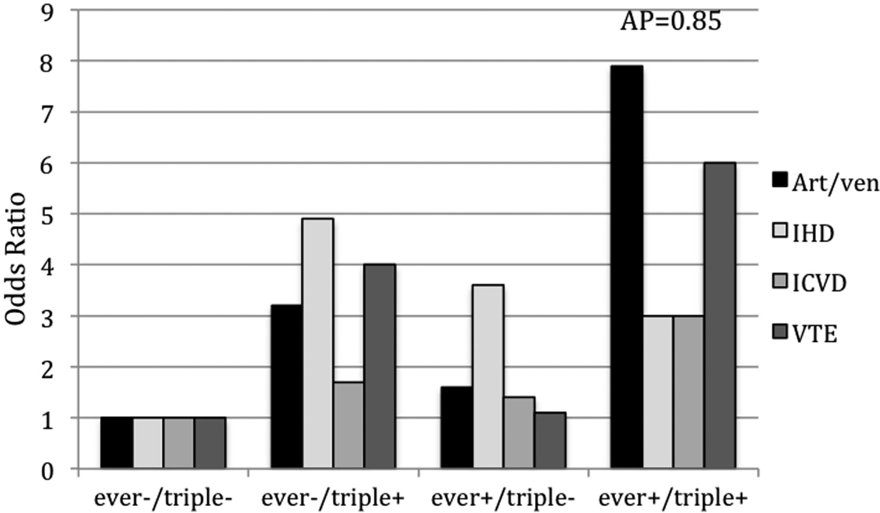
Due to uncertainties regarding the temporality between smoking and VE, and to gain power, we used ever smoking in the main analyses between smoking, aPL and VEs. The associations between aPL subtypes, ever smoking and previous VE are presented in online supplementary table S4. (For results regarding former and current smoking, see online supplementary tables, table S4A and B, respectively). In never smokers, presence of aPL, regardless of subtype, was not associated with the occurrence of previous VEs. In aPL-negative patients, ever smoking was moderately associated with arterial events, mainly with IHD. However, the combination of ever smoking and aPL had a stronger association with arterial and venous events. These associations were significant in combination with aCL IgG/A, aβ2GP1 IgG/A, LA and triple positivity (see online supplementary table S4). Stratification for age (>50 vs <50 years) did not change the results (data not shown). The additive interaction analysis demonstrated a significant interaction between ever smoking and LA (AP=0.80, 95% CI 0.5 to 1.0) (figure 2, see online supplementary table S4), and ever smoking and triple positivity (AP=0.85, 95% CI 0.6 to 1.0) (figure 3, see online supplementary table S4), regarding the association with presence of any VEs.

Ever smoking and LA in relation to previous vascular events. Art/ven, any arterial/venous event, IHD, ischaemic heart disease, ICVD, ischaemic cerebrovascular disease, VTE, venous thromboembolism, AP, attributable proportion due to interaction.

Ever smoking and triple positivity in relation to previous vascular events. Art/ven, any arterial/venous event, IHD, ischaemic heart disease, ICVD, ischaemic cerebrovascular disease, VTE, venous thromboembolism, AP, attributable proportion due to interaction.
Discussion
A positive association between ever, and above all former, regular cigarette smoking and presence of aPL is demonstrated among patients with SLE. Furthermore, a positive interaction between ever smoking and aPL was noted for the association with previous VEs.
The observation that former smoking status is associated with the more pathogenic aPL of IgG isotype as well as with LA positivity,18 while current smoking tended to be associated with aPL of IgM isotype, is new. If true, this pattern raises further questions about the origin of aPL and the interaction of smoking with the immune system. Whether smoking can trigger an immunological response with initial IgM production and a later persistent ‘IgG memory’, similar to infectious immunity needs to be addressed in prospective studies. Smoking cessation does not seem to reduce ‘aPL autoimmuny’ as years since smoking cessation did not affect aPL status.
Decreased total levels of serum IgG and IgM have been reported in current smokers,19 but in our study, total immunoglobulin levels did not differ by smoking status and are thus unlikely to have led to a skewing towards aPL of IgM isotype in current and IgG isotype in former smokers.
It is of particular note that smoking status was exclusively associated with aPL but not with other commonly occurring SLE-related autoantibodies, Rf or anti-CCP2 in our study. Previously, Rubin et al reported that former smokers had higher titres of anti-dsDNA IgG compared to current and never smokers. They hypothesised, supported by animal studies, that smoking suppresses IgG production, and that smoking cessation leads to an exacerbation of humoral autoimmunity with enhanced IgG production.20 By contrast with us, Freemer et al reported that current versus never smoking was associated with the occurrence of anti-dsDNA in SLE patients.4
Not much is known about the role of environmental factors for the occurrence of aPL. Some reports suggest that infections, trauma, vaccinations and drugs may be important.21 Likewise, APS-associated clotting events have been observed in aPL-positive patients after triggering ‘second hits’, such as infections or warfarin withdrawal.22 Hypothetically, smoking cessation may alter the immunological balance and cause either an aPL ‘class switch’ from IgM to more pathogenic IgG or an enhanced rebound IgG production, as suggested by Rubin et al.20 Smoking cessation could thus constitute a ‘triggering event’ in aPL-positive individuals. Further studies are needed to investigate these possibilities.
Antibodies to oxidised LDL (aoxLDL) cross-react with aPL of different specificities.23 Smoking was associated with higher titres of aCL IgG and of aoxLDL IgG in the general population.24 ,25 Cigarette smoke, a potent source of oxidants,26 may induce oxidative alterations in lipoproteins, such as LDL, or hypothetically in β2GP1, the major autoantigen in APS. Patients with APS have high levels of β2GP1 and a larger proportion of oxidised β2GP1 than controls.27 ,28 We recently demonstrated that there is a strong genetic component to the occurrence of aPL and VE in SLE.29 ,30 A possible scenario is that smoking may contribute to oxidative alterations, which, in genetically predisposed individuals, may give rise to an autoimmune response, epitope spreading and induction of aPL.
The association between former smoking and aPL was only present among patients with a previous VE. To investigate whether the patients who had stopped smoking after a VE explain this observation, we excluded the 18 patients (5%) who reported quitting smoking at the time of or after their first VE in an additional posthoc sensitivity analysis. The association between former smoking and aPL persisted in this setting, although attenuated. We cannot fully exclude the possibility that patients had stopped smoking earlier, advised to do so based on the presence of SLE, aPL or other cardiovascular risk factors. The present cross-sectional study does not allow us to further determine whether this reverse causation explains our findings and, therefore, results should be interpreted with this caveat in mind.
Among aPL-negative patients, ever smoking was predominantly associated with IHD. This observation is consistent with the fact that smoking is a strong risk factor for arterial events in the general population and in SLE.10 ,11 ,31 Although several studies recently demonstrated that smoking is a risk factor for VTE in the general population and in SLE,32–34 we did not find any association between ever smoking and history of VTE in aPL-negative patients in this study (see online supplementary table S4).
aPL positivity in patients who had not smoked regularly was not associated with previous VEs in this study, which contrasts with the recognised effect of these antibodies.8 The ORs for these analyses were, however, generally in the same direction (>1) (except for venous events, online supplementary table S4) with broad CIs, indicating that we may have been underpowered to detect a positive but weaker association.
Though the individual associations of smoking and aPL with VE were moderate, the combination of the two factors markedly increased the ORs for VE. These patterns of associations were consistent across investigated aPL (see online supplementary table S4) and are, to our knowledge, new. A significant interaction was found between ever smoking and LA, and triple positivity,9 for the association with any VE. Our observations are supported by Urbanus et al who reported a joint effect of aPL and smoking for the occurrence of MI and stroke.35
The strengths of this study are the relatively large and well-defined cohort of patients, the detailed information about smoking history and events, and the uniform measurements of aPL.
There are, however, also several limitations. We were not adequately powered in stratified and subgroup analyses and, despite detailed questions about the patient's smoking history we cannot exclude that recall bias has led to exposure misclassification. The interpretation of the interaction analysis is based on the assumption that smoking and aPL preceded VE. According to patient reports and records, only one of the reported VEs occurred prior to smoking start, and it is clinically reasonable, but not proven, to assume that aPL occurred before the VE. Finally, due to the cross-sectional design, aPL status was based on a single point in time, and we cannot exclude the possibility that aPL positivity was transient in some patients. However, aPL positivity, not APS, was the outcome in this study.
To conclude, we report that SLE patients who have smoked regularly are more often aPL-positive, and that former smokers were more prone to be positive for the more pathogenic LA and IgG aPL isotypes. We observed a strikingly high frequency of former VE among aPL-positive ever regular smokers. Further prospective and mechanistic studies need to address how smoking is related to the development of aPL, and how smoking and procoagulant autoantibodies interact to enhance the risk of thrombotic events.
Acknowledgments
We are grateful to Julia F Simard for statistical advice, Eva Jemseby, Gull-Britt Almgren, Julia Boström, Gloria Rostvall, for management of blood samples and to Anna-Britta Johansson for determination of aPL.
References
Supplementary materials
Supplementary Data
This web only file has been produced by the BMJ Publishing Group from an electronic file supplied by the author(s) and has not been edited for content.
Files in this Data Supplement:
- Data supplement 1 - Online supplement
Footnotes
Handling editor Tore K Kvien
Contributors JG acquired, analysed and interpreted the data and drafted the manuscript. IG acquired the data. HK analysed and interpreted the data and drafted the manuscript. SP coordinated the study and acquired the data. AZ and AV acquired the data. SM coordinated the study and acquired the data. JR and KE coordinated and acquired laboratory data and drafted the manuscript. ES designed the study, acquired, analysed and interpreted the data and drafted the manuscript. All authors critically reviewed and approved the final manuscript.
Funding This work was supported by Swedish Heart-Lung Foundation, Stockholm County Council and Karolinska Institutet (ALF), The King Gustaf V 80th Birthday Fund, The Swedish Rheumatism Association, The Swedish research council, The Åke Wiberg Foundation, Alex and Eva Wallströms Foundation, Karolinska Institutet's Foundations, and The Foundation in memory of Clas Groschinsky, The Swedish Society of Medicine. JF Simard is supported by the Strategic Research Program in Epidemiology at Karolinska Institutet, Sweden.
Competing interests None.
Ethics approval The local ethics committee of the Karolinska University Hospital approved the study protocol.
Provenance and peer review Not commissioned; externally peer reviewed.