Article Text

Abstract
Objectives To determine whether clinical tenderness can be considered a sign of inflammatory joint activity in patients with rheumatoid arthritis (RA), osteoarthritis (OA) or psoriatic arthritis (PsA) and to assess other possible factors associated with tenderness.
Methods Patients diagnosed with RA, PsA and OA underwent clinical and ultrasound examination of wrists and finger joints. Radiographs of the hands were scored for erosions, joint space narrowing (JSN), osteophytes and malalignment. A binary damage score (positive if ≥1 erosion, JSN and/or presence of malalignment) was calculated. Differences in grey scale signs of synovitis and power Doppler (PD) between tender non-swollen (TNS) versus non-tender non-swollen (NTNS) joints were calculated. Disease duration was assessed,<2 years was regarded as early and >5 years as long-standing arthritis.
Results In total, 34 patients (9 early and 14 long-standing) from patients with RA, 31 patients (7 early and 15 long-standing) with PsA and 30 with OA were included. We found equal frequencies of PD signal between TNS and NTNS joints in RA (p=0.18), PsA (p=0.59) or OA (p=0.96). However, PD had a significant association with tenderness in early arthritis both in RA (p=0.02) and in PsA (p=0.02). The radiographic damage score showed significant association with tenderness in RA (p<0.01), PsA (p<0.01) and OA (p=0.04).
Conclusion Tenderness might not always be a sign of active inflammation in RA, PsA and OA. While tenderness in early arthritis may be more related to inflammation, established disease is better explained by joint damage and malalignment.
- arthritis
- rheumatoid
- ultrasonography
- arthritis
- psoriatic
- osteoarthritis
- outcome assessment
- health care
Data availability statement
Data are available upon reasonable request. Deidentified, coded data will be made available from the corresponding author upon reasonable request.
Statistics from Altmetric.com
Key messages
What is already known about this subject?
Tender and swollen joint counts are part of disease activity scores for inflammatory arthritides.
Joint swelling is associated with synovitis and development of radiographic damage.
Whether tenderness in non-swollen joints can or should be regarded as a sign of inflammation, it is presently unclear.
What does this study add?
We found tenderness in early arthritis to be associated to inflammation, while in established disease, it is better explained by joint damage and malalignment.
How might this impact on clinical practice?
Tenderness in non-swollen joints in patients with long-standing rheumatoid arthritis or psoriatic arthritis should not automatically be regarded as a sign of active disease.
Introduction
Rheumatoid arthritis (RA) and psoriatic arthritis (PsA) are chronic inflammatory joint diseases, characterised by swelling, pain, stiffness of joints, as clinical signs of synovitis, and systemic inflammation also mirrored by elevated acute phase reactants.1–3 Synovitis leads to joint destruction, characterised by bone erosions and cartilage loss.4 5
Joint swelling as assessed by clinical examination is generally regarded to denote the presence of synovitis and causes joint damage.6 In contrast, tender joint counts (TJCs) exhibit a weaker association with radiographic progression and less signs of inflammation detected by various imaging modalities compared with swollen joints.7–9 At the same time, the TJC has better interobserver reliability and higher sensitivity to change compared with the swollen joint count (SJC).10 In the 28 joint disease activity score, the TJC even has a higher weight compared with the SJC.8 10–12 All currently used composite disease activity indices, remission criteria and inclusion criteria into clinical drug trials generally include TJCs.
In the early phases of highly suspicious RA (eg, presence of joint swelling in one or more joints) and for classification of the disease, tenderness is regarded as a feature of inflammatory joint affection.13 However, tenderness may have causes other than active inflammation, particularly in more established disease such as irreversible joint damage, psychological factors or central sensitisation in the medulla after long-lasting activation of pain signals.7 14
Similarly to RA, TJCs are part of most disease activity scores in PsA15 16 while arthralgia alone in patients with psoriasis is insufficient to diagnose PsA clinically.17 18 Tenosynovitis, synovitis and enthesitis were detected sonographically in patients with psoriasis with or without arthralgia and found to be associated with the development of clinically evident PsA.19 20
Hand osteoarthritis (OA) is characterised by loss of articular cartilage, joint pain and joint deformation as opposed to synovial swelling.21 Recent studies however showed that low grade inflammation triggered, for example, by mechanical stress is involved in the pathogenesis of OA.22 23 Also, osteoarthritic joints are prone to secondary inflammation, which might lead or increase tenderness.24
Musculoskeletal ultrasound (US) has been reported to be a sensitive tool for the evaluation of inflammatory joint activity in RA and PsA.25–29 Using US, joints can be assessed for signs of synovitis, that is, synovial hyperproliferation and synovial effusion detected by grey scale (GS) and hypervascularisation detected by colour or power Doppler (PD). In hand OA, synovitis detected by MRI and US was reported to be associated with pain and radiographic progression.30 31
It is presently unclear whether tenderness in non-swollen joints can or should be regarded as a sign of inflammation. The aim of this study was to evaluate, using US to detect subclinical inflammation, whether clinical tenderness in the absence of swelling may be considered a sign of inflammatory joint activity in patients with RA, PsA or OA. As a secondary aim, we wanted to detect other factors that may be associated with joint tenderness.
Methods
Patients
Patients with RA diagnosed according to the American College of Rheumatology/European League Against Rheumatism (ACR/EULAR) 2010 criteria,32 patients with PsA diagnosed according to the CASPAR criteria18 and patients with OA diagnosed according to the ACR 1990 criteria33 were recruited from our outpatient clinic. Patients were included, if they had at least one proximal interphalangeal (PIP) or metacarpophalangeal (MCP) joint, which was tender and not swollen. All recruited patients with RA and PsA additionally were included in an observational database where standard variables including joint counts are routinely assessed every 3–5 months and prospectively documented. Details of the database have been published elsewhere.34 35 For additional subanalyses, patients with RA and PsA were grouped according to disease duration as early disease (duration of less than 2 years) and long-standing disease (disease duration of more than 5 years).
We performed a sample size calculation to detect a difference of positive PD signals between tender non-swollen (TNS) joints and non-tender non-swollen (NTNS) joints of 8.3%. This cut-off was set according to the publication of Hammer et al,36 where TNS and NTNS were PD positive in 17.7% and 9.4%, respectively. Alpha was set at 0.05 and beta at 0.8 for the sample size calculation. Therefore, 263 joints for each disease would be necessary. We aimed to recruit 30 patients for each disease, assuming that 22 joints per patient would yield enough TNS and NTNS to detect a difference of 17.7% versus 9.4%.
Clinical examination
TJCs and SJCs of 22 joints (bilateral wrists, MCP joints 1–5 and PIP joints 1–5 were recorded of all patients by biometricians, health professionals with more than 5 years of experience performing daily joint counts in patients with arthritides, who were blind with regards to diagnosis as well as with regards to sonographic data. Particular care was taken to only count synovial swelling and not bony swelling. Additionally, a 28-joint count according to the Clinical Disease Activity Index (CDAI)37 was assessed in RA and a 66/68 joint count according to the CDAI for psoriatic arthritis (cDAPSA)38 was assessed in PsA. Joints which underwent operation or replacement were excluded from both the clinical and imaging analyses. The CDAI37 was calculated for each patient with RA and the cDAPSA38 was calculated for each patient with PsA to quantify disease activity. Furthermore, joints of patients with RA and PsA were tracked back for up to 12 years to identify the time point of last swelling of each joint.
Ultrasound examination
All patients underwent an US examination of the same 22 joints of both hands on the day of the clinical examination. The examinations were performed on an ESAOTE Mylab Twice ultrasound unit equipped with a high-frequency linear transducer (6–18 MHz) by two rheumatologist sonographers with 3 years of experience (>300 examinations) in musculoskeletal US (IG, MP), who were blinded to clinical diagnosis and examination as well as radiographic data. Scanning was performed according to the EULAR standardised procedures.39 Longitudinal and transverse scans were performed on the dorsal aspect of each joint using both B-mode (GS) and PD flow. The hand was examined palms down resting on the examination table. Wrist and MCP were examined in the neutral position, while the PIP joints were in slight flexion to ensure that the potentially present synovial hypertrophy is distinguished from the extensor apparatus attaching to the base of the middle phalanx. Copious amounts of gel were used and special care was taken not to apply too much pressure with the US transducer so as not to compress potential Doppler signal (online supplemental figure 1). Doppler gain was adjusted to the level just below random noise; pulse repetition frequency was set between 0.5 and 0.8 MHz.
Supplemental material
GS and PD were both scored semiquantitatively (0–3) and as a combined score (EULAR-OMERACT combined synovitis scoring system)40 according to the OMERACT definitions for sonographic pathology.41 In case of conflicting grades, the higher grade was selected.
Structural damage by radiography
X-rays of the hands at the time of the clinical and sonographic examination (±1 year) were evaluated for structural joint damage. Wrists, PIP and MCP joints were scored for erosions and joint space narrowing (JSN) according to the Sharp/van der Heijde method42 in patients with RA and PsA and according to the Interphalangeal Osteoarthritis Radiographic Simplified (iOARS) scoring system43 in patients with OA. Additionally, all wrists, PIP and MCP joints were scored for malalignment and osteophytes (presence/absence) in patients with RA, PsA as well as OA. Osteophytes were scored according to the iOARS scoring system.43 Malalignment was scored as present/absent according to a published atlas of radiographic images44 and defined as significant joint deviation. In order to assess the probability of tenderness in case of any joint damage, a binary damage score was used, in which a joint was regarded as damaged in case of either erosions (scored 1 or higher) or JSN (scored 1 or higher) or malalignment (presence).
Statistics
Patient characteristics are described as percentages and frequencies for categorical variables or mean with SD for normally distributed continual variables. Differences in patient characteristics were assessed with Student’s t-test or χ2 test, respectively. Differences in PD and GS signals and the EULAR-OMERACT combined US score between groups (RA vs OA, RA vs PsA, PsA vs OA) as well as TNS versus NTNS were calculated by χ2 test. To further increase the power, we combined RA and PsA patients and performed the same analysis.
Kaplan-Meier estimates for the occurrence of the last time point of swelling were compared between TNS and NTNS joints.
To assess interobserver reliability, the recorded, anonymised images of 15 patients each with RA, OA and PsA were independently reviewed after 4 weeks by two rheumatologist experts in musculoskeletal US and scored for GS (0–3) signs of synovitis and PD (0–3) signal. Interobserver agreement was assessed by intraclass correlation. A value of 0–0.4 was interpreted as poor; 0.49–0.59 as fair; 0.60–0.74 as good and 0.75–1.00 as excellent reliability.
A binary logistic regression analysis adjusted for age and sex was performed to assess the association of damage score, JSN, erosions, osteophytes and malalignment with tenderness in non-swollen joints. Furthermore, multivariable logistic regression using a block-wise forward stepwise conditional approach was used to assess tenderness in non-swollen joints. Independent variables included in the analyses were age, sex and disease duration (for RA and PsA) (block 1), erosions, JSN, osteophytes and malalignment (block 2) and GS and PD (block 3).
Additionally, we separately performed logistic analyses adjusted for age and sex assessing the value of PD for TNS in split patient groups according to a disease duration of <2 years and >5 years.
A p value of <0.05 was regarded as significant. No correction for multiple testing was performed. All analyses were performed using SPSS software, V.25 (IBM, Armonk, New York, USA). This study was conducted in accordance with the declaration of Helsinki and approved by the ethics committee of the Medical University of Vienna (no: 1415/2015).
Results
Patient characteristics
In total, 745 joints from patients with RA, 682 joints from patients with PsA and 657 joints from patients with OA were included in the study. The majority of RA patients (53.1%) was in moderate disease activity (CDAI of >10 and ≤22)45 at the time of the US and the clinical examination. Patients with RA were seropositive in 61.8% (21/34). Most patients with PsA (65.5%) were in high disease activity (cDAPSA of >28).38 The mean disease duration was 7.2±6.6 years for patients with RA and 7.4±6.3 years for patients with PsA (online supplemental table 1). Interobserver reliability for the US examination was excellent with 0.75 (95% CI 0.71 to 0.78) and 0.9 (95% CI 0.88 to 0.91) for GS and PD, respectively.
Sonographic characteristics of tender non-swollen (TNS) joints
In patients with RA, 155/745 (20.8%) joints were TNS; in PsA and OA, these were 32.2% (219/682) and 19.5% (128/657), respectively. No PD signals at all were observed in the majority of TNS, namely, 85.8% of the TNS in RA, 90.9% TNS in PsA and 89.8% of TNS in OA (p=0.25). No significant difference was seen between PD findings in TNS versus NTNS in RA (14.2% vs 10.2%, respectively; p=0.18), in PsA (9.1% vs 8%, respectively; p=0.59) or in OA (10.2% vs 9.3%, respectively; p=0.96) (figure 1). Similarly, the combined RA and PsA group revealed no significant difference in PD signals between TNS and NTNS (11.2% vs 9.3%, p=0.3) (online supplemental figure 2).

Power Doppler signals (grade 0, 1, 2 or 3) in tender non-swollen joints (tender) versus non-tender non-swollen joints (not tender) in rheumatoid arthritis (RA), psoriatic arthritis (PsA) and osteoarthritis (OA). Difference between tender non-swollen and non-tender non-swollen joints was calculated for each disease by χ2 test.
GS synovitis (any grade) was detected more often in TNS joints of patients with PsA as compared with those of patients with RA (64.8% vs 54.2%, respectively; p=0.04); in patients with OA, 65.6% joints showed signs of GS synovitis (OA vs RA: p=0.05; OA vs PsA: p=0.39). TNS showed higher scores compared with NTNS in OA (65.6% vs 58.1%, respectively; p<0.01) but similar scores in PsA (64.8% vs 59.9%, respectively; p=0.10) and RA (54.2% vs 48.4%, respectively; p=0.17) (figure 2).

Grey scale signals (grade 0, 1, 2 or 3) in tender non-swollen joints (tender) versus non-tender non-swollen joints (not tender) in rheumatoid arthritis (RA), psoriatic arthritis (PsA) and osteoarthritis (OA). Difference between tender non-swollen and non-tender non-swollen joints was calculated for each disease by χ2 test.
The EULAR-OMERACT score revealed higher scores in OA compared with RA (p=0.02) and PsA (p=0.02) but no difference between RA and PsA (p=0.19). Similar scores were found between TNS and NTNS in RA (TNS: 54.5% vs 48.4%, p=0.14), PsA (TNS: 65.4% vs 60.2%, p=0.08) while TNS had significantly higher EULAR-OMERACT scores compared with NTNS in OA (TNS: 65.6% vs 58.2%, p<0.01) (figure 3).

EULAR-OMERACT combined ultrasound score (grade 0, 1, 2 or 3) in tender non-swollen joints (tender) versus non-tender non-swollen joints (not tender) in rheumatoid arthritis (RA), psoriatic arthritis (PsA) and osteoarthritis (OA). Difference between tender non-swollen and non-tender non-swollen joints was calculated for each disease by χ2 test.
Tenderness as sign of past/preceding swelling
Kaplan-Meier analysis showed no difference in the time to last observed swelling between TNS and NTNS joints in patients with RA (62.1±3.3 vs 66.6±2 months, respectively; p=0.40) or PsA (101±6.2 vs 106.4±4.1 months, respectively; p=0.17) (online supplemental figure 3).
Tender non-swollen (TNS) joints and structural damage
In RA, the mere presence of damage was associated with tenderness in non-swollen joints (OR 1.76, 95% CI 1.16 to 2.66, p<0.01). The binary logistic regression adjusted for age and sex showed an association of TNS with JSN (OR 1.12, 95% CI 1.00 to 1.25; p>0.05), but this was not significant (figure 4A). In PsA, we observed a similar association between tenderness and damage (OR 2.01, 95% CI 1.31 to 3.10; p<0.01), in particular, with malalignment (OR 4.17, 95% CI 1.48 to 11.81; p<0.01) and erosions (OR 1.49, 95% CI 1.05 to 2.10; p=0.03) (figure 4B). Similarly, in OA, the damage score (OR 1.89, 95% CI 1.03 to 3.46; p=0.04) was associated with tenderness. Furthermore, osteophytes (OR 3.52, 95% CI 2.04 to 6.14; p<0.01), malalignment (OR 5.65, 95% CI 2.16 to 14.76; p<0.01), erosions (OR 1.85, 95% CI 1.17 to 2.92; p<0.01) and JSN (OR 1.24, 95% CI 1.02 to 1.49; p=0.03) all had an association with tenderness in OA (figure 4C).

Difference of detected erosions (0 vs ≥1), joint space narrowing (0 vs ≥1), malalignment (presence/ absence), osteophytes (presence/ absence) and damage score (0 vs ≥1) in patients with rheumatoid arthritis (A), psoriatic arthritis (B) and osteoarthritis (C). Association of erosions (semiquantitatively), joint space narrowing (semiquantitatively), malalignment, osteophytes and damage score with tenderness in non-swollen joints was calculated by age-adjusted and sex-adjusted binary logistic regression.
In swollen joints, the damage score showed neither association with tenderness in RA (OR 0.54, 95% CI 0.11 to 2.55; p=0.45) nor in PsA (OR 2.38, 95% CI 0.56 to 10.14; p=0.24).
Factors associated with tenderness in non-swollen joints
In non-swollen joints in RA, the block-wise multivariable regression analysis including age, sex and disease duration (block 1), erosions, JSN, osteophytes and malalignment (block 2) and GS and PD (block 3) resulted in exclusion of all variables except for sex (OR 0.40, 95% CI 0.19 to 0.84, p=0.02), JSN (OR 1.12, 95% CI 1.00 to 1.26, p=0.06) and disease duration (OR 1.03, 95% CI 1.00 to 1.07, p=0.07) with female sex being associated with tenderness. In PsA, only malalignment remained as a single variable after the analysis (OR 3.91 95% CI 1.29 to 11.87, p=0.02). In OA, osteophytes (OR 2.05, 95% CI 1.12 to 3.74, p=0.02), malalignment (OR=2.75, 95% CI 0.91 to 8.38, p=0.07), GS (OR 1.56, 95% CI 1.07 to 2.28, p=0.2) and PD (OR 1.05, 95% CI 0.46 to 2.38, p=0.91) remained as variables after the analysis (table 1).
Remaining variables in the block-wise forward conditional multivariable regression analysis including age, sex and disease duration (block 1), erosions, joint space narrowing (JSN), osteophytes and malalignment (block 2) and Grey scale (GS) and power Doppler (PD) (block 3) for tenderness as the dependent variable in non-swollen joints
Tender non-swollen (TNS) joints in early disease
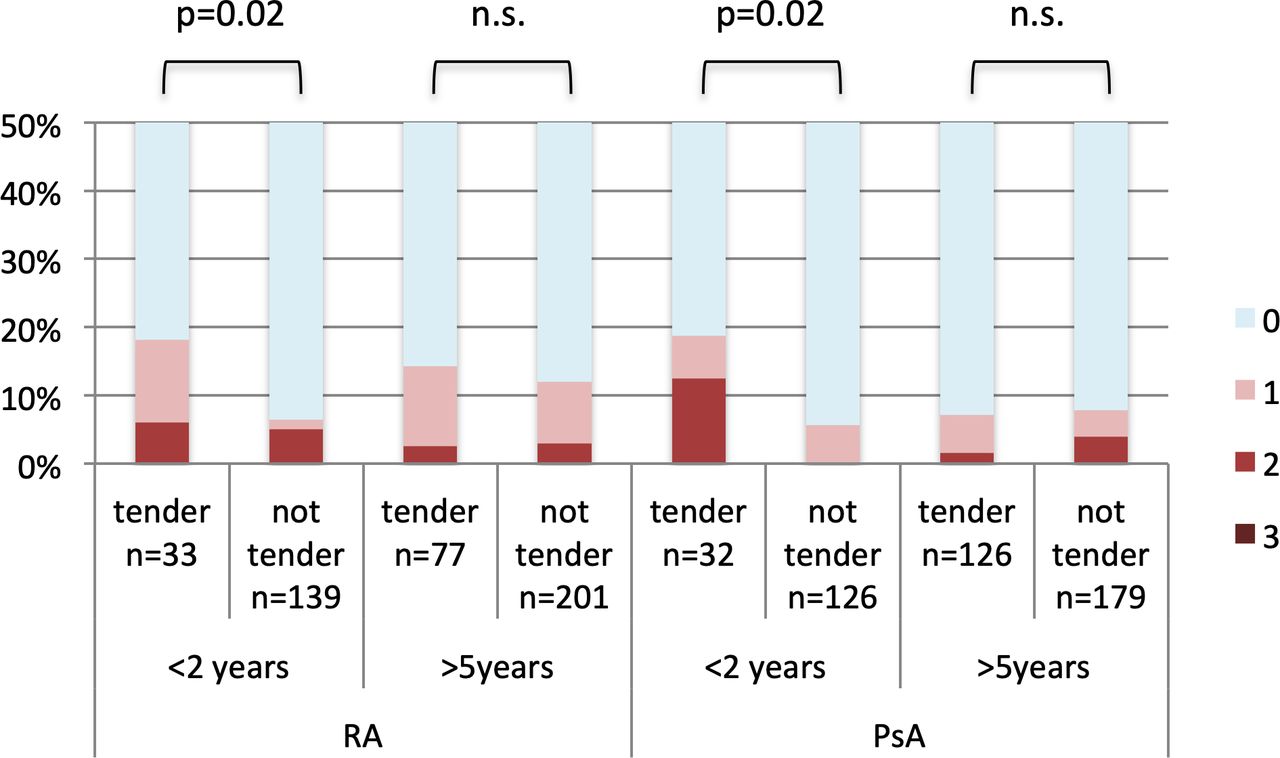
We included 174 joints in 9 patients with RA and 138 joints in 7 patients with PsA in the early disease group, respectively (duration of less than 2 years) and 278 joints in 14 patients with RA and 305 joints in 15 patients with PsA in the long-standing disease group, respectively (disease duration of more than 5 years). In patients with RA with disease duration of less than 2 years, presence of PD had a significant association with tenderness (OR 2.22, 95% CI 1.12 to 4.43, p=0.02) in a binary logistic regression adjusted for age and sex, in contrast to patients with RA with a disease duration of more than 5 years (OR 1.17, 95% CI 0.65 to 2.13, p=0.60). Similarly, in patients with PsA with a short disease duration of less than 2 years, higher PD coincided with a higher likelihood of tenderness (OR 3.26, 95% CI 1.21 to 8.81, p=0.02) while no significant results were found in patients with PsA with a disease duration of more than 5 years (OR 0.84, 95% CI 0.46 to 1.56, p=0.59) (figure 5).
Difference in power Doppler signal (0–3) between tender not swollen joints (tender) and not tender not swollen joints (not tender) in patients with rheumatoid arthritis (RA) and psoriatic arthritis (PsA) with a disease duration of less than 2 years (<2 years) and more than 5 years (>5 years). Age-adjusted and sex-adjusted logistic regression analysis was performed to determine the association of power Doppler signal on tenderness.
Discussion
In this study, we investigated the meaning of tenderness in RA, PsA and OA. Presence of tenderness was not associated with increased sonographic signs of synovitis in non-swollen joints in RA and PsA except for patients with a disease duration of less than 2 years. In contrast, radiographic damage had a significant association with tenderness in non-swollen joints. This suggests that tenderness might only be a sign of inflammation early in the disease course of RA and PsA, while damage has more association with tenderness in established disease.
Recently, several studies have reported poor to no association of tenderness with sonographic signs of synovitis on patient-level as well on joint-level in RA.36 46 47 Similarly, we found no difference in PD or GS in TNS as compared with NTNS in RA. We could show the same findings in PsA, where to our knowledge this relationship has not yet been evaluated.
Another possible reason for tenderness without swelling is structural damage. When performing this study, we deliberately chose to analyse different diseases in order to see whether we can find any distinctions between the three conditions with regards to joint tenderness. We chose to include patients with inflammatory arthritides (RA and PsA) as well as those with OA which, despite certain inflammatory features, is nonetheless seen as a primarily degenerative condition. While the three diseases are indeed distinct, the key pathologic findings with regards to structural damage and indeed the radiographic methods by which we assess such damage in each of these conditions are indeed overlapping.
Osteophytes in OA were shown to be associated with pain in multiple studies31 48–50 and secondary OA may cause pain in RA and PsA.51 In our study, osteophytes were more common in TNS as compared with NTNS joints in OA, but not in RA or PsA. These findings suggest that secondary OA likely does not explain tenderness in patients with RA or PsA.
JSN and erosions are part of various damage scores for PsA52 53 and RA.42 54 JSN and erosions are seen in OA and JSN is part of a radiographic damage score for OA.43 In addition to JSN and erosions, we wanted to assess malalignment to include more possible causes for tenderness. Malalignment or subluxation is caused by instability of the articular capsule and its ligaments as well as incongruity of articular surfaces and as is part of several radiographic damage scores in RA54 55 and in PsA52 and may also occur in OA43 as well. In order to assess whether the probability of tenderness in non-swollen joints is higher in case of any joint damage, we created a binary damage score, in which a joint was regarded as damaged in case of either erosions, JSN or malalignment. Indeed, this combined damage score consisting of these three components had a significant impact on tenderness in all three diseases, also after adjustment for age and sex. Interestingly, this difference was apparent only in non-swollen joints: structural damage was not found to be more common in tender swollen joints compared with TNS joints. In RA, the multivariable regression analysis including age, sex, disease duration, erosions, JSN, osteophytes, malalignment, GS and PD resulted in exclusion of all variables except for sex and JSN, with female sex being associated with tenderness. This underlines the greater impact of radiographic damage compared with synovitis on tenderness in RA. In PsA, only malalignment remained in the analysis, again supporting the hypothesis that damage has more impact than synovitis on tenderness.
TNS joints did not have a shorter time to last observed swelling as compared with NTNS joints. This suggests that in our study, pain memory due to joint swelling within the preceding year does not explain the occurrence of tenderness without swelling.
Interestingly, we saw a significant impact of PD on tenderness in RA and PsA with a disease duration of less than 2 years. Many patients who are ultimately diagnosed with RA have a prodromal phase dominated by pain before the development of synovitis.56 Sonographic57 58 and MRI59 signs of synovitis are seen before the onset of arthritis in patients with and without arthralgia. In an animal model for arthritis, histological signs of synovitis were seen before clinical arthritis.60 Our results suggest that early in the disease course inflammation and synovitis may explain tenderness.
Another important question regarding tender joints is their predictive value for radiographic progression. Some studies, mostly on patient level, reported that as compared with swelling, tenderness is not or only poorly associated with radiographic progression in RA.8 61 62 This underlines the findings of our study and other recent studies suggesting that tenderness without swelling may not be a sign of inflammation.7 34
Interestingly, in contrast to the results in RA and PsA, tender joints in OA were associated with sonographic signs of synovitis. This has been reported in several studies, although the strengths of such associations vary.31 63 However, the association of tenderness with osteophytes and malalignment was higher compared to that with GS and PD.
Out study is not without limitations. We did not assess distal interphalangeal joints, which are among the most commonly involved joints in OA and PsA, since we aimed to perform the same assessments in all three diseases. The binary joint damage score, whereby a joint is regarded as damaged, if it exhibits any of the above-mentioned structural changes, needs to be validated in further studies. An additional limitation of our study is its cross-sectional design, which did not allow us to evaluate the predictive utility of tenderness in radiographic progression. Furthermore, we did not assess extraarticular involvement such as enthesitis or tenosynovitis as well as other potential reasons for tenderness like fibromyalgia and other chronic pain syndromes.64 65 Furthermore, we did not assess fibromyalgia as a potential comorbidity in our study.
In conclusion, structural damage had a higher impact on tenderness in non-swollen joints in RA, PsA and OA. The results of this study suggest that an interpretation of tenderness in established inflammatory arthritides as sign of inflammation may not be appropriate. In early disease and possibly also for diagnostic purposes, tenderness may be used as a potential sign of inflammation.
Data availability statement
Data are available upon reasonable request. Deidentified, coded data will be made available from the corresponding author upon reasonable request.
Ethics statements
Patient consent for publication
References
Supplementary materials
Supplementary Data
This web only file has been produced by the BMJ Publishing Group from an electronic file supplied by the author(s) and has not been edited for content.
Footnotes
Handling editor David S Pisetsky
Twitter @Stiddyo
Deceased 27.07.2019
Correction notice This article has been corrected since it published Online First. The provenance and peer review statement has been included.
Contributors GI: substantial contributions to the conception or design of the work, or the acquisition, analysis or interpretation of data, drafting the work or revising it critically for important intellectual content. MP: substantial contributions to the conception or design of the work, or the acquisition, analysis or interpretation of data. VS: substantial contributions to the conception or design of the work, or the acquisition, analysis or interpretation of data. GS: substantial contributions to the conception or design of the work, or the acquisition, analysis or interpretation of data. TD: substantial contributions to the conception or design of the work, or the acquisition, analysis or interpretation of data. MD: substantial contributions to the conception or design of the work, or the acquisition, analysis or interpretation of data. MH: drafting the work or revising it critically for important intellectual content. ML: substantial contributions to the conception or design of the work, or the acquisition, analysis or interpretation of data. PS: drafting the work or revising it critically for important intellectual content. MZ, JSS: drafting the work or revising it critically for important intellectual content. DA: substantial contributions to the conception or design of the work, drafting the work or revising it critically for important intellectual content. PM: substantial contributions to the conception or design of the work, or the acquisition, analysis or interpretation of data, drafting the work or revising it critically for important intellectual content, final approval of the version published, agreement to be accountable for all aspects of the work in ensuring that questions related to the accuracy or integrity of any part of the work are appropriately investigated and resolved.
Funding The authors have not declared a specific grant for this research from any funding agency in the public, commercial or not-for-profit sectors.
Competing interests None declared.
Provenance and peer review Not commissioned; externally peer reviewed.
Supplemental material This content has been supplied by the author(s). It has not been vetted by BMJ Publishing Group Limited (BMJ) and may not have been peer-reviewed. Any opinions or recommendations discussed are solely those of the author(s) and are not endorsed by BMJ. BMJ disclaims all liability and responsibility arising from any reliance placed on the content. Where the content includes any translated material, BMJ does not warrant the accuracy and reliability of the translations (including but not limited to local regulations, clinical guidelines, terminology, drug names and drug dosages), and is not responsible for any error and/or omissions arising from translation and adaptation or otherwise.