Article Text

Abstract
Objectives Osteoarthritis is associated with cell death and extracellular matrix degradation in articular cartilage. Autophagy is an essential cellular homeostasis mechanism that was found to be deficient in ageing and osteoarthritic cartilage. This study determined whether pharmacological inhibition of the mammalian target of rapamycin (mTOR), a key inhibitor of autophagy, has disease-modifying activity in experimental osteoarthritis.
Methods Experimental osteoarthritis was induced by transection of the medial meniscotibial ligament and the medial collateral ligament in 2-month-old C57Bl/6 mice (n=36). Rapamycin (1 mg/kg weight/day) (n=18 mice) or dimethyl sulphoxide vehicle control (n=18 mice) was administered intraperitoneally for 10 weeks. Histopathological changes in articular cartilage and synovium were examined by using semiquantitative scoring systems. Rapamycin effects on mTOR signalling, autophagy, cartilage homeostasis and inflammation were analysed by immunohistochemistry and immunofluorescence staining.
Results Rapamycin affected the mTOR signalling pathway in mouse knee joints as indicated by the inhibition of ribosomal protein S6 phosphorylation, a target of mTOR and activation of LC3, a main marker of autophagy. The severity of cartilage degradation was significantly (p<0.01) reduced in the rapamycin-treated group compared with the control group and this was associated with a significant (p<0.05) decrease in synovitis. Rapamycin treatment also maintained cartilage cellularity and decreased ADAMTS-5 and interleukin-1β expression in articular cartilage.
Conclusions These results suggest that rapamycin, at least in part by autophagy activation, reduces the severity of experimental osteoarthritis. Pharmacological activation of autophagy may be an effective therapeutic approach for osteoarthritis.
Statistics from Altmetric.com
Osteoarthritis, the most common ageing-related joint pathology, is characterised by degradation of cartilage extracellular matrix (ECM) and reduced cartilage cellularity.1 While changes in the articular cartilage appear to be critical in osteoarthritis initiation and progression, other joint tissues are invariably involved.2 Chondrocytes, the only cell population of adult articular cartilage, are capable of responding to structural changes in the surrounding cartilage matrix but the capacity of the adult articular chondrocytes to regenerate the normal cartilage matrix architecture is limited and declines with ageing, due to cell death and abnormal responsiveness to anabolic stimuli.3 Previous attempts at treating established osteoarthritis, for example by inhibiting ECM-degrading enzymes, failed to show efficacy or were associated with adverse events in clinical trials.4 As an alternative approach, the protection of cells against failure of homeostasis mechanisms, which results in global changes in gene expression, may have potential for greater efficacy.
Autophagy is a cellular homeostasis mechanism that plays an essential role in energy and nutrient regulation, and in the removal of damaged and dysfunctional macromolecules and organelles.5 6 At the cellular level, failure of autophagy results in the increased production of reactive oxygen species (ROS), abnormal gene expression, and can lead to cell death.7 Consequences of autophagy failure at the tissue and organismal level are neurodegeneration, cardiomyopathies, abnormal skeletal development and premature death.8,–,10
Mammalian target of rapamycin (mTOR) is an important suppressor of autophagy, functioning upstream of the autophagy proteins and is centrally regulated by multiple upstream signalling pathways involving PI3-kinase/Akt and AMP-activated protein kinase.11,–,13 Imbalances in the mTOR pathway are also involved in obesity, diabetes, inflammatory diseases and cardiac hypertrophy, and pharmacological inhibition of mTOR has been proposed as a potential treatment for those conditions.14 Rapamycin, a lipophilic macrolide antibiotic, which is used as an immunosuppressive drug in solid organ transplantation, can induce autophagy in a variety of cell types.15 16 In addition, rapamycin treatment extends lifespan in mice,17 and protects against ageing-related pathology in brain and heart.18,–,21
In articular cartilage, which is characterised by a very low rate of cell turnover, autophagy would appear to be essential to maintain cell survival and function. Previously we demonstrated that autophagy is a constitutively active and apparently protective process for the maintenance of cartilage homeostasis. Reduced expression of autophagy regulators was observed in joint ageing and osteoarthritis in humans and mice, and this was accompanied by an increase in chondrocyte apoptosis.22
Collectively, these observations suggest that compromised autophagy may contribute to the development of osteoarthritis. The objective of this study was to establish proof of principle that pharmacological enhancement of autophagy may have disease-modifying activity in experimental osteoarthritis.
Materials and methods
Experimental osteoarthritis in mice
All animal experiments were performed according to protocols approved by the Institutional Animal Care and Use Committee at The Scripps Research Institute. Experimental osteoarthritis was induced in 2-month-old male C57Bl/6J mice by transection of the medial meniscotibial ligament and the medial collateral ligament in the right knee.23 The left knee was not subjected to surgery and was used as a control. The animals were killed 10 weeks after the knee surgery.
Rapamycin treatment
Two independent experiments were performed. Each experiment included a total of 18 mice (nine mice treated with rapamycin and nine mice receiving vehicle). Rapamycin was obtained from LC Laboratories (Woburn, Massachusetts, USA), dissolved in dimethyl sulphoxide (DMSO) at 25 mg/ml and stored at −20°C. For injection, the stock solution was diluted in phosphate buffered saline (PBS). Mice received daily intraperitoneal injections of rapamycin at 1 mg/kg body weight/dose in a total injection volume of 0.3 ml for 10 weeks and control animals received the DMSO vehicle at 0.4% in a total injection volume of 0.3 ml.
Histological analysis of mouse knee joints
Knee joints (total n=72) from mice with experimental osteoarthritis and vehicle treatment (n=18), experimental osteoarthritis and rapamycin treatment (n=18) and vehicle administration (n=18/each group) were harvested. The joints were fixed in 10% zinc-buffered formalin (Z-Fix; Anatech, Battle Creek, Michigan, USA)for 24 h, decalcified in TBD-2 (Shandon, Pittsburgh, Pennsylvania USA) for 48 h, followed by paraffin embedding. Serial sections (4 µm) were cut, stained with Safranin O-fast green, and examined for histopathological changes using a semiquantitative scoring system.24 In this system the scores are defined as follows: 0, normal cartilage; 0.5, loss of proteoglycan with an intact surface; 1, superficial fibrillation without loss of cartilage; 2, vertical clefts and loss of surface lamina (any percentage of joint surface area); 3, vertical clefts/erosion to the calcified layer for 1–25% of the quadrant width; 4, lesion reaches the calcified cartilage for 25–50% of the quadrant width; 5, lesion reaches the calcified cartilage for 50–75% of the quadrant width; 6, lesion reaches the calcified cartilage for over 75% of the quadrant width.
Histological analysis of inflammation
Synovium from mice with experimental osteoarthritis and vehicle treatment (N=16) and rapamycin treatment (N=11) was examined using a grading system for synovial inflammation.25 Three parameters, hyperplasia/enlargement of synovial lining cell layer, activation of resident cells/synovial stroma and inflammatory cell infiltration were graded as absent (grade 0), slight (grade 1), moderate (grade 2) or severe (grade 3).
Cartilage cellularity
Knee joint sections were stained with H&E. In cartilage from knees with experimental osteoarthritis (vehicle and rapamycin-treated mice), three pictures were taken under 40× magnification, representing the centre of the femoral condyle that is not covered by the menisci as well as the medial and lateral femoral condyles. The total number of cells in each section was counted.26
Immunohistochemistry
Sections from paraffin-embedded joints were first deparaffinised in the xylene substitute Pro-Par Clearant (Anatech) and rehydrated in graded ethanol and water. For antigen unmasking, sections in 10 mM sodium citrate buffer (pH 6.0) were heated in a microwave oven and kept at 80–85°C for 1.5 min. Slides were cooled for 20 min at room temperature after antigen unmasking. After washing with PBS, sections were blocked with 5% serum for 30 min at room temperature. Antibodies to the phosphorylated form of ribosomal protein S6 (Cell Signalling Technology, Boston, Massachusetts, USA; 1:100 dilution), ADAMTS-5 (Abcam, Cambridge, Massachusetts, USA; 5 μg/ml) and interleukin (IL)-1β (Santa Cruz Biotechnology, Santa Cruz, California, USA; 1:100) were applied and incubated overnight at 4°C. After washing with PBS, sections were incubated with biotinylated goat antirabbit secondary antibody for 30 min at room temperature and then incubated using Vectastain ABC-AP alkaline phosphatase (Vector Laboratories, Burlingame, California, USA) for 30 min. Slides were washed, and sections were incubated with alkaline phosphatase substrate for 20–30 min.
Immunofluorescence
Paraffin-embedded samples were deparaffinised in the xylene substitute Pro-Par Clearant (Anatech) and rehydrated in graded ethanol and water. After washing with PBS, sections were blocked with 5% serum for 30 min at room temperature and then incubated with rabbit anti-LC3 polyclonal antibody (1:50) (Abgent, San Diego, California, USA) overnight at 4°C. After washing with PBS, sections were incubated with Alexa Fluor 488 antirabbit immunoglobulin G (IgG) for 30 min. Finally, slides were washed and mounted with Prolong Gold Antifade Reagent (Invitrogen, Carlsbad, California, USA).
Quantification of ADAMTS-5-positive cells
In cartilage from mice subjected to experimental osteoarthritis and treated with vehicle or rapamycin, three pictures were taken under 40× magnification, showing the centre of the femoral condyle that is not covered by the menisci as well as the medial and lateral femoral condyles. The total number of chondrocytes was counted,26 and then the total number of ADAMTS-5-positive cells was counted in each section. Results are expressed as the percentage of ADAMTS-5-positive cells.
Statistical analysis
Statistically significant differences between the two groups were determined with t tests. The results are reported as mean±S.D. p Values less than 0.05 were considered significant.
Results
Systemic administration of rapamycin modulates mTOR signalling and autophagy in mouse knee joints
First, we evaluated the effect of intraperitoneal rapamycin on the mTOR signalling pathway in mouse knee joints by determining the phosphorylation levels of ribosomal protein S6, a downstream target of mTORC1.27 Rapamycin treatment suppressed rpS6 phosphorylation in articular cartilage and menisci in the knee joints compared with vehicle-treated mice (figure 1A). To determine whether the mTOR inhibition by rapamycin promotes autophagy, knee joint sections were stained with LC3 antibody. We found an increase in LC3 expression after rapamycin treatment. This increase was correlated with an increase in LC3 puncta, indicating strong activation of autophagy in articular cartilage (figure 1B). These results indicate that the intraperitoneal administration of rapamycin caused the expected effects of inhibiting mTOR signalling and autophagy activation in knee joints.

Systemic administration of rapamycin (rapa) modulates mammalian target of rapamycin (mTOR) signalling and autophagy in mouse knee joints. (A) Knee joints from C57Bl/6J mice were collected 10 weeks after knee destabilisation or and treatment with rapamycin or vehicle (n=3/each). Contralateral knees were used as a control. Sections were analysed by immunohistochemistry for phosphorylation of ribosomal protein rpS6. Magnification: ×10 and ×40. (B) Knee joints from 2-month-old C57Bl/6J mice after osteoarthritis (OA) surgery treated with vehicle (n=4) or rapamycin (n=4) were analysed by immunofluorescence for LC3. Magnification: 10× and100×.
Rapamycin reduces severity of experimental osteoarthritis
To investigate the role of mTOR in the development of osteoarthritis, we performed surgery on knee joints of C57Bl/6J mice to destabilise the medial meniscus by transection of the medial meniscotibial ligament and the medial collateral ligament. One day after the surgery, daily intraperitoneal injections of rapamycin (1 mg/kg body weight/day; injection volume 0.3 ml) or vehicle (DMSO 0.4%; injection volume 0.3 ml) were given for 10 weeks. We performed two independent studies, each including nine mice in the rapamycin group and nine mice in the vehicle group (n=36 total mice). Mouse knee joints in the vehicle group exhibited significant cartilage degeneration, with proteoglycan depletion, loss of surface lamina and fibrillations. Rapamycin treatment decreased the severity of these osteoarthritis-like changes (figure 2A). We did not observe any structural changes in the control knees in vehicle or rapamycin-treated mice. Analysis of osteoarthritis pathology by a semiquantitative scoring system indicated a significant decrease in the severity of the osteoarthritis-like changes after rapamycin treatment (p<0.05) compared with vehicle-treated mice in both studies (figure 2B, C). Comparison of the combined scores from the two experiments showed a significant decrease in cartilage pathology (p<0.01) by approximately 50% after rapamycin treatment compared with vehicle control (figure 2D).

Rapamycin (rapa) reduces severity of experimental osteoarthritis (OA). Two-month-old C57Bl/6J mice were subjected to osteoarthritis surgery in the right knee. The left knee was not subjected to surgery and was used as a control. Two separate experiments were performed. Each experiment included 18 mice, with nine receiving rapamycin and nine receiving vehicle. (A) Knee joints were analysed by staining with Safranin O. Magnification: 10× and 40×. (B–C) Histological scores for the two separate experiments. Values are mean±SD. *p<0.01 versus control vehicle and control rapamycin; **p<0.01 versus osteoarthritis vehicle. (D) Histological scores after combining both experiments (n=36) showed a significant decrease in osteoarthritis scores after rapamycin treatment. Values are mean±SD. *p<0.01 versus osteoarthritis vehicle.
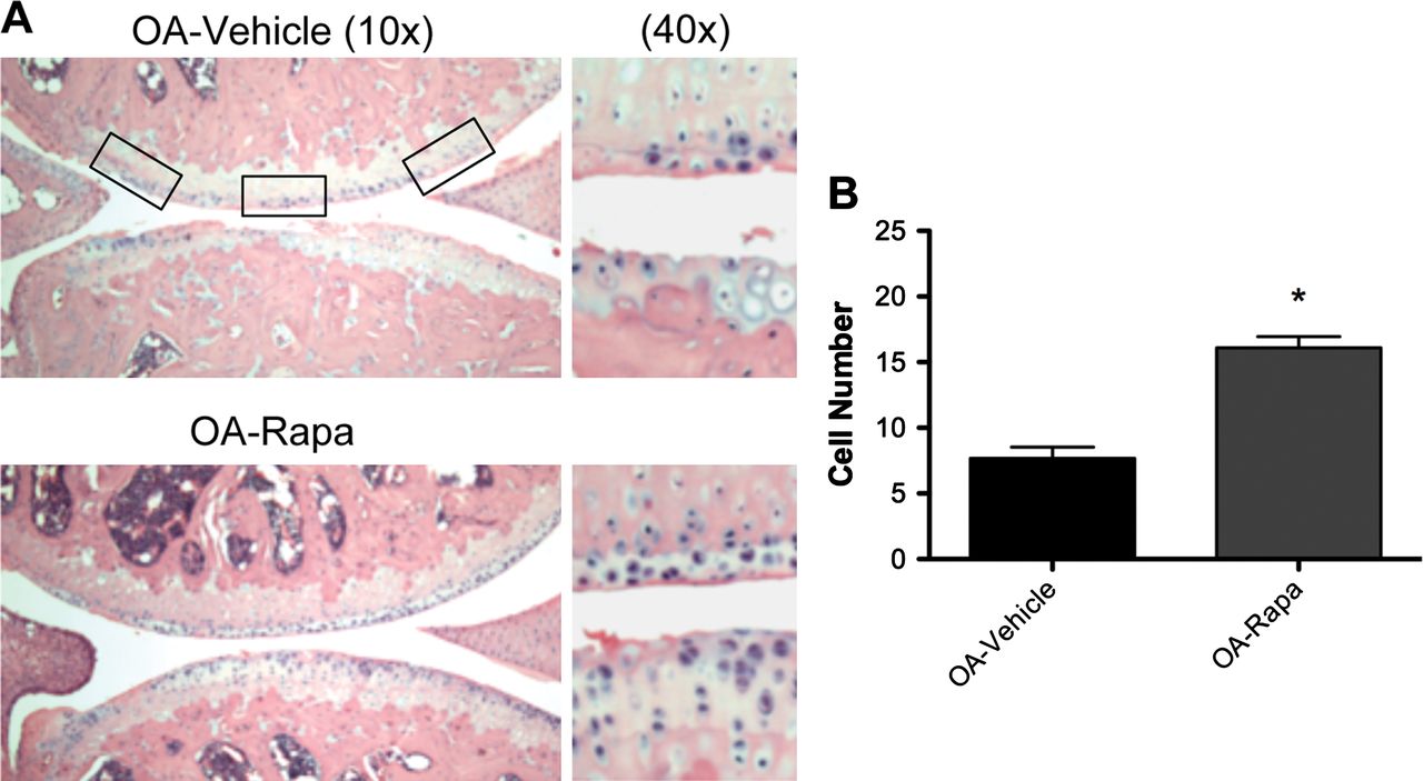
Rapamycin maintains cartilage cellularity and reduces ADAMTS-5 expression in experimental osteoarthritis
To determine the mechanism of action of rapamycin in experimental osteoarthritis, we analysed cartilage cell density in mouse knee joints. We observed preservation of cellularity after rapamycin treatment (figure 3A). This difference was significant (p<0.001) compared wtih vehicle-treated mice (figure 3B).

Rapamycin (rapa) prevents reduction in cartilage cellularity during experimental osteoarthritis (OA). (A) Knee joint sections from C57Bl/6J mice with or without osteoarthritis surgery under treatment with rapamycin or vehicle (n=6 each) were stained with H&E. Magnification: 10× and 40×. (B) Quantitative analysis of cell number showed a significant increase of cellularity after rapamycin treatment compared with vehicle treatment. Values are the mean±SD. *p<0.001 versus osteoarthritis vehicle.
Immunohistochemistry was performed for ADAMTS-5, a main proteinase responsible for aggrecan degradation in articular cartilage.28 ADAMTS-5 was expressed by a lower number of chondrocytes in rapamycin-treated mice compared with vehicle-treated mice with experimental osteoarthritis (figure 4A). This result was significant (p<0.05) compared with vehicle-treated knee joints (figure 4B). These findings indicate that rapamycin protects against cell loss and ECM damage by reducing ADAMTS-5 expression.

Rapamycin (rapa) reduces ADAMTS-5 expression. (A) Knee joints from C57Bl/6J mice were collected 10 weeks after osteoarthritis (OA) surgery and treatment with rapamycin or vehicle (n=5 each). Sections were analysed by immunohistochemistry for ADAMTS-5. Magnification: 10× and 40×. (B) Quantitative analysis of ADAMTS-5-positive cells. Total cell number in three fields and ADAMTS-5-positive cells were counted and the percentage of ADAMTS-5-positive cells was calculated. Results show a significant decrease in ADAMTS-5 after rapamycin treatment compared with vehicle. Values are mean±SD. *p<0.05 versus osteoarthritis vehicle.
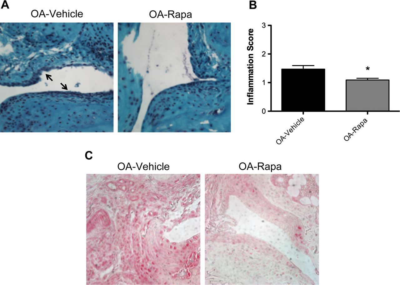
Effect of rapamycin on inflammation in experimental osteoarthritis
To study the effect of rapamycin on inflammation in the experimental osteoarthritis model, we examined histopathological changes using a semiquantitative grading system to measure inflammation in synovial tissue. The results indicate a decrease in synovial inflammation in response to rapamycin treatment compared with vehicle-treated mice (figure 5A). The decrease in the severity of inflammation after rapamycin treatment compared with vehicle-treated mice was significant (p<0.05) (figure 5B). Furthermore, IL-1β expression in synovial tissue was reduced after rapamycin treatment (figure 5C). Therefore, in addition to protective effects on articular cartilage, rapamycin also reduces the severity of synovitis and the synovial tissue level of IL-1β in mouse knees with experimental osteoarthritis.

Effect of rapamycin (rapa) on inflammation in experimental osteoarthritis (OA). (A) Synovium from mice after experimental osteoarthritis and vehicle (N=16) or rapamycin (N=11) treatment was analysed by staining with Safranin O. Magnification: 40×. (B) The histological score for inflammation was significantly decreased after rapamycin treatment. Values are mean±SD. *p<0.05 versus osteoarthritis vehicle. (C) Synovium sections from vehicle or rapamycin-treated mice (N=5 each) were analysed by immunohistochemistry for interleukin-1β. Magnification: 40×.
Discussion
Substantial progress has been achieved in understanding pathogenesis pathways in established osteoarthritis, and a large number of drug candidates were effective in osteoarthritis animal models. However, thus far no clinical trials showed disease-modifying activity of a large number of drug candidates tested.4 A potential explanation is that therapeutic agents were highly specific. For example, inhibiting one of a large number of ECM-degrading enzymes may not provide sufficient impact to change the overall course of the disease. Recent advances in understanding mechanisms in other ageing-related diseases have led to the concept that the failure of cellular homeostasis mechanisms, such as autophagy,5 6 leads to global changes in gene expression and can ultimately cause cell death and ECM destruction, two main features of osteoarthritis-affected cartilage. Based on this notion, we previously analysed human osteoarthritis joints and joints from experimental osteoarthritis models and observed a reduced expression in autophagy regulators that was associated with increased cell death.22
These observations motivated the present study to test whether the activation of autophagy can reduce the severity of experimental osteoarthritis. For this study we selected rapamycin, a specific and widely used inhibitor of the mTOR signalling pathway, which regulates autophagy initiation. Rapamycin, a lipophilic macrolide antibiotic used as an immunosuppressive drug, induces autophagy in a variety of cell types, extends lifespan and protects against ageing-related diseases in mice.15,–,21
Our results show that the systemic administration of rapamycin inhibited the mTOR signalling pathway in articular cartilage, as reflected by the reduced phosphorylation of ribosomal protein S6, which integrates processes of protein translation with cell growth and cell proliferation.29 Treatment of cells with rapamycin blocks S6K phosphorylation.30 Furthermore, rapamycin activated autophagy as indicated by increased LC3-II expression.31 32 These results confirmed the effect of systemically administered rapamycin on mTOR and autophagy pathways in articular cartilage.
In the experimental osteoarthritis model, we observed that rapamycin treatment caused a significant reduction in osteoarthritis severity by approximately 50%. The principal parameter measured in the osteoarthritis scoring system is the depth of the cartilage lesions and thus reflects ECM degradation. To address the potential mechanism by which rapamycin mediates this beneficial effect, we examined cartilage cellularity and levels of ADAMTS-5, a major aggrecan-degrading enzyme.33 The number of chondrocytes in knees with experimental osteoarthritis in rapamycin-treated mice was significantly increased compared with vehicle-treated mice. Furthermore, ADAMTS-5 expression was significantly decreased in cartilage from rapamycin-treated mice with osteoarthritis. The observed reduction in cartilage lesion depth and width could be mediated by a reduction in the expression of ADAMTS-5.
Osteoarthritis in humans and in experimental models is associated with inflammatory changes in synovium.34 In the present study, rapamycin also reduced the severity of synovitis. This was associated with a reduction in IL-1β expression after rapamycin treatment in synovium. These results are consistent with previous studies in which rapamycin significantly decreased synovial inflammation and protected against bone loss and cartilage destruction in an inflammatory arthritis model.35 36
The observed effects of rapamycin treatment on cartilage degradation, ADAMTS-5 and IL-1β expression, and synovial inflammation are a consequence of mTOR inhibition. Rapamycin is a highly specific inhibitor of mTOR and there is no evidence that it has off-target effects on other enzymes.37 38 It is well established that mTOR inhibition induces autophagy, but as direct and indirect consequences of mTOR inhibition other signalling pathways are affected, such as the PI3K/Akt signalling pathway.39 With regard to the role of autophagy activation in the effect observed in the present study, it is possible that rapamycin restores the suppressed autophagy, which we previously observed in experimental and human osteoarthritis,22 and as a result, inhibits cell death. The induction of cell death is a well-known effect of defective autophagy,40 and the preservation of cartilage cellularity observed in the present study can be due to autophagy activation.
The reduction of IL-1β and ADAMTS-5 expression observed after rapamycin treatment in the osteoarthritis model could be due to the removal of aggregation-prone proteins that stimulate the production of ROS. Both ROS accumulation and mTORC1 activation are associated with accelerated ageing and the development of ageing-associated pathologies.41 42 Conversely, a reduction of ROS levels by antioxidants or as a result of mTORC1 inhibition causes an extension of lifespan.43
One important limitation of rapamycin as a therapeutic molecule in humans is its immunosuppressive effect. The clinical feasibility of using mTOR inhibitors for the treatment of osteoarthritis is supported by recent advances in the development of new and safer rapamycin analogues. The treatment with rapamycin analogues with a short half-life might reduce systemic exposure to mTOR inhibitors and may show an improved safety profile compared with the lead compound.44
The present study is the first to establish the efficacy of rapamycin in an animal model of osteoarthritis. We used young mice subjected to surgical knee stabilisation. This is a widely used model for this purpose. For further preclinical development of autophagy activators it will be necessary also to test other animal species and older animals to model the human condition more realistically.
In summary, we report that rapamycin, at least partly by autophagy activation, reduces the severity of experimental osteoarthritis. These results suggest that the pharmacological inhibition of mTOR by rapamycin may be a potentially effective therapeutic approach for osteoarthritis.
Acknowledgments
The authors acknowledge Diana Brinson, Jean Valbracht and Lilo Creighton for technical assistance.
References
Footnotes
-
Funding This study was supported by National Institutes of Health grants AG007996, AR058954, RR027577 and the Sam and Rose Stein Endowment Fund. BC was supported by postdoctoral fellowship ‘Anxeles Alvariño’, Secretaria Xeral I+D+i, Xunta de Galicia, Spain.
-
Competing interests None.
-
Ethics approval This study was conducted with the approval of the Institutional Animal Care and Use Committee at The Scripps Research Institute.
-
Provenance and peer review Not commissioned; externally peer reviewed.