Article Text

Abstract
Objective Congenital heart block may develop in the fetuses of Ro/SSA-positive and La/SSB-positive mothers. Recurrence rates of only 10–20% despite persisting maternal antibodies indicate that additional factors are critical for the establishment of heart block. The authors investigated the influence of other maternal and fetal factors on heart block development in a Swedish population-based cohort.
Methods The influence of fetal gender, maternal age, parity and time of birth on heart block development was analysed in 145 families, including Ro/La-positive (n=190) and Ro/La-negative (n=165) pregnancies.
Results There was a recurrence rate of 12.1% in Ro/La-positive women, and no recurrence in Ro/La-negative women. Fetal gender and parity did not influence the development of heart block in either group. Maternal age in Ro/La-positive pregnancies with a child affected by heart block was, however, significantly higher than in pregnancies resulting in babies without heart block (p<0.05).Seasonal timing of pregnancy influenced the outcome. Gestational susceptibility weeks 18–24 occurring during January–March correlated with a higher proportion of children with heart block and lower vitamin D levels during the same period in a representative sample of Swedish women and a corresponding higher proportion of children with heart block born in the summer (p<0.02). Maternal age or seasonal timing of pregnancy did not affect the outcome in Ro/La-negative pregnancies.
Conclusion This study identifies maternal age and seasonal timing of pregnancy as novel risk factors for heart block development in children of Ro/La-positive women. These observations may be useful for counselling when pregnancy is considered.
Statistics from Altmetric.com
Congenital complete heart block without cardiac malformation is a rare disease, affecting 1 in 15 000–20 000 births in the general population. However, association with the presence of maternal autoantibodies to Ro/SSA and/or La/SSB is well established,1 2 and the risk of complete congenital heart block is 1–2% in Ro/SSA-positive pregnancies.3,–,6 Furthermore, the reported risk of giving birth to a second child with complete heart block for anti-Ro/SSA-positive mothers ranges from 12% to 20%,7,–,9 despite the persistence of maternal autoantibodies.10 This indicates that additional factors are critical for establishing heart block. Fetal genetic susceptibility has been suggested as a potential risk factor,11 12 and polymorphisms in the gene encoding tumor growth factor β have been implicated in the development of heart block.13 14 Variations in the intrauterine environment between pregnancies have also been suggested to contribute to the penetrance of the disease. Maternal disease severity has been investigated as such a potential risk factor but was not found to contribute to the development of congenital heart block.15
Given the rarity of congenital heart block occurrence in the general population, it is difficult to investigate potential risk factors associated with the disease. In particular, there is very little information on the influence of maternal age and parity on pregnancy outcome in anti-Ro/La-positive mothers. In an effort to address these questions in a reasonable cohort, we identified patients with heart block in the Swedish population via national healthcare and hospital registries and investigated maternal and fetal factors associated with heart block in 145 families. Variables analysed included maternal serological status, age and parity, as well as fetal gender and season of birth.
Patients and methods
Patients and families
Identification and enrolment of patients had been described in detail previously.16 Briefly, patients with heart block diagnosed before 15 years of age were identified using the Swedish National Patient Register (http://www.socialstyrelsen.se) and the Swedish Pacemaker and ICD Register (http://www.pacemakerregistret.se), as well as by local clinical registers and through a network of paediatric and adult cardiologists and rheumatologists at six university hospitals in Sweden. Four additional heart block cases with autoantibody-positive mothers were diagnosed after 15 years of age (at 18, 18, 30 and 36 years, respectively). Patients with cardiac structural abnormalities, as well as patients with postoperative or infection-induced block, were excluded from the study. Patients and their families were contacted and offered to take part in the study and to donate blood samples. Blood samples were collected from the mothers between 2007 and 2009. Families for whom (a) a blood sample from the mother and (b) information on the dates of birth of the mother and all siblings were available were included in the present study. In total, 145 families—including 149 individuals with heart block born between 1949 and 2009, 145 mothers and 208 siblings without heart block—were included in the study. Information on the definite time for heart block diagnosis could be retrieved from patient records for 82/84 cases with an autoantibody-positive mother and for 40/65 cases with an autoantibody-negative mother. For cases with an autoantibody-positive mother, 72/82 (88%) were congenital, as defined by Brucato et al17 (ie, diagnosed in utero or during the 27 first days of life), while 8/40 (20%) cases with an autoantibody-negative mother were diagnosed congenitally. Of all cases that could be defined as congenital (n=80), 72/80 (90%) had autoantibody-positive mothers. Excluding the four cases diagnosed at >15 years, the median age for the non-congenital heart block diagnosis was 4.0 years (range 0.25–15 years). The study was approved by the regional ethical committee of Karolinska Institutet. All participants—or the parents (if the individual was <18 years)—gave informed written consent.
Serological analyses
Autoantibodies were detected by Inno-Lia ANA (Innogenetics, Cambereley, UK) according to the manufacturer's instructions, and antibodies to Ro52 were further confirmed by ELISA.18
Vitamin D levels
For evaluation of vitamin D levels, data were obtained from female Swedish individuals (n=1068) who were selected as controls from the national population register, as described previously.19 The mean age of this group of women was 34.9 years (n=1064; information on age was not available in four cases). Vitamin D levels were measured as serum 25(OH) vitamin D by chemiluminescent assay, which recognises 100% of both serum 25(OH) D2 and serum 25(OH) D3) (Liason; DiaSorin, Saluggia, Italy).
Statistical analyses
Statistical analyses for fetal gender and time of birth were performed using χ2 test, and statistical analyses for recurrence rates were performed using Fisher's exact test. The difference in maternal age between antibody-positive and antibody-negative pregnancies leading to the birth of a child with or without heart block was evaluated by Kruskal–Wallis test, followed by Dunn's post test. The difference in maternal age at the birth of the first child was evaluated by Mann–Whitney U test. The OR of developing heart block associated with each maternal age category, with and without adjustment for parity, together with 95% CI, was calculated by logistic regression. The correlation between vitamin D levels for each month of the year and the ratio of the number of heart block pregnancies to the number of healthy pregnancies for which the 21st week fell in that particular month was assessed by non-parametrical Spearman correlation test. A p value of <0.05 was considered significant. Statistical analyses were performed using GraphPad Prism V.5.01 for Windows (GraphPad Software, San Diego, California, USA; http://www.graphpad.com), Statistica 8.0 (StatSoft, Tulsa, Oklahoma, USA) and SAS software for Windows V.9.1.
Results
Distinction of families with or without maternal autoantibodies
One hundred and forty-five families with at least one child diagnosed as having heart block were included in the study. In 80 families (55%), the mother carried autoantibodies to Ro and/or La; in 65 families (45%), the mother had no detectable anti-Ro/La autoantibodies.16 The presence versus the absence of maternal antibodies has been suggested to define two distinct groups of patients with heart block in terms of pathogenic mechanisms, risk of recurrence and age at diagnosis.20 We therefore investigated separately the maternal and fetal factors potentially associated with heart block in Ro/La-positive (n=190) and Ro/La-negative (n=165) pregnancies.
Recurrence of heart block
Of the 80 families enrolled in the study where the mother tested positive for anti-Ro/La antibodies, 33 (41%) had additional children after the birth of a child with heart block. Of the 33 pregnancies immediately following the birth of the first affected child, four resulted in the birth of another child with heart block, generating a recurrence rate of 12.1% (figure 1). The overall familial recurrence rate was 8.9%, with four affected pregnancies out of a total of 45, including all pregnancies occurring after the birth of a child with heart block.

Family size and birth order of heart block cases. Children with heart block are represented by solid black circles, whereas children without heart block are represented by open circles. A solid grey circle represents twins without heart block.
Of the 65 families where the mother was negative for the presence of anti-Ro/La antibodies, 36 (55%) included one or more siblings born after a child with heart block. However, none of the pregnancies following the birth of the first heart block case was affected, generating a recurrence rate of 0% in this group (figure 1). The difference between the recurrence rates in the groups of antibody-positive and antibody-negative women was statistically significant (p=0.047).
Fetal gender of patients with heart block versus healthy siblings
The influence of fetal gender on the development of heart block was investigated next. Female gender was observed in 52% of affected children and 56% of healthy children born to anti-Ro/La-positive mothers, indicating that fetal gender did not predict the occurrence of heart block (p=0.661). Similarly, no significant skewing of gender distribution was observed in the group of children born to anti-Ro/La-negative mothers, with 57% of the affected children and 53% of the healthy children being female (p=0.580).
Maternal age, but not parity, influences pregnancy outcome in Ro/La-positive mothers
While the risk of pregnancy complications generally increases with the age of the mother, little is known about the influence of maternal age or parity on pregnancy outcome and heart block development in Ro/La-positive women. We therefore investigated the influence of these maternal factors on the development of heart block.
We first analysed if there was any difference in the age of the mothers in the autoantibody-positive and autoantibody-negative groups. The age of the mother at the birth of a child with heart block was significantly higher in anti-Ro/La-positive women than in antibody-negative women: mean (95% CI), 29.5 (28.4 to 30.6) years versus 26.9 (25.8 to 27.9) years, respectively (p<0.01) (figure 2A). Since women with anti-Ro/La antibodies represent a group where the prevalence of autoimmune diseases, such as Sjögren's syndrome and systemic lupus erythematosus, will be much higher than in the group of anti-Ro/La-negative women, it is possible that the observed difference in maternal age results from a less favourable health status of antibody-positive women and delayed maternity in general. The difference in maternal age at the birth of the first child between antibody-positive and antibody-negative women was, however, not statistically significant (p=0.069, Mann–Whitney U test; figure 2B) and almost negligible when only non-affected children were considered (p=0.606).

Development of heart block in Ro/La-positive pregnancies is associated with increased maternal age but is independent of parity. (A) Maternal age at the birth of a child with or without AVB in anti-Ro/La-positive or anti-Ro/La-negative pregnancies. (B) Maternal age at the birth of the first child for antibody-positive and antibody-negative women. (A,B) Data shown as box plots (25th–75th percentiles), with line at median and with whiskers at minimum and maximum. (C) Age of antibody-positive women at the birth of their children, grouped by parity (first child to sixth child) and outcome (AVB or no AVB). Line at mean. (D) Maternal age at the birth of a child depending on antibody status (anti-Ro/La-positive or anti-Ro/La-negative) and pregnancy outcome (AVB or no AVB), presented as the mean with 95% CI. (E) Graphic representation of OR with 95% CI for giving birth to a child with heart block in autoantibody-positive women, dependent on maternal age and adjusted for parity. The reference group corresponds to maternal age of ≤24 years (n=16 AVB, n=35 no AVB); 25–29 years (n=28 AVB, n=36 no AVB); 30–34 years (n=24 AVB, n=26 no AVB); ≥35 years (n=16 AVB, n=11 no AVB). *p<0.05, **p<0.01. AVB, atrioventricular block.
We next compared the age of the mothers in anti-Ro/La-positive pregnancies resulting in a child with or without heart block. Anti-Ro/La-positive mothers were significantly older at the time of a pregnancy resulting in a child with heart block than at the time of birth of a non-affected baby: mean (95% CI), 29.5 (28.4 to 30.6) years versus 27.5 (26.5 to 28.5) years, respectively (p<0.05) (figure 2A). An obvious correlate of increasing age is increasing parity. To clarify the effect of parity on heart block development, we analysed the risk of having a child with heart block in relation to parity and maternal age. Stratifying the different pregnancies by parity, we observed that anti-Ro/La-positive mothers who gave birth to a child with heart block were consistently older than antibody-positive women who gave birth to a healthy child, independent of whether it was the first, second, third or higher-birth-order pregnancy (figure 2C,D). This trend was not observed in the group of antibody-negative mothers (figure 2D). Analysis of the influence of maternal age and parity on the risk of having a child with heart block in antibody-positive women revealed that only maternal age had a significant effect (p=0.01; logistic regression model analysis), while parity did not significantly affect pregnancy outcome (p=0.35).
To estimate the increased risk due to increasing maternal age, we calculated the OR for giving birth to a child with heart block in anti-Ro/La-positive women in relation to age (figure 2E and table 1). Increasing OR with increasing maternal age was observed: OR (95% CI), 2.3 (1.0 to 5.5, p=0.05) and 4.2 (1.4 to 11.9, p=0.01) for women 30–34 and ≥35 years of age, respectively, compared to women <25 years of age. In addition, we did not find any evidence of an interaction between maternal age and parity in terms of increased OR for the risk of heart block (p=0.21). No significant influence of maternal age or parity was observed in the group of anti-Ro/La-negative women (table 1).
OR of fetal heart block in relation to maternal age
Season of birth influences the development of congenital heart block
Evidence that the time of birth, as defined by season or month, may influence the development of autoimmune diseases later in life has emerged lately.21 Heart block in anti-Ro/La-positive pregnancies usually develops between 18 and 24 weeks of pregnancy, indicating that the pathogenic mechanisms leading to conduction defects in the fetal heart are initiated before and/or within this period of pregnancy. The 18–24 weeks of pregnancy taking place during the late Swedish winter months (January–March) correspond to a birth occurring during the Swedish summer (June–August), and we hypothesised that events linked to the winter season may affect the outcome of pregnancy and the ratio of healthy births to affected births during the summer, as compared to the rest of the year.
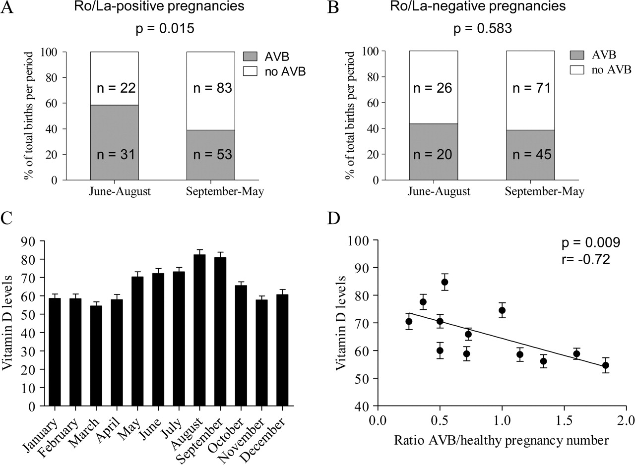
Analysis of the number of births during the summer (June–August) and the rest of the year revealed a significant difference between affected children and healthy siblings in the Ro/La-positive pregnancy group, with the births of children affected by heart block representing 58.5% of all births in the summer and only 39.0% of all births during the rest of the year (p=0.015; figure 3A). This skewed distribution towards an increase in the proportion of affected births in the summer was not observed in the Ro/La-negative pregnancy group (p=0.583; figure 3B).
Development of heart block shows a season-of-birth pattern and correlates to vitamin D levels during the susceptibility weeks (weeks 18–24) of pregnancy. (A,B) Proportions of affected (grey fields) and healthy (white fields) births during summer and the rest of the year for anti-Ro/La-positive (A) and anti-Ro/La-negative (B) mothers, represented as percentage of the total number of births during the respective periods. The number (n) of births in each group is indicated. (C) Vitamin D levels (mean, SEM) in a group of healthy Swedish women (n=1068), pooled by month according to blood sampling time. (D) Correlation between the average vitamin D levels for each month (mean±SEM) and the ratio of the number of heart block pregnancies to the number of healthy pregnancies for which week 21 of pregnancy falls in that particular month. p Value is calculated with the Spearman correlation test. AVB, atrioventricular block.
The significant increase in the proportion of children with heart block born in the summer, as compared to the rest of the year, supported our hypothesis that events linked to the winter season and occurring during susceptibility weeks 18–24 may affect the outcome of the pregnancy. Such a possible factor is the marked decrease in light exposure during winter in Nordic countries like Sweden that may affect vitamin D levels. We therefore analysed the variation of vitamin D levels over the year based on samples from 1068 Swedish women that had been taken at different times of the year. Vitamin D levels were higher during the summer, with the highest mean value observed in August, than during the winter months, with the lowest mean level recorded in March (figure 3C). We found that average vitamin D levels for each month were significantly correlated to the ratio of heart block to healthy pregnancies for which the median susceptibility week (defined as week 21 of pregnancy) fell in that particular month (p=0.009), with low vitamin D levels corresponding to a significantly higher proportion of heart block pregnancies (figure 3D).
Discussion
Isolated complete congenital heart block is well known to be associated with the presence of maternal anti-Ro/SSA and La/SSB antibodies. A recurrence rate of 12–20%, however, indicates that additional factors are involved in the pathogenic process. The objective of the present study was to investigate the influence of variables such as maternal age, parity, time of birth and fetal gender on the development of heart block. More specifically, we assessed the potential association of these factors with heart block by comparing heart block cases to healthy siblings in a cohort of families with autoantibody-positive mothers and also by analysing the same variables in a group of patients with heart block and healthy siblings born to autoantibody-negative mothers. The presence or the absence of maternal autoantibodies has indeed been suggested to define two distinct groups of patients with heart block differing in pathogenic mechanism, recurrence rate, prognosis and age at diagnosis.15 20 Congenital heart block is almost always associated with maternal anti-Ro/La seropositivity, whereas heart block detected later in infancy or during childhood is commonly not, as reflected also in the patients included in our study.
In the anti-Ro/La-positive mothers, we observed a recurrence rate of 12.1% in 33 pregnancies immediately following the birth of a child with heart block, and an overall recurrence rate of 8.9% in all siblings born after an affected child. While low recurrence rates have already been reported,22 recurrence rates of 17% in pregnancies immediately following the birth of a child with congenital heart block have been observed in large cohorts in two independent studies.8 23 However, recurrent cases in both of these studies may be over-represented, as the cohorts were formed either by specifically recruiting families with cases of neonatal lupus23 or based on registers from tertiary referral centres.8 By contrast, in this study, we analysed a population-based cohort gathered from national health registers in an effort to minimise the bias towards the recruitment of families with multiple heart block cases. It is also possible that the different recurrence rates observed between our cohort, including index cases from the Swedish population, and the cohort used by Julkunen and Eronen8, including cases from the Finnish population, are due to genetic or other differences between the two studied populations.
Fetal gender was not associated with development of heart block in Ro/La-positive or Ro/La-negative pregnancies, in accordance with previous reports.9 23 A trend towards higher maternal age for mothers who give birth to babies with heart block, compared to mothers who give birth to healthy babies, was previously reported by Skog et al.24 In the present study, we found that the risk of fetal heart block in Ro/La-positive pregnancies increased significantly with increasing maternal age, but was not influenced by parity. This finding was specific for autoantibody-associated heart block, as neither maternal age nor parity influenced the outcome of Ro/La-negative pregnancies.
Complications during pregnancy have been associated with increasing maternal age, affecting both the mother and the child (reviewed by Montan25). Disorders such as placental growth retardation and placental abruption are known to increase in frequency with age, mainly due to insufficient uteroplacental perfusion. However, there is little information about placental function in relation to maternal age. Increased maternal age might be associated with less effective placental function, potentially leading to episodes of low oxygenation, which may in turn influence the inflammatory reaction initiated in the fetal heart by the maternal antibodies. Hypoxic conditions have indeed been suggested to amplify the deleterious effects of maternal anti-Ro/La antibodies on the fetal heart,26 and placental function may also influence the outcome in twins, who have been reported to be both concordant and discordant for heart block.8 27 Hypothyroidism, which is known to be associated with ageing, may also be another factor of interest as anti-Ro-positive women with hypothyroidism have been shown to be at higher risk for having a child with complete congenital heart block compared to women with autoantibodies alone.28 It is also possible that the increasing risk for heart block with increasing maternal age reflects the appearance and/or increased serum levels of anti-Ro/La autoantibodies in women over time. This hypothesis could, however, not be addressed in the present study, as blood samples from mothers were only collected after the birth of a child with heart block.
Seasonal variation in the time of birth has recently been associated with the development of autoimmune diseases later in life (eg, multiple sclerosis).21 We speculated that events such as low wintertime vitamin D levels at the critical period of potential heart block development in fetuses of women with anti-Ro/La autoantibodies might affect the outcome of the pregnancy. This specific time window (18–24 weeks of gestation) during the winter corresponds to birth in the summer. In the present study, we found that the distribution of heart block births over the year was skewed towards an increased proportion of affected children born in the summer. Additionally, we found that vitamin D levels, which varied in a Swedish female population over the year in relation to the seasons, were inversely correlated to the number of heart block pregnancies in autoantibody-positive women. Although the vitamin D levels used in this study originated from a population of healthy Swedish women and not from the specific mothers of our cohort, it is reasonable to believe that these women would follow the same general trend. Despite the well-documented immunomodulatory role of vitamin D (reviewed by Mora et al29), its potential relation to the development of heart block in utero remains to be elucidated. While the association between the winter season, decreased sun exposure and vitamin D levels comes readily to mind, other events linked to the winter season, such as viral infections, might also influence the development of heart block. An increased frequency of viral infections during the winter season, when the vitamin D levels are low, could potentially augment the risk for heart block to develop. Although no link between infection and development of heart block has been shown to date, it may be a risk factor worth investigating further.
In summary, we report a recurrence rate of 12.1% in anti-Ro/La-positive pregnancies and demonstrate a significant association of increased maternal age and birth in the summer with development of heart block in antibody-positive pregnancies. These associations were not observed in children of antibody-negative women. Our data support the idea of different pathogenic mechanisms of heart block in anti-Ro/La-positive and anti-Ro/La-negative women and are also of interest to improve the counselling of antibody-positive women contemplating pregnancy.
Acknowledgments
The authors thank Drs Paul Blomqvist (Department of Medicine, Karolinska Institutet) and Solveig Wållberg-Jonsson (Department of Rheumatology, Umeå University Hospital) for valuable discussions and case contribution, respectively.
References
Footnotes
AA and SS contributed equally (shared first authorship).
-
Funding Financial support for this study was obtained from KIRCNET (Karolinska Institutet Circulation and Respiratory Research Network), the Magn. Bergvalls Foundation, the Jerring Foundation, Stiftelsen Samariten, the Karolinska Institute, The Royal Swedish Academy of Sciences, the Swedish Research Council, the Göran Gustafsson Foundation, the Torsten and Ragnar Söderberg Foundation, the King Gustaf Vth 80-Year Foundation, the Swedish Foundation for Strategic Research, the Heart–Lung Foundation and the Swedish Rheumatism Association.
-
Competing interests None.
-
Patient consent Obtained.
-
Ethics approval Regional ethical committee of Karolinska Institutet, Stockholm.
-
Provenance and peer review Not commissioned; externally peer reviewed.