Article Text

Abstract
Background Idiopathic thrombocytopenic purpura (ITP) may play a role in early-stage systemic lupus erythematosus (SLE). The incidence of SLE in patients with ITP and the potential relationship between them is still unclear. This study was performed to provide epidemiological evidence regarding the relationship between ITP and SLE occurrence.
Methods In this population-based retrospective cohort study, the risk of SLE was analysed in a cohort of patients newly diagnosed with ITP between 2000 and 2013. Controls were selected at a 1:2 ratio through propensity score matching (PSM) using the greedy algorithm. The Cox proportional hazard model was used to analyse the association between ITP and SLE incidence. There were four different Cox regression models, and the sensitivity analyses were implemented to evaluate the HR of SLE after exposure with ITP.
Results In the age-matched and sex-matched ITP and non-ITP cohort, the average follow-up time was about 80 months in this study. There were 34 (4.70%) and 27 (0.19%) incident cases of SLE in ITP and non-ITP group. The incidence rates were 62.0 (95% CI 44.3 to 86.8) and 2.10 (95% CI 1.44 to 3.06), respectively. The adjusted HR of incidental SLE in the ITP group was 25.1 (95% CI 13.7 to 46.0). The other risk factors for SLE were female sex and Sjogren’s syndrome. After PSM, the incidence rate and Kaplan-Meir curves of SLE were consistent with the results for the age-matched and sex-matched population, the HR 17.4 (95% CI 5.28 to 57.4) was estimated by conditional Cox model.
Conclusion This cohort study demonstrated that patients with ITP have a higher risk of SLE. Clinically, patients with ITP should be monitored for incidental lupus.
- systemic lupus erythematosus
- autoimmune diseases
- epidemiology
Statistics from Altmetric.com
Key messages
What is already known about this subject?
Idiopathic thrombocytopenic purpura (ITP) may play a role in early-stage systemic lupus erythematosus (SLE). The incidence of SLE in patients with ITP and the potential relationship between them is still unclear.
What does this study add?
There were literatures about ITP and lupus. However, most of them are either small sample size, nor short duration. For example, Moutsopoulos et al showed that haematological abnormalities such as thrombocytopaenia in ANA positive individuals are not associated with an evolution to SLE after a follow-up of 2–5 years. Olivera et al described four cases of immune thrombocytopenic purpura diagnosed from 7 months to 5 years (average 3 years) before juvenile SLE became evident.
Our study aimed to investigate this association by using a population-based database with 1 million population and 14 years follow-up. We believed this big data approach might reduce the selection bias and strengthen the grade of evidence. Moreover, by this big database, we can provide more information about ITP and SLE, including cumulative incidence, age stratification, as well as interactions with comorbidities.
How might this impact on clinical practice or future developments?
This cohort study demonstrated that patients with ITP have a higher risk of SLE. Clinically, patients with ITP should be monitored for incidental lupus.
Introduction
Idiopathic thrombocytopenic purpura (ITP) is an idiopathic bleeding disorder that is characterised by thrombocytopaenia and normal or increased numbers of bone marrow megakaryocytes. The pathogenesis of primary ITP remains unknown. Secondary ITP can be related to autoimmune disease, immunodeficiency and infection.1 The premature destruction of platelets by the reticuloendothelial system occurs due to the binding of autoantibodies to several platelet surface antigens.2 ITP is often accompanied by an increase in bone marrow megakaryocyte number.3 Clinically, ITP is mainly diagnosed based on the following criteria: (1) the reduced platelet count is detected at least twice, and the blood cell morphology is normal; (2) the spleen is generally small; (3) the number of megakaryocytes in the bone marrow is normal or increased, accompanied by maturation disorders and (4) other secondary thrombocytopaenia is excluded. Clinical manifestations of ITP are chills, fever and even sudden spontaneous mucosal bleeding in the skin.4
Systemic lupus erythematosus (SLE) is a chronic inflammatory autoimmune disease involving multiple systems, with a remitting or relapsing course.5 6 It is characterised by the production of various autoantibodies and by various clinical manifestations, although the more common early manifestations are arthritis, glomerulonephritis and haemocytopenia.7 Platelets play a crucial role in immunity, not only in the defence against pathogens,8 9 but also in the development of autoimmune diseases.10 Some studies had indicated that the platelet system is activated during SLE.11 Olivera et al described four cases of immune thrombocytopenic purpura diagnosed from 7 months to 5 years (average 3 years) before juvenile SLE became evident.12 On the contrary, in a retrospective study, Moutsopoulos et al showed that haematological abnormalities such as thrombocytopaenia in anti-nuclear antibody (ANA) positive individuals are not associated with an evolution to SLE after a follow-up of 2–5 years.13 Most of previous studies are either small sample size, nor short duration. Hence, our study aimed to investigate this association by using a population-based database with 1 million population and 14 years follow-up, and to analyse the incidence of ITP in SLE, including data on cumulative incidence, and age stratification, as well as interactions with comorbidities.
Patient and methods
Data sources
Data from the National Health Insurance Research Database (NHIRD) of Taiwan were used in this study, which is maintained by the National Research Agency for research purposes. The NHIRD includes the data of more than 99% of Taiwan’s population. The database contains information on diagnoses, hospitalisations, examinations and prescriptions. The informed consent was waived because of the retrospective nature of observational study and the anonymous datasets were used.
Study participants and patient inclusion and exclusion criteria
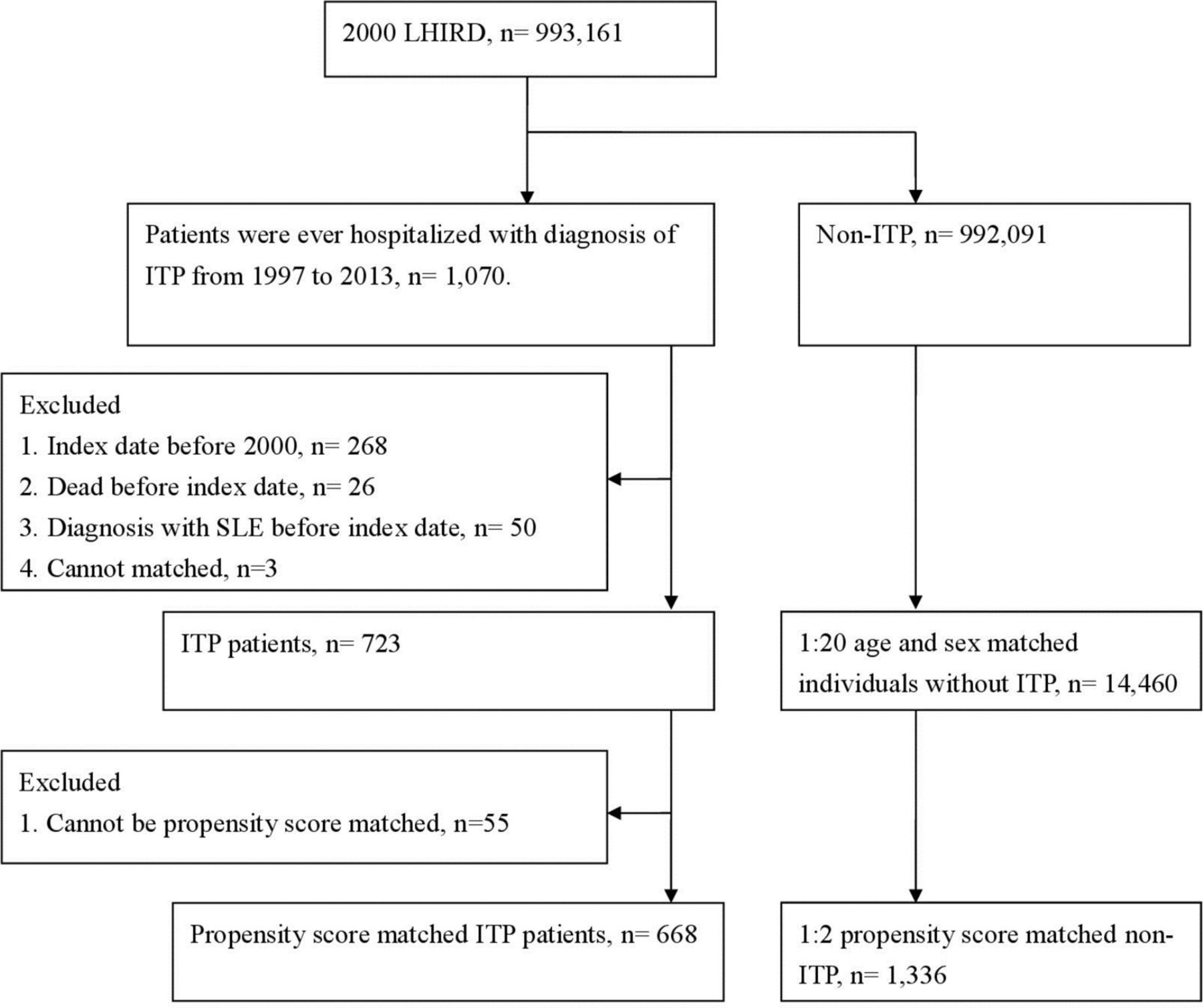
As shown in figure 1, Taiwan’s NHIRD enrolled a total of 993 161 patients. Diseases were defined according to International Classification of Diseases, Ninth Revision (ICD-9) codes. The participants of the present study were hospitalised patients diagnosed with ITP (ICD-9-Clinical Modification (CM) code 287.3) during 1997–2013 (n=1070). The index date was defined as the first date of admission with ITP +28 days. The exclusion criteria were as follows: (1) index date before 2000 (n=268); (2) died before the index date (n=26); (3) SLE diagnosis before the index date (n=50) and (4) no age-matched and sex-matched controls (n=3). Finally, 723 patients were included in this cohort. Follow-up started on the index date and ended at SLE occurrence, death or end of the study (31 December 2013).

Flow chart of the study design. ITP, idiopathic thrombocytopenic purpura; SLE, systemic lupus erythematosus.
Non-ITP controls
We then matched ITP patients at a ratio of 1:20 to 14 460 controls by sex and age. We performed propensity score matching (PSM) at a 1: 2 ratio to minimise the potential confounding effects of sex, age and selected comorbidities on the incidence of SLE. In order to make the frequency of selected variables uniform in the study and control cohort. Then we included PSM to identified 668 IPT patients and 1336 individuals without ITP.
There were 992 091 patients who had never hospitalised for ITP in Longitudinal Health Insurance Database (LHID) 2000. We then matched patients with ITP to 14 460 controls at a 1:20 ratio by sex and age at the index date, while the controls were all at risk at index date.
Outcome and relevant variables
Patients diagnosed with SLE (ICD-9-CM code 710.0) with at least three outpatient visits or one hospital admission within 1 year were defined as the main outcome for this study. The comorbidities analysed in this study were rheumatoid arthritis (ICD-9-CM code 714.0), Sjogren’s syndrome (ICD-9-CM code 710.2), systemic sclerosis (ICD-9-CM code 710.1), vasculitis (ICD-9-CM code 443.0), hypertension (ICD-9-CM codes 401–405), diabetes mellitus (ICD-9-CM code 250), hyperlipidaemia (ICD-9-CM codes 272.0–272.4), coronary artery disease (ICD-9-CM codes 410–414), osteoporosis (ICD-9-CM code 733), cerebral vascular accident (ICD-9-CM codes 430–438), asthma (ICD-9-CM code 493), chronic obstructive pulmonary disease (ICD-9-CM codes 490–492 and 493–496), chronic kidney disease (ICD-9-CM code 585), chronic liver diseases (ICD-9-CM codes 571 and 573), hyperthyroidism (ICD-9-CM code 242), thyroiditis (ICD-9-CM code 245), pancreatitis (ICD-9-CM codes 5770 and 5771), affective psychosis (ICD-9-CM code 296) and ankylosing spondylitis (ICD-9-CM code 720.0), inflammatory bowel disease (ICD-9-CM codes 555–556), human immunodeficiency virus infection (ICD-9-CM codes 042–044, V08) and antiphospholipid antibody syndrome (ICD-9-CM codes 289.8).
Statistical analysis
The absolute standardised difference (ASD) was used to evaluate the balance of baseline characteristics in the age-matched and sex-matched and propensity score-matched populations. Generally, ASD <0.1 is considered a small difference. The incidence rate (per 100 000 person-months) of SLE was calculated, and the crude relative risk (and its 95% CI).14 was estimated through Poisson regression. The Kaplan-Meier curves were plotted to determine the cumulative incidence of SLE since the index date. The log-rank test was used to compare the difference in cumulative incidence. Multiple Cox proportional hazard regression was used to estimate the adjusted HR (aHR) of SLE in the patients with ITP after controlling for covariates (listed in table 1). We used four models to explore the HR of SLE for ITP exposure and covariates,14 the models including (1) model 1, the predictor is ITP exposure alone, (2) model 2, ITP exposure +demographic variables, (3) model 3, ITP exposure +demographic variables+medical utilisation and comorbidities at baseline in age-matched and sex-matched population. (4) Model 4, the conditional Cox model with ITP exposure alone in propensity score-matched population, to estimate the effect of ITP on the risk of SLE. The sensitivity analysis was implemented to evaluate the HR of SLE after exposure with ITP. Statistical significance was defined as p<0.05. All statistical analyses were performed using SAS V.9.4 software (SAS Institute).
Baseline characteristics among ITP group and non-ITP group
Results
Demographic characteristics and comorbidities
Given that SLE has a low incidence, the control group was first matched by sex and age at a ratio of 1:20. Eligible study participants were 723 patients in the ITP group and 14 460 age-matched and sex-matched individuals in the control (comparison) group. Further, for comparison, PSM was conducted at a ratio of 1:2 (only this ratio was selected, and a higher ratio could not provide a suitable control). Finally, 668 patients with ITP and 1336 controls after PSM (figure 1) were included. Although number of women was slightly more (57%) than men, the difference in age and sex between the two groups was non-significant (table 1). The prevalence of ITP comorbidities in the age-matched and sex-matched population was high, as shown in table 1. Following PSM, most baseline characteristics were balanced. The mean (SD) follow-up time of the ITP and control groups was 76.85 (52.00) and 86.20 (50.83) months, respectively.
Comparison of SLE incidence and HR between ITP and SLE and comparison groups
Before PSM, the incidence of SLE in the ITP group and the control group was 62.0 and 2.10 per 100 000 person-years, respectively. After PSM at a 1:2 ratio, the incidence of SLE in the ITP group and the control group was 56.5 and 2.60, respectively. Before PSM (age and sex matching at a 1:20 ratio), the crude relative risk of SLE in the ITP group was 29.6 (95% CI 17.9 to 49.0) compared with the control group (table 2). After PSM at a 1:2 ratio, the crude relative risk of SLE for the ITP group was 21.7 (95% CI 6.6 to 71.2). The survival curve (figure 2) revealed that the cumulative incidence of SLE was higher in the ITP group than in the comparison group (p<0.0001).

The cumulative incidence of SLE for patients with and without ITP, (A) age-matched and sex-matched population. (B) Propensity score-matched population. ITP, idiopathic thrombocytopenic purpura; PSM, propensity score matching; SLE, systemic lupus erythematosus.
Incidence of SLE in PSM study group
Proportional hazard regression used to estimate the aHR of SLE
Table 3 indicated the results of Cox regressions, the HRs of SLE for ITP exposure were 28.7 (95% CI 17.3 to 47.6), 28.5 (95% CI 17.2 to 47.3), 25.1 (95% CI 13.7 to 46.0) and 17.4 (95% CI 5.28 to 57.4) through model 1 to model 4. The consistent findings of significant higher risk of SLE in ITP patients were found across four different regression models. In model 4, we also found female had increased risk (aHR=20.5, 95% CI 4.94 to 84.9) of SLE compared with male. The patients who had comorbidity with Sjogren syndrome (aHR=6.02, 95% CI 1.33 to 27.3) got significantly excess relative risk of SLE.
Cox proportional hazard regressions for estimation of adjusted HRs on SLE
Sensitivity analysis for estimating the aHR of SLE
Table 4 provided four scenarios to test the stability of HR of SLE in different definition of ITP exposure and SLE event. In NHIRD, there was no laboratory report to identify the cases of SLE. We try to develop three scenarios, listed in the table 4, to improve the accuracy of diagnosis with SLE. We found the aHRs were 25.1 (95% CI 13.7 to 46.0), 32.0 (95% CI 16.8 to 61.1) and 39.7 (95% CI 19.1 to 82.4) in scenarios 1, 2 and 3. The higher association (aHR) was found between ITP exposure and risk of moderate or severe SLE. Furthermore, we excluded the cases potentially had secondary ITP, in scenario 4, showed the aHR=27.0 (95% CI 14.4 to 50.5) of SLE for ITP exposure.
Sensitivity analysis in the estimation of the SLE risk for ITP exposure in age-matched and sex-matched population
Discussion
This study is the first retrospective cohort study that used nationwide population-based data to assess the risk of SLE in patients with ITP. The present study demonstrated that the patients with ITP had a 26 times higher risk of new-onset SLE compared with the control population. Furthermore, the consistent findings of significant higher risk of SLE in patients with ITP were found across four different regression models. we also found that the men had a decreased risk of SLE compared with the women. The patients who had comorbidity with Sjogren syndrome got significantly excess relative risk of SLE.
Previous studies revealed that thrombocytopaenia is a common clinical manifestation and is one of the haematological criteria of SLE,15 and it is the first manifestation in 5% of the patients with SLE, occurring in 20% of the patients.16 Moreover, 1%–5% of adult patients with ITP develop SLE later.17 SLE and ITP have similar clinical manifestations, and it has been reported that 3%–16% of patients with ITP may develop SLE.18 Zhao et al reported that 12.8% of patients with thrombocytopaenia in SLE at the first episode were diagnosed with ITP.19 Keita et al found that initial haematological abnormalities may be the first clinical manifestation of SLE.20 Our study was consistent with previous studies and had provided more information on long-term cumulative incidence and interaction with comorbidities.
The possible mechanisms of ITP in SLE remain unknown. Several studies from different countries have assessed the association between ITP and SLE risk but have not yet arrived at a consensus.21 22 Severe organ involvement in lupus is associated with thrombocytopaenia.19 Thrombocytopaenia indicates a poor prognosis of SLE.23 Thrombocytopaenia is an independent risk factor of death and has been found to be associated with higher disease activity and greater damage in different SLE cohorts.24 Thrombocytopaenia is associated with more serious diseases and has a negative effect on the survival of patients with SLE.25 Thrombocytopaenia has been shown to be a predictor in patients with higher disease activity, likelihood of end-organ damage and mortality.19 26 An obvious negative correlation was found between platelet and B-cell percentage, confirming the functional role of B cells in the pathogenesis of autoimmune thrombocytopaenia.27 Min et al confirmed that patients with ITP BAFF and CD4+ T-cell expression of peripheral blood have significantly higher than healthy individuals.28 Thai et al 29 observed that BAFF could enhance the survival rate of spleen plasma cells and induce B-cell resistance during ITP cell failure treatment. These findings about ITP prove the link between BAFF and CD4+ T cells, and that ITP can induce B-cell resistance.
Our present study is noteworthy for several reasons. First, this was the national study on the association between ITP and the subsequent development of SLE. Second, this was a nationwide study in the Taiwanese population; this study provided a sufficiently large sample size to avoid selection bias. Third, the 16-year long-term population data were extracted from the LHID from 1997 to 2013. Therefore, the results are considered more comprehensive.
This study has several limitations. First, the cause–effect relationship was not established. Second, we included patients with ITP from the LHID 2000 database. Thus, some patients who may not have been adequately diagnosed with ITP were included in the cohort. Furthermore, a similar study on the relationship between ITP and SLE should include patients from other races and countries. There was no study provide the validation of diagnosis with SLE or ITP, however, in the sensitivity analysis (table 4), we observed consistent results in different definition of SLE and ITP.
Conclusion
This study demonstrated that patients with ITP are at a higher risk of SLE. Clinically, patients with ITP should be educated and monitored for the risk of incidental SLE.
Acknowledgments
The authors thank all patients and staffs who made this study possible.
References
Footnotes
Handling editor Josef S Smolen
Contributors F-XZ analysed the data, generated figures and wrote the manuscript. J-YH, ZY and Q-QW performed bioinformatics analysis and wrote the manuscript. JC-CW made substantial contributions to the design of the study, conducted data analysis and figure generation, and wrote the manuscript. All authors read and approved the final manuscript.
Funding The present study was supported by the Programme of Scientific and Technology Project (Guilin Science Research and Technology Development; grant no. 2016012706–2).
Competing interests None declared.
Patient and public involvement Patients and/or the public were not involved in the design, or conduct, or reporting, or dissemination plans of this research.
Patient consent for publication Not required.
Ethics approval This study was approved by Chung Shan Medical University Hospital (IRB, CS15134).
Provenance and peer review Not commissioned; externally peer reviewed.
Data availability statement All data relevant to the study are included in the article or uploaded as online supplementary information. Please fix the following issues then click Save or Save & Continue:Permissions—images is a required field.