Article Text

Abstract
Objectives The release of neutrophil extracellular traps (NETs) represents a novel neutrophil effector function in systemic lupus erythematosus (SLE) pathogenesis. However, the molecular mechanism underlying NET release and how NETs mediate end-organ injury in SLE remain elusive.
Methods NET formation and NET-related proteins were assessed in the peripheral blood and biopsies from discoid lupus and proliferative nephritis, using immunofluorescence, immunoblotting, quantitative PCR and ELISA. Autophagy was assessed by immunofluorescence and immunoblotting. The functional effects of NETs in vitro were assessed in a primary fibroblast culture.
Results Neutrophils from patients with active SLE exhibited increased basal autophagy levels leading to enhanced NET release, which was inhibited in vitro by hydroxychloroquine. NETosis in SLE neutrophils correlated with increased expression of the stress-response protein REDD1. Endothelin-1 (ET-1) and hypoxia-inducible factor-1α (HIF-1α) were key mediators of REDD1-driven NETs as demonstrated by their inhibition with bosentan and L-ascorbic acid, respectively. SLE NETs were decorated with tissue factor (TF) and interleukin-17A (IL-17A), which promoted thrombin generation and the fibrotic potential of cultured skin fibroblasts. Notably, TF-bearing and IL-17A-bearing NETs were abundant in discoid skin lesions and in the glomerular and tubulointerstitial compartment of proliferative nephritis biopsy specimens.
Conclusions Our data suggest the involvement of REDD1/autophagy/NET axis in end-organ injury and fibrosis in SLE, a likely candidate for repositioning of existing drugs for SLE therapy. Autophagy-mediated release of TF-bearing and IL-17A-bearing NETs provides a link between thromboinflammation and fibrosis in SLE and may account for the salutary effects of hydroxychloroquine.
- neutrophils
- autophagy
- lupus
- fibrosis
- thromboinflammation
This is an open access article distributed in accordance with the Creative Commons Attribution Non Commercial (CC BY-NC 4.0) license, which permits others to distribute, remix, adapt, build upon this work non-commercially, and license their derivative works on different terms, provided the original work is properly cited, appropriate credit is given, any changes made indicated, and the use is non-commercial. See: http://creativecommons.org/licenses/by-nc/4.0/.
Statistics from Altmetric.com
Key messages
What is already known about this subject?
In systemic lupus erythematosus (SLE), neutrophils display excessive cell death by forming extracellular chromatin traps (the so-called neutrophil extracellular traps (NETs)) but the mechanism underlying their release and the resultant tissue injury are not known.
What does this study add?
Excessive NET production by SLE neutrophils is driven by autophagy, a process normally involved in degradation and recycling of cellular components.
Lupus serum induces in neutrophils autophagy and NETosis by upregulating the hypoxia and stress-response protein DDIT4/REDD1.
NETs from active SLE neutrophils show abundant expression of bioactive tissue factor and interleukin-17A, which promote thromboinflammation and fibrosis in target tissues such as kidneys and skin.
Endothelin-1 and hypoxia inducible factor-1α are key mediators of neutrophil-driven end-organ injury in SLE, through the REDD1/autophagy axis.
How might this impact on clinical practice?
Targeting the REDD1/autophagy axis or its mediators by existing agents through drug repositioning or other novel agents may alleviate neutrophil-mediated inflammation in SLE.
Introduction
Genome-wide association studies and gene expression analyses have implicated neutrophils and deregulated autophagy in systemic lupus erythematosus (SLE).1–7 Specifically, neutrophils have emerged as key players in the disease pathogenesis through putative effector functions including neutrophil extracellular traps (NETs).8 NETs are networks of extracellular fibres, comprised of extruded nuclear DNA and associated granular components, histones and cytoplasmic proteins.9 However, the molecular mechanism underlying NET release and how NETs mediate end-organ injury in SLE are unknown.
Recent data suggest that the autophagic pathway—a homeostatic catabolic mechanism involving degradation of cell components—may be required for NETs release.10 11 NETs represent a common denominator across different disorders; however, depending on the inflammatory context of each pathophysiological condition, neutrophils may express and release through NETs distinct bioactive proteins involved in different biological processes.12 To this end, the protein composition of NETs in SLE and their contribution to tissue injury have not been explored. Recently, focus has shifted on the role of NETs-expressing tissue factor (TF), the main in vivo initiator of the coagulation, as a mediator of thromboinflammation.11 In addition, interleukin-17A (IL-17A), a proinflammatory cytokine implicated in SLE and lupus nephritis (LN),13 promotes NET-dependent lung fibrosis.14
Although the presence of NETs in SLE has been associated with type I interferon production and vasculopathy,15 16 the underlying mechanism that regulates their release and their role in SLE inflammation and fibrosis remain unknown. Here in, we demonstrate that the inflammatory microenvironment of active SLE upregulates the expression of hypoxia-response and stress-response protein DDIT4/REDD1 (herein after referred as REDD1) in neutrophils, leading to autophagy induction and formation of TF-decorated and IL-17A-decorated NETs. We also demonstrate thromboinflammatory NETs in kidney and skin sections derived from patients with active SLE, linking them with end-organ injury and fibrosis.
Materials and methods
Patients and sampling
Peripheral blood neutrophils and sera were isolated from six patients with SLE during active (SLEDAI-2K>8) and then inactive (SLEDAI-2K<3) disease. SLE was diagnosed according to the 1997 Update of the 1982 American College of Rheumatology classification criteria.17 Six sex-matched/age-matched healthy individuals served as controls (online supplementary table 1). Kidney biopsies were obtained from six patients with active proliferative LN. Kidney biopsies, obtained from a patient with renal carcinoma, a patient with minimal change disease and a patient with membranous nephropathy served as controls. Skin biopsies were obtained from three patients with active discoid lupus, both from the active lesion and the normal skin of each patient. Unaffected skin tissue from three healthy individuals was used as control. Human skin specimens from healthy individuals were used to isolate primary human skin fibroblast (HSFs).18 Written informed consent was obtained from all participants. The study protocol was in accordance with the Helsinki Declaration. Detailed information for all methods can be found in the online supplementary materials and methods. Results from each patient are provided in online supplementary figures 8–11.
Supplemental material
Results
Increased basal autophagy of peripheral neutrophils from patients with SLE is correlated with NETosis
Patients with SLE are characterised by a strong neutrophil and deregulated autophagy gene signature.3–7 19 Since SLE neutrophils exhibit ex vivo increased NET release15 20–22 and autophagy is a key mechanism regulating NET generation,10 11 we assessed autophagy levels in ex vivo isolated, unstimulated neutrophils from patients with SLE. Neutrophils from patients with active SLE (active SLE neutrophils) demonstrated increased basal autophagy levels as compared with neutrophils from healthy individuals (control neutrophils) and patients with inactive SLE (inactive SLE neutrophils), as evidenced by immunofluorescence for the autophagy protein LC3B (figure 1A,B) and immunoblotting for both the lipidated LC3B-II (figure 1C) and the consumption of p62/SQSTM1 (figure 1D).

Peripheral blood neutrophils isolated from patients with active systemic lupus erythematosus (SLE) are characterised by increased basal autophagy levels. (A) Autophagy induction assessed with LC3B staining (confocal microscopy; red: LC3B, blue: 4′,6-diamidino-2-phenylindole (DAPI)/DNA, 45 min incubation) in active SLE neutrophils compared with inactive SLE or control neutrophils. (B) LC3B puncta/cell are depicted. (C) LC3B-I/ΙΙ (45 min incubation) and (D) p62/SQSTM1 (90 min incubation) immunoblotting. For (C) and (D) integrated optical density (IOD) ratio of LC3BII/glyceraldehyde 3-phosphate dehydrogenase (GAPDH) and p62/GAPDH relative to control. For (B)–(D), data presented as mean±SD, ***p<0.001. For (A)–(D), one representative experiment of 6 is shown, n=6 patients.
To address whether increased autophagy levels may be driven by inflammatory mediators present in SLE serum, control neutrophils were stimulated in vitro with serum derived from patients with active (active SLE serum) or inactive (inactive SLE serum) SLE. Active SLE serum induced increased autophagy levels in contrast to inactive SLE serum, as demonstrated by immunofluorescence for LC3B (online supplementary figure 1A) and diminished p62/SQSTM1 immunoblotting (online supplementary figure 1B).
Supplemental material
Next, we observed that active SLE neutrophils exhibited increased NET release compared with control or inactive SLE neutrophils (figure 2A,B), myeloperoxidase (MPO)-DNA complex ELISA in ex vivo cell culture supernatants (figure 2C) and MPO-DNA complex ELISA measured directly in serum derived from patients with SLE (figure 2D). Inhibition of autophagy with early-stage or late-stage autophagy inhibitors, wortmannin or hydroxychloroquine (HCQ), respectively, significantly attenuated this effect (figure 2A–C). Similar findings were observed in control neutrophils cultured with active SLE serum in the presence of autophagy inhibitors (wortmannin, HCQ, AKT activator or Bafilomycin A1) (online supplementary figure 2A-C) or with low dose of rapamycin (online supplementary figure 2C). Together, these findings suggest that lupus inflammatory microenvironment induces NETs in an autophagy-dependent manner, which can be reversed by autophagy inhibitors.
Supplemental material

Neutrophil extracellular trap (NET) formation is increased in peripheral blood neutrophils of patients with active systemic lupus erythematosus (SLE) and is mediated by autophagy. (A) NET formation (3 hours incubation) in isolated peripheral neutrophils from patients with active SLE compared with inactive SLE or control subjects. Wortmannin (30 min pretreatment) was used as an early stage autophagy inhibitor, while hydroxychloroquine (HCQ, 30 min pr-treatment) as a late stage autophagy inhibitor (confocal microscopy; green: neutrophil elastase (NE), red: citrullinated histone H3 (CitH3), blue: 4′,6-diamidino-2-phenylindole/DNA). (B) Percentage of NET-releasing neutrophils (3 hours incubation). Myeloperoxidase-DNA complex measured (C) in NET structures released from patients’ neutrophils or (D) directly in blood serum derived from patients with active SLE compared with patients with inactive SLE or control subjects. For (B)–(D), data presented as mean±SD, ***p<0.001. For (A)–(D), one representative experiment of 6 is shown, n=6 patients.
NETs from patients with active SLE are decorated with TF and IL-17A
The protein composition of NETs plays a crucial role in disease pathogenesis.12 We focused on two inflammatory mediators which have been previously implicated in NET-associated inflammatory injury, namely, TF and IL-17A. TF/thrombin axis has a key role in thromboinflammation11 23–25 which often complicates SLE.26 IL-17A is a proinflammatory cytokine implicated in SLE pathogenesis and severe LN.27–30 Indeed, we have demonstrated that IL-17A-bearing NETs have a potent fibrotic role.14 Accordingly, we investigated whether TF and IL-17A are externalised via SLE NETs and could represent a link between increased thrombogenicity and fibrosis observed in the disease.
We observed that NETs from patients with active SLE (active SLE NETs) expressed TF (figure 3A,B) and IL-17A (figure 3C,D), as assessed by immunofluorescence and immunoblotting on NET structures. Importantly, TF on active SLE NETs was bioactive, since NET structures increased thrombin levels in healthy platelet-poor plasma, as assessed by thrombin-antithrombin assay (figure 3E). This effect was TF-dependent as shown by inhibition of TF with an anti-TF neutralising antibody (figure 3E). NETs from patients with inactive SLE (inactive SLE NETs) demonstrated reduced thrombin generation when compared with active SLE NETs (figure 3E). These findings were further supported in vitro, where active SLE serum upregulated intracellular TF and IL17A mRNA expression in control neutrophils (online supplementary figure 3A-B) and induced NETs bearing functional TF (online supplementary figure 3C-D) and IL-17A (online supplementary figure 3E). Thus, the inflammatory microenvironment of SLE induces NETs decorated with TF and IL-17A.
Supplemental material

Patients with active systemic lupus erythematosus (SLE) release tissue factor (TF) and interleukin (IL)-17A-bearing neutrophil extracellular traps (NETs). (A) Localisation of TF on NETs released by isolated peripheral neutrophils from patients with active SLE compared with inactive SLE (confocal microscopy; green: TF, red: neutrophil elastase (NE), blue: 4′,6-diamidino-2-phenylindole (DAPI)/DNA). (B) TF expression in purified NET proteins from peripheral neutrophils from patients with active SLE. (C) Localisation of IL-17A on NETs released by isolated peripheral neutrophils from patients with active SLE compared with inactive SLE (confocal microscopy; green: IL-17A, red: NE, blue: DAPI/DNA) and (D) IL-17A expression in purified NET proteins. (E) Thrombin levels in control plasma incubated with NET structures (thrombin-antithrombin assay). Neutralising anti-TF antibody was used to inhibit TF-mediated thrombin generation. A mouse immunoglobulin (Ig)G antibody was used as isotype control. For (B) and (D), integrated optical density (IOD) relative to control, one representative experiment of 4 is shown, n=4 patients. For (B), (D), and (E), data presented as mean±SD, ***p<0.001. For (A), (C) and (E), one representative experiment of 6 is shown, n=6 patients. For (A)–(E), neutrophils were harvested after 3 hours of incubation.
Hypoxia-inducible factor-1α (HIF-1α) and endothelin-1 (ET-1) induce REDD1 expression in SLE neutrophils that activates the REDD1/autophagy pathway mediating NET release
Next, we sought to identify pathways that drive the autophagy-mediated NET release in SLE. Since we have recently shown that the stress-induced protein REDD1 regulates NET release through autophagy induction,31 we examined the involvement of REDD1/autophagy pathway in the formation of SLE NETs. Active SLE neutrophils demonstrated increased REDD1 mRNA and protein expression as compared with control or inactive neutrophils (figure 4A,B). Active SLE serum was also able to induce REDD1 in control neutrophils (figure 4C,D).

Hypoxia factor-1α (HIF-1α) and endothelin-1 (ET-1) are involved in REDD1-driven neutrophil extracellular trap (NET)osis in active systemic lupus erythematosus (SLE). (A) REDD1 mRNA (45 min incubation) and (B) REDD1 protein (45 min incubation) levels in active SLE neutrophils, compared with inactive SLE or control neutrophils. (C) REDD1 mRNA levels and (D) REDD1 (45 min incubation) or (E) p62/SQSTM1 (90 min incubation) immunoblotting in control neutrophils stimulated with active SLE serum in the presence of specific inhibitors (30 min pretreatment); L-ascorbic acid (10 mM), a HIF-1α inhibitor or bosentan (10 μM), an ET-1 receptor antagonist. (F) MPO-DNA complex in isolated NET structures. For (B), (D) and (E), integrated optical density (IOD) relative to control. For (A)–(F), data presented as mean±SD, ***p<0.001, one representative experiment of 6 is shown, n=6 patients.
To define the mechanism of increased REDD1 in SLE neutrophils, we focused on the upstream mediators HIF-1α and ET-1. HIF-1α, an oxygen sensitive transcription factor affecting numerous immune cells,32 involved in the mechanistic target of rapamycin (mTOR) system33 and an upstream regulator of REDD1 signalling34 is expressed in the kidneys and is found increased in urine of patients with LN.35–37 To this end, control neutrophils were pretreated with L-ascorbic acid, a HIF-1α inhibitor or a specific HIF-1α inhibitor (C26H29NO5) resulting in significant reduction in REDD1 levels (figure 4C,D, online supplementary figure 4A), autophagy induction (figure 4E, online supplementary figure 4B) and subsequent NET release (figure 4F, online supplementary figure 4C). ET-1, a potent vasoconstrictor involved in the mTOR pathway,38 is increased in SLE sera39 40 and correlates with disease activity and LN.41–43 Thus, control neutrophils were treated with the ET-1 receptor antagonist bosentan or a neutralising antibody against ET-1 prior to stimulation with active SLE serum. Both agents abolished REDD1 upregulation (figure 4C,D, online supplementary figure 4A), resulting in a significant reduction of autophagic levels (figure 4E, online supplementary figure 4B) and subsequent NET formation (figure 4F, online supplementary figure 4C). Importantly, recombinant ET-1 alone in concentration similar to that present in sera from patients with active SLE39 40 neither upregulated REDD1 expression nor induced autophagy and NETs in control neutrophils (online supplementary figure 5A-C). However, combination of recombinant ET-1 with inactive SLE serum upregulated REDD1 expression, enhanced autophagy and led to NET release (online supplementary figure 5A-C) in control neutrophils, similar to stimulation with active SLE serum. These findings suggest that ET-1 may synergise with HIF-1α or/and other mediators within the lupus microenvironment to mediate the phenomenon.
Supplemental material
Supplemental material
Taken together, these data demonstrate the regulatory role of REDD1/autophagy pathway in the formation of NETs in SLE and point to ET-1 and HIF-1α as emerging key mediators of neutrophil-driven thromboinflammation in SLE, through REDD1 axis.
REDD1-mediated NETs bearing TF and IL-17A activate and differentiate human fibroblasts in vitro
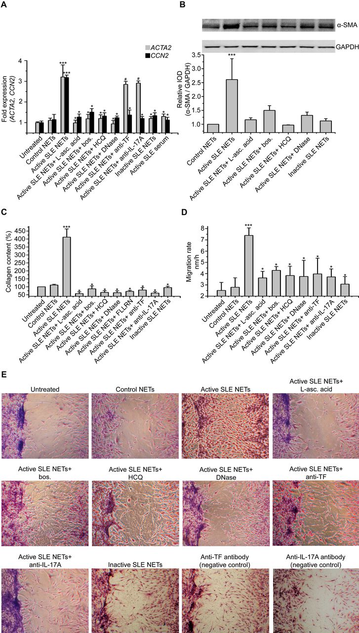
To investigate the effect of TF-bearing and IL-17A-bearing NETs on tissue resident cells, human skin fibroblasts (HSF) were incubated with NET structures induced by active SLE serum (active SLE NETs). This stimulation resulted in the activation of fibroblasts, as evidenced by α-smooth muscle actin (α-SMA) mRNA (ACTA2) and protein overexpression (figure 5A,B), compared with untreated HSF. Treatment of HSF with active SLE NETs also enhanced CCN2 expression (figure 5A), a matricellular protein implicated in fibrosis, collagen production (figure 5C) and proliferation/migration rates (figure 5D,E).

Active systemic lupus erythematosus (SLE) neuttrophil extracellular traps (NETs) bearing tissue factor (TF) and interleukin (IL)-17A promote the fibrotic activity in human skin fibroblasts (HSF) in vitro. (A) ACTA2 (24 hours incubation) and CCN2 (48 hours incubation) mRNA expression; (B) α-SMA protein expression (26 hours incubation); (C) collagen production (48 hours incubation) and (D–E) migration rate (18 hours incubation) in HSF treated with active SLE NET structures in the presence or absence of specific inhibitors (30 min pre-treatment). For (A), treatment of HSF with active SLE serum instead of NETs was used as negative control. For (B), integrated optical density (IOD) relative to control. For (E), anti-TF or anti-IL17 antibody directly on HSF was used as negative control. For (A)–(D), data presented as mean±SD, ***p<0.001 compared with respective control; *p<0.001 compared with respective ‘active SLE NETs’ and ♯non-significant compared with respective ‘active SLE nets’. For (A)–(E), one representative experiment of 6 is shown, n=6 patients.
Contrary to NETs, direct stimulation of HSF with active SLE serum did not induce their activation/differentiation towards a fibrotic phenotype (figure 5A), suggesting a specific effect of NETs. In the same context, dismantling of NETs with DNaseΙ or inhibition of autophagy with HCQ further abolished the fibrotic potential of HSF (figure 5A–E).
To confirm that HIF-1α and ET-1 are involved in REDD1-mediated active SLE NET release and subsequent HSF activation/differentiation, neutrophils were treated with either L-ascorbic acid or bosentan prior to incubation with active SLE serum. A significant attenuation of HSF activation/differentiation, collagen production and proliferation/migration rate were observed (figure 5A–E). A similar finding was also detected on treatment of active SLE serum with a specific HIF-1α inhibitor or with an ET-1 neutralising antibody (online supplementary figure 4D). Moreover, NETs mediated by the combination of recombinant ET-1 and inactive SLE serum induced enhanced collagen production by HSF (online supplementary figure 5D).
Next, we tried to elucidate the NET components responsible for the activation/differentiation of HSF and particularly the role of TF and IL-17A that were found to decorate active SLE NETs. As assessed by ACTA2 expression, TF or IL-17A neutralisation on NETs did not mediate HSF activation/differentiation (figure 5A). Conversely, their neutralisation affected the fibrotic potential of HSF, as evidenced by the marked decrease in CCN2, collagen production and proliferation/migration rate (figure 5A–E). To further investigate TF-thrombin axis involvement and considering that thrombin signals through protease-activated receptors (PAR)-1, HSF were pretreated with FLLRN, a PAR-1-specific peptide. A significant reduction in the fibrotic potential of differentiated HSF was noted (figure 5C).
Collectively, these results demonstrate that active SLE NETs contribute to the activation/differentiation of HSF, while TF and IL-17A present on SLE NETs enhance the fibrotic activity of differentiated HSF. These findings suggest multiple potential targets for therapeutic interventions in end-organ injury in SLE.
TF-bearing and IL-17A-bearing NETs are present within the kidneys and skin biopsies of patients with SLE
To gain further insights into the role of NETs in end-organ injury in SLE, we studied NET deposition in kidney biopsies of patients with proliferative LN and skin biopsies from active lesions and non-affected skin areas of patients with discoid lupus. Even in the absence of intact neutrophils in the kidneys, remnants of neutrophil activation (such as NET structures) were prominent in the glomerular (online supplementary figure 6A) and tubulointerstitial compartment close to the Bowman’s capsule and adjacent to renal tubular cells (figure 6A, online supplementary figure 6A and C). These NET structures were co-localised with TF and IL-17A (figure 6A). Conversely, neutrophils/NETs were absent in kidney biopsies from patients with renal carcinoma, minimal change disease or membranous nephropathy (online supplementary figure 6B-C).
Supplemental material

Neutrophil extracellular traps (NETs) expressing tissue factor (TF) and interleukin (IL)-17A are identified in kidney and skin biopsy specimens from patients with active systemic lupus erythematosus (SLE). (A) NETs visualised in kidney specimens from a patient with proliferative lupus nephritis (LN), as extracellular structures by staining with neutrophil elastase (NE) and citrullinated histone H3 (CitH3) (confocal microscopy; green: NE, red: CitH3, blue: 4′,6-diamidino-2-phenylindole (DAPI)/DNA), expressing both TF (confocal microscopy; green: TF, red: NE, blue: DAPI/DNA) and IL-17A (green: IL-17A, red: NE, blue: DAPI/DNA). Representative data from six patients. (B) NETs were identified in skin biopsy specimens from patients with active discoid lupus (DLE) by staining with NE and CitH3, compared with normal tissue obtained either from the same patient with SLE or from healthy subject (control) (confocal microscopy; green: NE, red: CitH3, blue: DAPI/DNA). (C) Presence of TF (confocal microscopy; green: TF, red: NE, blue: DAPI/DNA) and IL-17A (confocal microscopy; green: IL-17A, red: NE, blue: DAPI/DNA) on NET structures observed in skin specimens from patient with active DLE. For (B) and (C), representative data from four patients. (D) Tissue specimen stained with isotype control antibodies.
NETs were also detected in skin biopsies obtained from lesions of patients with active discoid lupus, as observed by the extracellular localisation of elastase and citrullinated histone 3 (figure 6Β). Skin biopsies obtained either from non-inflamed skin of the same patients with SLE or from healthy subjects (controls) did not demonstrate the presence of NETs (figure 6B). Similar to kidney specimens, NETs present in skin lesions were decorated with TF or IL-17A (figure 6C).
Together, these findings indicate the presence of NET-derived components, such as TF and IL-17A, in skin or renal biopsies, suggesting their involvement in the thromboinflammatory and fibrotic aspects of SLE.
Discussion
Herein, we implicate for the first time the REDD1/autophagy pathway in neutrophil-mediated end-organ injury in SLE. Serum from patients with active SLE—a surrogate of the inflammatory microenvironment in SLE—through ET-1 and HIF-1α, upregulates neutrophil REDD1 expression, resulting in autophagy induction and subsequent NET release. Bioactive IL-17A- and TF-decorated NETs, detected in active lupus kidney and skin, activate tissue resident cells mediating inflammation and fibrosis.
Autophagy is enhanced in lupus T cells44 and B cells.45 Herein, to elucidate the mechanism underlying NET release in SLE, we investigated autophagy in SLE neutrophils and its association to NET release. We demonstrate that active SLE neutrophils display increased basal autophagy levels mediated by inflammatory mediators within active SLE sera. We link NETs with autophagy in SLE and provide evidence that HCQ, a late-stage autophagy inhibitor, has a key role in NETs reduction through autophagy inhibition. Our data extend previous observations suggesting that chloroquine abrogated NET formation in lupus low-density granulocytes46 and may account, at least in part, for the beneficial effects of HCQ in various organ manifestations in SLE, including skin and kidneys.
Upstream regulatory molecules, governing autophagy and NET release, remain elusive. To date, SLE pathogenesis was partially attributed to impaired clearance of NETs, due to decreased serum DNaseI activity.20 21 The stress-induced protein REDD1 represents a ‘gate’ to inflammation by linking environmental triggers to cell response through autophagy.47 48 We show that increased autophagy in SLE neutrophils and subsequent NET release are mediated by REDD1 upregulation, induced by lupus inflammatory mediators. We also provide novel insights into the disease pathogenesis by demonstrating that the REDD1/autophagy pathway is critically involved in SLE NETosis and show that this pathway represents a shared mechanism between autoinflammatory31 and autoimmune disorders.
ET-1 and HIF-1α are potent mediators of the REDD1/autophagy pathway in SLE. HIF-1α inhibition by L-ascorbic acid or ET-1 inhibition with bosentan, used for treatment of pulmonary hypertension and scleroderma, reduces REDD1 overexpression and abrogates autophagy and subsequent NET release in vitro. Of note, pharmacological inhibition of HIF-1α activity also blocked NET formation by LPS-stimulated neutrophils.49 Importantly, HIF-1α or ET-1 inhibition, prior to stimulation of neutrophils with active serum, ameliorated the activation/differentiation of HSF to myofibroblasts, indicating the importance of NETs in the activation/differentiation of fibroblasts. These findings are in accordance with our previous in vitro data demonstrating the involvement of autophagy-dependent NET release in pulmonary fibrosis through lung fibroblasts activation and differentiation.14 Although further studies are needed to identify additional triggers, our findings demonstrate that ET-1 and HIF-1α could be therapeutically targeted as mediators of the REDD1/autophagy pathway that regulates NET release in SLE.
Disease-specific bioactive proteins on NETs could contribute to different biological processes and histological phenotypes in various diseases.50–53 We therefore searched for SLE-specific proteins on NETs and asked whether increased autophagy is associated with their expression on NETs. We demonstrate that active SLE serum upregulates the expression of the thromboinflammatory TF and profibrotic IL-17A in neutrophils and mediates their expression on NETs in an autophagy-dependent manner. We demonstrate that these proteins on NETs are bioactive, inducing thrombin generation and activation/differentiation of HSF to collagen-producing myofibroblasts. We also demonstrate that the NET scaffold is essential for these proteins to exert their function and provide evidence that TF-expressing and IL-17A-expressing NETs represent a link between increased thromboinflammation and fibrosis in patients with active SLE.
Since NETs are associated with lupus nephritis,16 20 54 we reasoned that NETs may be involved in tissue inflammation and fibrosis via the TF/thrombin axis and IL-17A, respectively. We demonstrate the presence of TF-decorated and IL-17A-decorated NETs within end-organ tissues of SLE, in the absence of intact neutrophils. In the kidneys of patients with proliferative LN, TF- and IL-17A-decorated NETs are found within glomeruli whereas TF-decorated NETs are observed within the tubulointerstitial compartment close to the Bowman’s capsule, suggesting their possible involvement in capsule rapture and crescent formation (features of rapidly progressive glomerulonephritis).
Pathogenic events at more easily accessible organs in SLE may mirror pathogenic processes in the kidney.55 NETs have been identified in the skin of patients with SLE56 57; however, the disease-associated proteins externalised on NETs were remained unknown. Thus, we analysed skin biopsies from patients with active SLE and found TF-decorated and IL-17A-decorated NETs within affected skin areas. We further demonstrated that the blockade of TF and IL-17A on NETs attenuated the activation/differentiation, collagen production and proliferation/migration in HSF. Accordingly, we provide evidence supporting the important role of TF-bearing and IL-17A-bearing NETs in end-organ injury in SLE, suggesting NETs as a connecting link between the thromboinflammatory and fibrotic aspects of the disease. To this end, agents targeting the IL-17A pathway—currently used for treatment of psoriasis, psoriatic arthritis and ankylosing spondylitis—and/or thrombin inhibitors or PAR blockers could potentially attenuate tissue injury in SLE.
In summary, our findings identify upstream regulators and downstream molecules that mediate NET release in human SLE linking immunometabolism, thromboinflammation and fibrosis towards end-organ injury. ET-1 and HIF-1α in active SLE serum activate the REDD1/autophagy pathway to induce NETs. Active SLE NETs represent scaffolds with high concentration of bioactive IL-17A and TF that remain in end-organ tissues even in the absence of intact neutrophils, activating resident cells and promoting thromboinflammation and fibrosis. To this end, we propose a multistep model for end-organ injury in SLE that can be targeted at multiple levels by repositioning of available drugs to ameliorate tissue injury (online supplementary figure 7). Accordingly, ET-1 receptor antagonists (eg, bosentan) and HIF-1α inhibitors (eg, L-ascorbic acid) could disrupt the ‘pre-NETotic’ step; autophagy inhibitors (eg, hydroxychoroquine) could prevent the ‘NETotic’ step, and finally, agents targeting the IL-17A pathway (eg, secukinumab) and/or TF/thrombin axis (eg, thrombin inhibitors or PAR blockers) could offset the ‘post-NETotic’ deleterious effects in SLE.
Supplemental material
Supplemental material
![[annrheumdis-2018-213181-supp1.JPEG]](https://ard.bmj.com/content/annrheumdis/78/2/238/DC9/embed/inline-supplementary-material-9.jpg?download=true)
Supplemental material
Supplemental material
Supplemental material
Supplemental material
Acknowledgments
The authors would like to thank Eleni Sertaridou, Antonis Fanouriakis, Aggelos Banos, Dionysis Nikolopoulos, Maria Grigoriou, G Rapsomaniki, Themis Alissafi, Eirini Apostolidou, Victoria Tsironidou, Charalambos Papagoras, Pelagia Kriki and Ploumis Pasadakis for their assistance in clinical specimen collection, processing and help with the assays used. Immunofluorescence experiments were performed at the CIBIT facilities of Democritus University of Thrace, Alexandroupolis, Greece and at the Biomedical Research Foundation of the Academy of Athens, Athens, Greece.
References
Footnotes
EF and AC contributed equally.
KR and DTB contributed equally.
Handling editor Josef S Smolen
Contributors EF and AC designed and conducted experiments, acquired and analysed the data and wrote the manuscript. AM, KK, SA, IA, AA and HG performed experiments and contributed to data analysis. PV and GB reviewed and analysed the data. DB and KR conceived, designed, wrote and critically reviewed the data and they equally cosupervised this manuscript. All authors have approved the submitted manuscript.
Funding This study was supported by grants from the Aristeia program of the Greek Secretariat of Research and Technology; the BTCure IMI program (EU and EFPIA); the Hellenic Society of Rheumatology; and the Scientific Committee of Democritus University of Thrace. Dr Akrivi Chrysanthopoulou was supported by the Bodossaki Foundation. This project has received funding from the European Research Council (ERC) under the European Union’s Horizon 2020 research and innovation programme (grant agreement No 742390).
Competing interests None declared.
Patient consent Obtained.
Ethics approval Institutional Review Board of the ‘Attikon’ University Hospital.
Provenance and peer review Not commissioned; externally peer reviewed.