Article Text

Abstract
Background: Leflunomide is now an approved agent for the management of adult rheumatoid arthritis (RA). Its active metabolite A771726 inhibits de novo pyrimidine biosynthesis. Although considered to be an immunosuppressive agent, its mechanism of action remains obscure.
Objectives: Evaluation of the leflunomide active metabolite A771726 (LEF) effect on interleukin 1β (IL1β), tumour necrosis factor (TNFα), nitric oxide (NO), and stromelysin (metalloproteinase-3 (MMP-3)) production by activated human synovial tissue in culture.
Methods: Synovial tissue was obtained during surgery from patients undergoing total knee replacement owing to RA or osteoarthritis (OA), cut into small pieces, and cultured in Petri dishes with test materials as previously described. IL1β, TNFα, NO, and MMP-3 were measured in the culture media after 48 hours incubation with different doses of LEF by methods previously described.
Results: LEF (0.3, 3, and 9 μg/ml) inhibited IL1β production in the presence of lipopolysaccharide (LPS; 3 μg/ml) in a dose dependent manner (p<0.01) at LEF 0.3 μg/ml. TNFα production in the presence of IL1β (1 ng/ml) was also inhibited in a dose dependent manner (p<0.05 at LEF 0.3 μg/ml). NO and MMP-3 production in the presence of LPS (3 μg/ml) was inhibited as well (p<0.01 at LEF 1 μg/ml and at LEF 0.3 μg/ml, respectively). Synovial cell viability evaluated by the tetrazolium salt XTT was unaffected by the LEF concentration used. There was no qualitative difference in the response of OA and RA synovial tissue.
Conclusion: Leflunomide may modulate the rheumatoid articular process by inhibition of local production of IL1β, TNFα, NO, and MMP-3.
- leflunomide
- tumour necrosis factor α
- interleukin 1β
- nitric oxide
- matrix metalloproteinase-3
- ELISA, enzyme linked immunosorbent assay
- FCS, fetal calf serum
- IL1β, interleukin 1β
- iNOS, inducible nitric oxide synthase
- LEF, active leflunomide metabolite, A771726
- LPS, lipopolysaccharide
- MMP-3, matrix metalloproteinase-3
- NO, nitric oxide
- OA, osteoarthritis
- RA, rheumatoid arthritis
- TNFα, tumour necrosis factor α
Statistics from Altmetric.com
- ELISA, enzyme linked immunosorbent assay
- FCS, fetal calf serum
- IL1β, interleukin 1β
- iNOS, inducible nitric oxide synthase
- LEF, active leflunomide metabolite, A771726
- LPS, lipopolysaccharide
- MMP-3, matrix metalloproteinase-3
- NO, nitric oxide
- OA, osteoarthritis
- RA, rheumatoid arthritis
- TNFα, tumour necrosis factor α
L eflunomide is a new immunomodulating agent acting as a disease modifying drug in rheumatoid arthritis (RA).1,2 It is a pro-drug, exerting its therapeutic effect through its major metabolite, A771726.3 Although the exact mechanism by which leflunomide exerts its effect in RA is largely unknown, it appears to have several potential sites of action. A771726 inhibits dihydro-orotate reductase impeding de novo pyrimidine synthesis, resulting in decreased lymphocyte proliferation.4,5 A771726 also interacts with primary and secondary signalling events by interference with the phosphorylation of tyrosine kinase.6,7 In addition, A771726 may reduce the local concentration of inflammatory mediators by inhibiting the release of histamine from mast cells and of reactive oxygen species from white blood cells.8
In vivo, leflunomide has been shown to directly affect synovial tissue, by decreasing expression of adhesion molecules and metalloproteinase-1.9 However, other aspects of the direct effect of A771726 in synovial cells are not well known.
RA is characterised by synovial proliferation (pannus formation) and by local release of proinflammatory cytokines and metalloproteinases, leading to the destruction of cartilage and other articular components.10 In all of them, the synovial mesenchymal cell has a central role, both as a proliferating cell as well as producer of proinflammatory cytokines and presenter of antigen to T cells.11 In vitro studies of synovial fibroblast function and its modulation may be useful for the understanding of pharmacological agents in vivo.12
We report herein the effects of A771726, the active metabolite of leflunomide, on the production of interleukin 1β (IL1β), tumour necrosis factor (TNFα), nitric oxide (NO), and stromelysin (metalloproteinase-3 (MMP-3)) in human synovial cells and synovial cultures.
MATERIALS AND METHODS
Specimen selection and culture conditions
Synovial tissue was obtained during surgery from patients undergoing total knee replacement owing to RA (two female patients aged 58 and 64) or osteoarthritis (OA; two female patients aged 62 and 68).
Synovial cells derived from small pieces (2 mm in diameter) of human synovial tissue were grown after trypsinisation in monolayers in tissue culture flasks, by methods described by Castor et al.13 Synovial cell lines were thus obtained and experiments performed in passages 2–4 after the first trypsinisation. The culture medium consisted of RPMI 1640, supplemented with l-glutamine (2 mM), penicillin (100 U/ml), and streptomycin sulphate (100 μg/ml) and 10% fetal calf serum (FCS). Experiments were performed in 96 well plates containing 15–20×103 cells/well in 1% FCS. Incubation time with different additives was 48 hours at 37°C (in 5% CO2).
Synovial tissue was incubated with different doses of A771726. At the end of the incubation supernatants were removed for evaluation and the results of four separate experiments performed in duplicate were expressed per milligram of tissue in percentage of “activated” (by IL1 or lipopolysaccharide (LPS)) cultures (n=2×4=8). The absolute values for controls and stimulated cultures are reported in the figure legends.
IL1β, TNFα, NO, and MMP-3 determinations
IL1β and TNFα in culture media were determined by the Quantikine Human Immunoassay (R&D Systems Inc, USA), which employs the quantitative sandwich enzyme linked immunoassay (ELISA) technique.
NO was determined as previously described by Ashab et al.14 NO2 and NO3 were determined after the reduction of NO3 to NO2 by 90 minutes’ incubation in a tilting bath (37%) using nitrate reductase from Escherichia coli and β-nicotinamide adenine dinucleotide phosphate as cofactor. The presence of NO2 was determined with the Griess reagent. Sensitivity of the procedure was 3 μmol/l.
MMP-3 (stromelysin) was measured by an ELISA (Chemicon International Inc, USA).
Cell viability and toxicity determination
Cell viability and toxicity in the presence of the active leflunomide metabolite (LEF) in the doses used in these experiments were determined in human synovial monolayer cultures by the tetrazolium salt XTT assay (Beit Haemek, Israel).15
LEF was obtained from the Aventis Company.
Statistical analysis
In synovial cell cultures, absolute values of IL1β, TNFα, NO, and MMP-3 were expressed per well. Statistical significance was performed using the Student’s t test.
RESULTS
IL1β production
The leflunomide active metabolite (LEF) significantly inhibited LPS (3 μg/ml) stimulated release of IL1β in the medium of human synovial tissue cultures in a dose dependent manner (p<0.01 at LEF 0.3 μg/ml) (fig 1) by 78%, 82%, and 85% at doses of 0.3 μg/ml, 3 μg/ml, and 9 μg/ml respectively.

Effect of the leflunomide active metabolite (LEF) on IL1β levels in the medium of synovial tissue cultures in the presence of LPS (3 μg/ml). Bars show the mean (SEM) of four separate experiments (two RA and two OA, n=8). The absolute mean (SEM) levels of IL1β in control and LPS stimulated cultures were 1.38 (0.012) and 5.16 (0.08) pg/mg synovia, respectively.
TNFα production
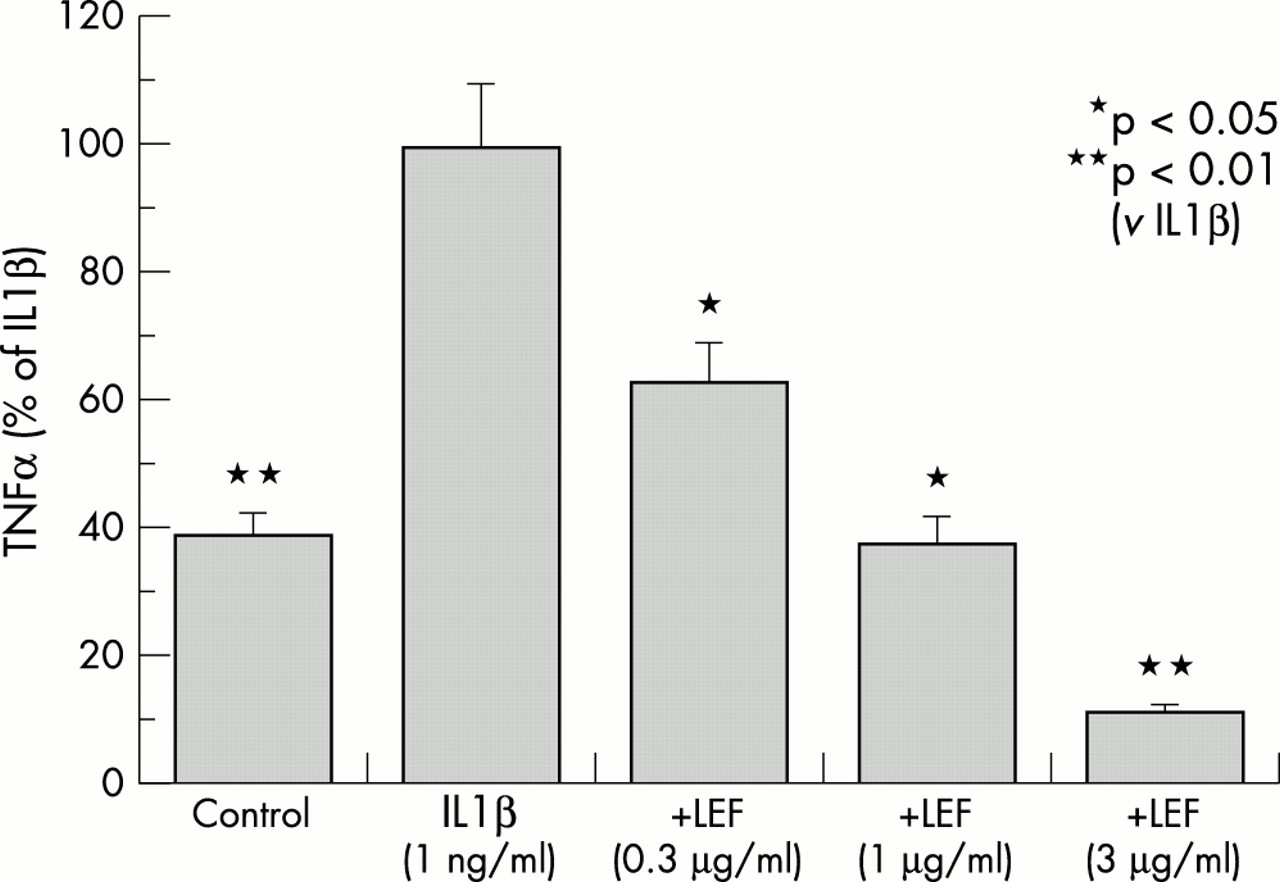
The production of TNFα in the medium of IL1β (1 ng/ml) stimulated synovial tissue cultures was significantly inhibited by LEF in a dose dependent manner (p<0.05 at LEF 0.3 μg/ml) (fig 2) by 40%, 60%, and 90% at doses of 0.3 μg/ml, 1 μg/ml, and 3 μg/ml, respectively.

Effect of leflunomide active metabolite (LEF) on TNFα levels in levels in the medium of synovial tissue cultures in the presence of IL1β (1 ng/ml). Bars show the mean (SEM) of four separate experiments (two RA and two OA, n=8). The absolute mean (SEM) levels of TNFα in control and IL1β stimulated cultures were 2.33 (0.23) and 4.95 (0.49) pg/mg synovia, respectively.
NO and MMP-3 production
NO release in the medium of LPS (3 μg/ml) stimulated cultures was inhibited by LEF in a dose dependent manner at LEF 3 μg/ml (p<0.01), while the inhibition achieved at a dose of 0.3 μg/ml was not significant (fig 3).

Effect of leflunomide active metabolite (LEF) on nitric oxide (NO) levels in the medium of synovial tissue cultures in the presence of LPS (3 μg/ml). Bars show the mean (SEM) of four separate experiments (two RA, n=4). The absolute mean (SEM) levels of NO in control and LPS stimulated cultures were 1.08 (0.01) and 2.45 (0.245) μmol/mg synovia, respectively.
Release of LPS stimulated MMP-3 was significantly inhibited by the different doses of LEF (p<0.01 at LEF 0.3 μg/ml, 1 μg/ml, 3 μg/ml, and 9 μg/ml) (fig 4).

Effect of LEF on stromelysin (MMP-3) levels in the medium of synovial tissue cultures in the presence of LPS (3 μg/ml). Bars show the mean of four separate experiments (two RA, n=4). The absolute mean (SEM) levels of MMP-3 in controls and LPS stimulated cultures were 1929 (83) and 5394 (197) ng/mg synovia, respectively.
There was no qualitative difference in the response of RA and OA synovial cultures to LEF.
Cell viability and toxicity
Evaluation of toxicity to LEF in the concentrations used in these experiments as measured in human synovial cell cultures by the tetrazolium salt XTT assay showed no toxicity (fig 5).

Effect of LEF on cell viability. Bars represent the percentage of viable cells incubated with LEF in comparison with control. LEF at a concentration of 0.3–9 μg/ml did not show any deleterious effect on cell viability, which remained >90% in comparison with controls.
DISCUSSION
In this study we have shown that A771726, the active metabolite of leflunomide, used in doses within the therapeutic range,16 causes a dose dependent reduction of production of IL1β, TNFα, NO, and MMP-3 release in synovial tissue culture media. No qualitative difference in the response of RA and OA synovial cultures to LEF was found. This might be related to the selection of patients with RA, who where end stage patients requiring surgery, or to the cytokine profile studied.
Although the pathogenetic mechanism of RA remains elusive, great advances in molecular biology and clinical research have identified a complex orchestration of immune subset cells, cell surface markers, soluble cell products such as cytokines, and other inflammatory products.17 Inflammation and subsequent degradation of the synovial tissue is probably induced by the influx of lymphocytes, triggered by an unknown antigen. In an oversimplified schema, activated T cells stimulate plasma cells, macrophages, and synovial fibroblasts to produce TNFα and IL1β, which are key agents in the process of inflammation.18 LEF suppresses proliferation of lymphocytes and may thus modulate the inflammatory process by reducing the influx of immune cells to the synovia.4 This may be the subcellular mechanism for the observed inhibition of TNFα, IL1β, and MMP-3, whereas inhibition of NO may be due to direct suppression of inducible nitric oxide synthase (iNOS) activation in fibroblasts by leflunomide through inhibition of the MEK/MAP pathway.19
The reduced production of TNFα and IL1β by synovial fibroblasts shown in our study is in concordance with the observation of Kraan et al, who reported a reduced expression of TNFα and IL1β in synovial tissues of patients with RA treated with leflunomide as well as a decreased expression of adhesion molecules and MMP-1.9
Metalloproteinases seem to have an important role in joint inflammation and in articular degeneration.20 MMP-3 levels in serum and synovial fluid are increased in patients with active RA21 and correlate with the degree of joint erosion, suggesting its involvement in joint inflammation and articular erosions.22 The reduction of MMP-3 produced in synovial cells culture by A771726 may explain the disease modifying effect of leflunomide in patients with RA and the deceleration in joint erosions seen in clinical trials.23
Our study shows that leflunomide produces a significant dose dependent suppression of NO production by synovial cells. Studies performed during the past years clearly indicate an important role for NO in inflammation.24 Nitric oxide is a pleiotropic inflammatory mediator overproduced in joints affected by arthritis. iNOS is found in both the synovial tissue and cartilage and plays a part in the pathogenic process that occurs in the pannus of RA.24 NO has been implicated in the development of both central25 and peripheral pain.26,27 It has been implicated as one of the important mediators of articular damage, in part through modulation of production of metalloproteinases by the rheumatoid cells28 and apoptosis.29 Several drugs, including non-steroidal anti-inflammatory drugs,30 tetracyclines,31 corticosteroids,32 and immunosuppressive drugs such as cyclosporin A and mycophenolate mofetil, attenuate the activation of NO.33
It has recently been shown that treatment of patients with RA with anti-TNFα monoclonal antibody significantly reduces iNOS type 2 protein expression and iNOS enzyme activity, the changes in iNOS activity correlating clearly with the degree of improvement in the number of tender joints.34 Leflunomide’s metabolite A771726 has been found to cause a dose dependent decrease of NO production in interferon gamma stimulated astrocytes19 and fibroblasts.35 Specific inhibition of MAP, PD 98058, but not unselective protein kinase inhibitor, completely mimicked the cell type specific and stimulus specific NO inhibitory action of leflunomide, suggesting that the suppression of iNOS activation in fibroblasts by leflunomide is through inhibition of the MEK/MAP pathway.19
In conclusion, our study indicates that A771726, the active metabolite of leflunomide inhibits production of TNFα, IL1β, NO, and MMP-3 by human synovial cells, in vitro. Inhibition of TNFα, IL1β, NO, and MMP-3 is an important mechanism by which leflunomide may affect pain, articular inflammation, and joint damage. Further studies are needed to establish the in vivo significance of these findings.