Article Text

Abstract
Objectives: To investigate the possible association of interleukin 1α autoantibodies (IL1α aAb) with the long term course of joint erosion in patients with rheumatoid arthritis (RA).
Methods: Serum samples from 176 patients with RA included in a prospective study over 30 years were analysed for IL1α aAb by binding to human [125I]IL1α. Erosions of 19 diarthrodial joints were radiographically scored by the Larsen method.
Results: The relative risk (RR) of early IL1α aAb positive patients developing at least 30% of maximum radiographic joint destruction was significantly lower than for IL1α aAb negative patients, RR=0.29 (p=0.04). In rheumatoid factor positive patients RR was only 0.18 (p=0.02). Patients who seroconverted more than two years after the onset of RA showed the most aggressive development of joint erosion, with a relative risk of at least 40% of maximum radiographic joint destruction of 2.56 (p=0.048)
Conclusions: The progression of radiographic joint destruction in patients with RA is associated with, and perhaps modified by, circulating IL1α aAb, suggesting that IL1α or IL1α aAb, or both, have a role in the erosive processes. IL1α aAb appear to be of prognostic significance in RA.
- autoantibodies
- cytokine
- interleukin 1α
- rheumatoid arthritis
- aAb, autoantibodies
- IL, interleukin
- ra, receptor antagonist
- RA, rheumatoid arthritis
- TNF tumour necrosis factor
Statistics from Altmetric.com
- aAb, autoantibodies
- IL, interleukin
- ra, receptor antagonist
- RA, rheumatoid arthritis
- TNF tumour necrosis factor
Cytokines constitute a large group of intercellular signal peptides regulating growth and development of many cells. Inflammatory cytokines, such as interleukin 1 (IL1)α, IL1β, and tumour necrosis factor (TNF), have been implicated as important mediators in inflammatory diseases1–5—for instance, in patients with rheumatoid arthritis (RA) and in animal models of RA.1,4,6–9 The three primary members of the IL1 gene family are IL1α, IL1β, and IL1 receptor antagonist (ra). IL1α and IL1β are agonists, and IL1ra is a specific receptor antagonist. IL1α has biological properties similar to those of IL1β, but unlike IL1β, remains mostly cell associated. The mature forms of both cytokines trigger the same extracellular receptor. The exact proportion of effects exerted by IL1α and IL1β is not known. Most evidence relates to IL1β. Infusion of IL1β induces and exacerbates arthritis in animal models.10–12 However, Feige et al showed that continuous infusion for two weeks of IL1α in rabbit knees induced arthritis changes similar to RA 13 indicating a separate effect of IL1α. Plasma IL1β is associated with disease activity in RA,14 but, at present, measurement of IL1α in plasma offers little of clinical value, although plasma IL1α15 and the surface expression of IL1α16 are both correlated with the erythrocyte sedimentation rate.
In certain strains of IL1ra deficient mice a chronic inflammatory polyarthropathy develops spontaneously. The joints show prominent synovial and periarticular infiltration of inflammatory cells, osteoclast activation, and structural erosion associated with the presence of granulation tissue.9 Overall, the joints appear similar to those from humans with RA. This strongly suggests that endogenous IL1ra functions to suppress inflammation by blocking the IL1 receptor.
It is known that sera of patients with various immunoinflammatory diseases contain antibodies to certain cytokines,17 and that aAb reacting with natural cytokines can be induced in patients treated with recombinant cytokines.18,19
Naturally occurring IL1α aAb were first identified in 1989.20 These aAb are found in 10–60% of normal adults depending on age and assay sensitivity.21 They are polyclonally derived and bind with high affinity to IL1α, but not to IL1β or to the IL1ra, neutralising IL1α receptor binding in vitro and, therefore, the biological activity of the cytokine.18,20,22 An IL1α aAb has recently been cloned.23 It is an IgG4/k antibody binding exclusively to human IL1α with a kDa of 10−10 M. The presence of somatic mutations in the variable regions suggests that this aAb was induced as the result of an antigenic stimulus. Naturally occurring IL1β aAb have been reported by a few investigators, but at present there is no general agreement about their existence.24
In patients with various forms of arthritis, those positive for IL1α aAb have been shown to have relatively mild disease manifestations.25–27 Experimental studies on mice have shown that IL1 antibodies ameliorated both early and full blown collagen induced arthritis.28 Furthermore, treatment of patients with RA with recombinant IL1ra reduced inflammatory variables and radiographic progression compared with placebo.29,30
Because of increasing evidence that IL1 blockade modifies the disease processes in RA, we investigated the possible influence of naturally occurring IL1α aAb on the long term course of RA. Serum IL1α aAb were measured in consecutive deep frozen serum samples from a cohort of patients with RA followed up prospectively for up to 30 years. The prognostic significance was investigated in patients positive for IL1α aAb during the first two years after onset of RA, in patients converting from a negative state to a positive state during the course of RA, and in positive patients with unknown time of seroconversion.
METHODS
From 1966 to 1978, 685 Danish white patients with RA classified according to the 1958 ARA criteria31 were admitted to the Department of Rheumatology, Aarhus University Hospital. At present, 456 of these patients have been included in a database. Of these, 176 (119 female, 57 male) with available deep frozen serum samples were included in the present study. A later reclassification according to the 1987 criteria showed that 171 patients fulfilled four criteria or more, three patients fulfilled three criteria, one patient two criteria, and one patient one criterion. The patients were assessed clinically and biochemically at admission, and when possible, subsequently at least once a year. Radiographs of the hands and feet were obtained at baseline and at intervals of one to three years.
The median duration of RA at the time of the first visit was one year (range 0–18). The median follow up period was 13 years (1–43) after the onset of RA. The median age at the onset of RA was 41 years (range 15–70). To investigate for selection bias, the 176 included patients with available sera were compared with the remaining 280 patients with RA from the database. The local ethics committee approved the study.
Blinded radiographic evaluations of joint destruction were used as the main outcome measure. We have previously correlated radiographic scores of different combinations of diarthrodial joints (one hand, two hands, one foot, two feet, one hand and one foot) with all remaining diarthrodial joints and found the highest correlation (r=0.95) when comparing the scores of the left hand combined with the left foot with the scores of the remaining joints (unpublished data). Consequently, we included the left hand and the left foot in the assessment programme, altogether 19 joints: the 10 left finger joints (metacarpophalangeal joints, interphalangeal 1, and proximal interphalangeal 2–5 joints) and six left toe joints (interphalangeal 1 and metatarsophalangeal 1–5), left wrist joint, left ankle joint, and the left tarsal joint.
The radiographic score was used to indicate the degree of radiographic joint destruction and was defined for each complete examination as the sum of the grading of affected joints. Joint changes were classified by comparison with standard reference films according to the method of Larsen32 and graded 0–5 (0, normal; 5, the most severe changes). Joints which had been operated on were evaluated as having grade 5 changes. Only definite erosive changes corresponding to grades 2–5 were considered when calculating the R score (maximum score 19×5=95). A radiographic outcome event was defined as the occurrence of at least 10% (RE10), 20% (RE20), 30% (RE30), and 40% (RE40) of maximal radiographic score (scores 10, 19, 29, and 38 of 95)33 during the observation period of up to 43 years.
To evaluate the possible influence of a variable on a disease it is important that the duration of the disease is comparable between the included patients. Furthermore, one must know at which time the variable first occurred in each patient. The prognostic value of IL1α aAb could be determined in 73 patients with an available serum sample within the first two years after onset of RA (the inception cohort).
Among the 103 patients with a disease duration of more than two years at inclusion (the late cohort), the significance of seroconversion could be evaluated in eight patients known to be negative at the inclusion, but positive at a later evaluation (the seroconversion group). In the remaining 95 patients, it was not possible to establish whether the positive fraction of patients (n=19) had been positive during the early course of RA or whether they seroconverted at a later time.
IL1α aAb were measured in three consecutive serum samples from each of 125 patients. Two samples were available in 18 patients, and only one sample was available in the remaining 33 patients. Human recombinant IL1α (Dainippon, Osaka, Japan) was 125I labelled as previously described.34 All determinations were made in duplicate. Screening of samples for [125I]IL1α complexes was done by 15% polyethylene glycol 6000 (Merck, Darmstadt, Germany) precipitation of 20% serum samples preincubated for 18 hours at 4°C in phosphate buffered saline. Samples with more than five times background activity were tested 10-fold diluted for protein G binding of [125I]IL1α.21 Binding of >7% of the added tracer (>2 × background) was considered positive, and this was confirmed by displacement of binding by excess, unlabelled IL1α. A reference solution of pooled normal human IgG (State Serum Institute, Copenhagen, Denmark) was run in parallel with the samples. This showed an intra- and interassay variation below 10% during the seven day period used to complete the determinations. Results are expressed as percentage bound of total cpm [125I]IL1α.34
Statistical analysis
Frequency distributions between two groups were compared by χ2 test. Unpaired comparisons between two groups were performed by the Mann-Whitney test. Patients with radiographic events up to 30 years after onset are shown as Kaplan-Meier plots and statistical comparisons calculated by means of the log rank test. The Cox proportional hazard model was used to calculate the relative risks. Correlation coefficients were calculated as Spearman's correlation (rs). Data are presented as frequencies or median and range.
RESULTS
No difference was found in the radiographic outcome between the 176 included patients and the 280 not included: RE10, p=0.64; RE20, p=0.50; RE30, p=0.78; RE40, p=0.93.
The overall prevalence of IL1α aAb was 44/176 (25%). In the inception cohort, 17/73 (23%) were positive for IL1α aAb and in the late cohort, 27/103 (26%) were positive.
In the inception cohort (n=73) 5/17 (29%) IL1α aAb positive patients had morning stiffness compared with 36/56 (64%) IL1α aAb negative patients (p=0.024), and 13/17 (76%) had erosions compared with 52/56 (93%) (p=0.06). The number of ARA criteria were borderline, with fewer in the IL1α aAb positive patients: 5 (1–7) v 6 (2–7) (p=0.06).
In the late cohort (n=103) no differences were seen in the prevalence of the 1987 ACR criteria or number of patients with morning stiffness between IL1α aAb positive and IL1α aAb negative patients.
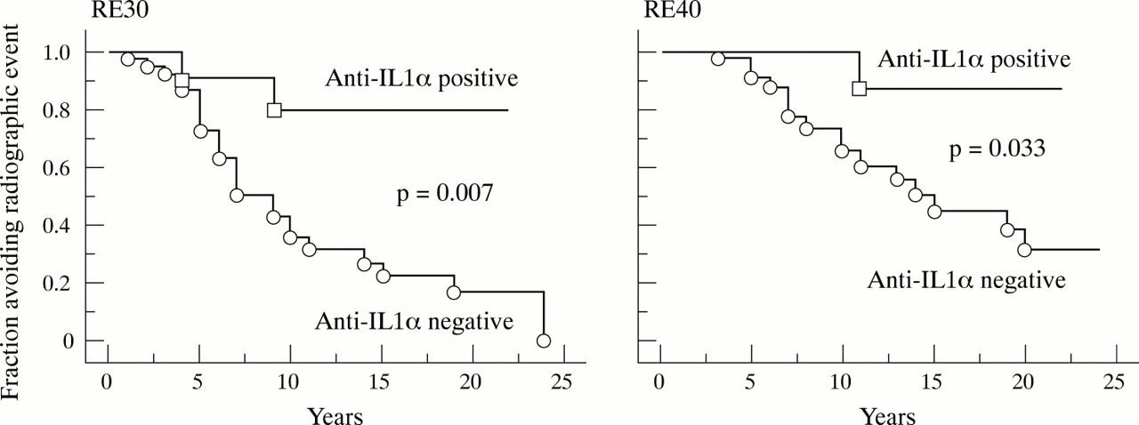
In the inception cohort (n=73) the IL1α aAb positive patients (n=17) showed a borderline better prognosis: RE10, p=0.14; RE20, p=0.27; RE40, p=0.10, or significantly better prognosis: RE30, p=0.027 than patients without IL1α aAb (n=56) (fig 1). The relative risk of IL1α aAb positive patients developing radiographic events was 0.60 for RE10 (p=0.17), 0.65 for RE20 (p=0.30), 0.29 for RE30 (p=0.04), and 0.32 for RE40 (p=0.13).

Effect of early IL1α aAb positivity on joint erosions in patients with RA. Kaplan-Meier plots of radiographic events defined as 30% and 40% of maximal radiographic destruction (RE30, RE40). The two patient groups are defined by the presence and absence of IL1α aAb during the first two years after onset of the disease. p Value: log rank test.
In a subanalysis of rheumatoid factor positive patients (n=57) the prognosis for IL1α aAb positive patients (n=14) was even better: RE10, p=0.05; RE20, p=0.11; RE30, p=0.007; RE40, p=0.033 (fig 2). The corresponding relative risks were 0.51 for RE10 (p=0.08), 0.51 for RE20 (p=0.14), 0.18 for RE30 (p=0.02), and 0.15 for RE40 (p=0.07).

Effect of early IL1α aAb positivity on joint erosions in rheumatoid factor positive patients with RA. Kaplan-Meier plots are as in fig 1. The two patient groups are defined by the presence and absence of IL1α aAb during the first two years after onset of the disease. p Value: log rank test.
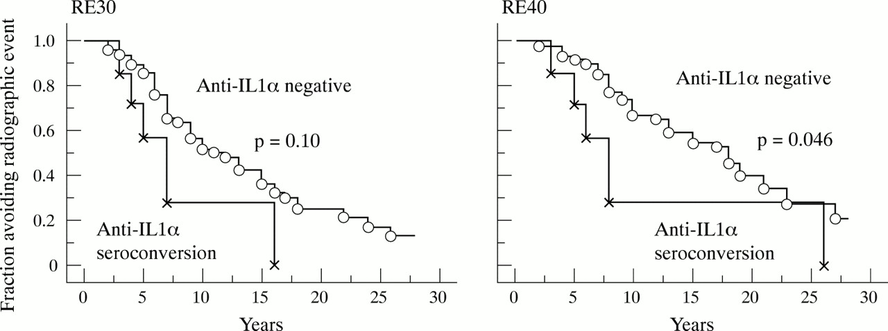
In the late cohort (n=103), the seroconversion group (n=8) had a worse course than those permanently negative (n=76) (fig 3). The relative risk of IL1α aAb seroconverting patients developing radiographic events was 1.20 for RE10 (p=0.60), 1.87 for RE20 (p=0.12), 1.94 for RE30 (p=0.15), and 2.56 for RE40 (p=0.048) (Cox proportional hazard model). Those positive at the first test (n=19) had a long term prognosis comparable with those who were permanently negative (n=76): RE10, p=0.51; RE20, p=0.33; RE30, p=0.76; RE40, p=0.20.

Effect of late IL1α aAb seroconversion on joint erosions. Kaplan-Meier plots are as in fig 1. The two patient groups are defined as those permanently negative for IL1α aAb and those initially negative who seroconverted more than two years after onset of the disease. p Value: log rank test.
DISCUSSION
In contrast with IL1β, IL1α is not commonly found in the circulation or in body fluids except during severe disease, in which case the cytokine may be released from dying cells. Consequently, it is less easy to get an impression of the role of IL1α in the pathogenesis of inflammatory disease. At present, the bulk of published work indicates that IL1β is important for the pathogenesis of RA, whereas there are few studies on IL1α.1,35 However, studies of experimental inflammatory bowel disease showed a better correlation of disease severity with colonic tissue levels of IL1α than of IL1β,36 presumably owing to the cell associated nature of IL1α. This indicates that IL1α may be at least as important as IL1β in the pathogenesis of RA and that selective blocking of IL1α might be of value in the treatment of RA.
The present cohort was followed up prospectively since 1966 with consecutive isolation of sera for later analyses. The decision to measure IL1α aAb in these serum samples was taken in 1999. This study is the first to show that the prognostic significance of IL1α aAb in RA may depend on the time of appearance of the autoantibody in the blood. Although the results were not statistically significant for all radiographic events (RE10-RE40), the trends were identical. Thus IL1α aAb present early in the disease predicts a mild course as measured by radiographically visible joint destruction (figs 1 and 2). Furthermore, fewer IL1α aAb positive patients had morning stiffness. In contrast, the development of IL1α aAb after the first two years of onset of RA is associated with a poor prognosis (fig 3).
Hypothetically, the present finding of an association of IL1α aAb with a good prognosis in early RA might be explained if IL1α aAb occurred particularly in rheumatoid factor negative patients, who are known to have a better prognosis. However, this was not the case. Furthermore, the association was more significant when investigated in the rheumatoid factor positive subgroup.
It is likely that more patients with RA with severe or permanent symptoms will continue to seek medical attention compared with patients with milder or fluctuating symptoms. Therefore, there will be many mild cases in an inception cohort. In contrast, patients included at a later stage of the disease will be part of a major cohort from which mild cases may have dropped out. Consequently, a group of patients with established RA and varying disease duration at the time of inclusion into a study, as the late cohort, would be more homogeneous than those in an inception cohort. This may in part explain the difference in prognosis between inception cohort patients and late cohort patients positive for IL1α aAb. An additional explanation might be that the fraction of IL1α aAb positive patients in the late cohort is a mixture of patients who were positive for IL1α aAb at the beginning of RA (with a good prognosis, see figs 1 and 2) and of patients who developed IL1α aAb during the course of RA (with a severe prognosis, see fig 3).
Jouvenne et al found an increased frequency of IL1α aAb in a group of unselected patients with arthritis compared with healthy controls (18.9% v 9.7%).26 However, the frequency of IL1α aAb positive patients with RA was comparable with the controls (10.9%), whereas the frequency in groups with milder arthritis varied between 18.5% and 27.3%. The relative risk of IL1α aAb negative patients developing RA was 2.2—that is, the relative risk of IL1α aAb positive patients developing RA was 0.45, or similar to the risk of patients with RA developing radiographic events as defined in our study. Furthermore, IL1α aAb were present in 25.5% of patients with non-destructive arthritis, but in only 13% of patients with destructive arthritis. Consequently, in IL1α aAb positive patients with arthritis, the risk of being classified as having RA and the risk of developing destructive arthritis were relatively low.
Hansen et al also found similar frequencies of IL1α aAb in RA (36%) and healthy controls (38%).27 Furthermore, there were no clinical differences between IL1α aAb positive and negative patients. However, the study of Hansen et al was a cross sectional study of only 42 patients and without radiographic evaluation of the joints.
Contrary to the above studies, Suzuki et al found a higher frequency of IL1α aAb in Japanese patients with RA than in healthy controls (16.6% v 5.6%).25 There is no immediate explanation for this discrepancy, apart from possible ethnic differences. Furthermore, Suzuki et al found fluctuations of the titres of IL1α aAb in parallel with disease activity estimated by tender joints and erythrocyte sedimentation rate. Radiographic joint destruction was not investigated. In another Japanese study of a non-inception cohort of patients with RA, Maniwa et al found a higher frequency of IL1α aAb in patients with RA and interstitial lung disease than in controls with other lung diseases and no RA (42.9% v 15%). There was no separate analysis of patients with early arthritis. They suggested that IL1α aAb are generated in response to the immunoinflammatory process of interstitial lung disease in RA, and that IL1α aAb may neutralise and regulate the IL1α activity.37
Together with TNFα, IL1α and IL1β have been implicated as important mediators of tissue damage in RA.1,4,6,7 Anti-TNFα treatments have been introduced successfully in the management of patients with RA on the basis of several studies showing significant improvement in measures of disease activity38–40 and radiographic joint destruction.41 IL1 blockade is not yet available as a treatment principle, but recent studies showed that recombinant IL1ra significantly lowered disease activity in RA and, in addition, reduced the progression of erosion.29,30 Although there is a general association between measures of activity and joint erosion,42 these variables may occur uncoupled. For example, prevention of cartilage and bone destruction in murine type II collagen induced arthritis was obtained by IL1α and IL1β blockade, whereas TNFα blockade only ameliorated joint inflammation.7,43 Consequently, IL1α and IL1β blockade may generally be as effective as anti-TNFα treatment, and in some cases more effective than anti-TNFα in preventing joint damage. Even if selective blockade of IL1α in patients with RA may contribute to slow the progression of joint erosion (fig 1), late emergence of IL1α aAb is associated with a severe prognosis (fig 3). We have no explanation for this finding, but it may be a consequence of early severe inflammation and overproduction of IL1α inducing early severe and irreversible joint damage. If true, the induction of IL1α aAb may in these cases be a suitable reaction limiting joint damage—that is, the mechanism that Maniwa et al suggested might be disease modifying in patients with RA with interstitial lung disease.37 The irreversible joint damage, however, will not improve and therefore these patients will be marked by joint damage for the rest of their lives. This does not exclude the possibility that the later course would be less serious. However, the present small group of seroconverting patients were not followed up for long enough to establish whether there was a slower disease progression after seroconversion.
In analogy with this hypothesis, IL1ra plasma levels are high in patients with RA44–46 and the levels correlate with disease activity, 44,46,47 but still, treatment with IL1ra improves the symptoms of RA.29 The possibility that late seroconversion may decrease disease progression is supported by the fact that, in a randomised design, the positive effect of recombinant IL1ra was seen also in patients with a disease duration longer than two years.29
In conclusion, the progression of radiographic joint destruction in patients with RA is associated with, and perhaps modified by, circulating IL1α aAb. When present early in RA, IL1α aAb are a marker of mild disease. Late seroconversion is associated with fast progression of joint destruction and may reflect a severe and uncontrolled inflammatory reaction.
Acknowledgments
We thank Susanne Meldgaard, Marianna Thomsen, and Lone Bredahl for skilled technical assistance and Dainippon for the donation of IL1α.
The study was supported by the Danish Rheumatism Association, the Danish Insurance Association, and the Danish Biotechnology programme.