Article Text

Abstract
Objective: To describe the associations between age, knee cartilage morphology, and bone size in adults.
Methods: A cross sectional convenience sample of 372 male and female subjects (mean age 45 years, range 26–61) was studied. Knee measures included a cartilage defect five site score (0–4 respectively) and prevalence (defect score of ⩾2 at any site), cartilage volume and thickness, and bone surface area and/or volume. These were determined at the patellar, medial, and lateral tibial and femoral sites using T1 weighted fat saturation MRI. Height, weight, and radiographic osteoarthritis (ROA) were measured by standard protocols.
Results: In multivariate analysis, age was significantly associated with knee cartilage defect scores (β = +0.016 to +0.073/year, all p<0.01) and prevalence (OR = 1.05–1.10/year, all p<0.05) in all compartments. Additionally, age was negatively associated with knee cartilage thickness at all sites (β = −0.013 to −0.035 mm/year, all p<0.05), and with patellar (β = −11.5 μl/year, p<0.01) but not tibial cartilage volume. Lastly, age was significantly positively associated with medial and lateral tibial surface bone area (β = +3.0 to +4.7 mm2/year, all p<0.05) and patellar bone volume (β = +34.4 μl/year, p<0.05). Associations between age and tibiofemoral cartilage defect score, cartilage thickness, and bone size decreased in magnitude after adjustment for ROA, suggesting these changes are directly relevant to OA.
Conclusion: The most consistent knee structural changes with increasing age are increase in cartilage defect severity and prevalence, cartilage thinning, and increase in bone size with inconsistent change in cartilage volume. Longitudinal studies are needed to determine which of these changes are primary and confirm their relevance to knee OA.
- MRI, magnetic resonance imaging
- OA, osteoarthritis
- ROA, radiographic osteoarthritis
- knee
- cartilage defects
- volume
- thickness
- bone area
- age
Statistics from Altmetric.com
Osteoarthritis (OA) is a major public health problem. It is well established that age1,2 is a strong risk factor for knee OA, but the underlying mechanism remains obscure. Whereas tensile stiffness of knee and hip, and to a lesser extent, ankle articular cartilage3,4 and proteoglycan content,5 decrease with age, advanced glycation end products6 and cartilage turnover markers7,8 increase with age. These biomechanical and biochemical changes in articular cartilage may have a role in age related OA, but age related morphological alterations in articular cartilage and subchondral bone are potential further explanations for OA.
Radiographic assessment as a surrogate measure of articular cartilage has provided inconsistent results. Knee joint space area9 and width10 appear to decrease with age, while incident knee joint space narrowing in middle aged women11 and joint space width in normal knees is inconsistently associated with age.9,12 This inconsistency may reflect what actually happens or reflect the two dimensional nature of radiographic measurement and the variability in measurement introduced by factors such as joint position. Furthermore, the radiographic joint space consists not only of articular cartilage but also menisci.
Magnetic resonance imaging (MRI) can visualise joint structure directly and is recognised as a valid, accurate, and reproducible tool to measure articular cartilage defects,13–,15 volume, thickness, and bone surface area.16–,19 However, the results from early MRI studies on the association between age and knee cartilage volume and thickness are contradictory, possibly owing to the small sample size in these studies.20–,23 The largest of these studies23 reported a substantial decrease in thickness of cartilage in the knee at all sites, although this did not reach statistical significance at the tibia. Furthermore, there is little information on the association between age, knee cartilage defects, and knee bone size (which can be measured as tibial cross sectional area and patella bone volume). This study, therefore, aimed at describing the association between age, knee cartilage defects, volume, thickness, and bone size in a large convenience sample of adult men and women.
PATIENTS AND METHODS
Subjects
The study was carried out in Southern Tasmania, primarily in the capital city of Hobart, from June 2000 until December 2001. The primary aim was to examine the genetic mechanisms of knee OA using a matched design. It was approved by the Southern Tasmanian Health and Medical Human Research Ethics Committee and all subjects provided informed written consent.
A convenience sample was used for this study. Subjects were selected from two sources. Half of the subjects were the adult children of subjects who had had a knee replacement performed for primary knee OA at any Hobart hospital in the years 1996–2000 (offspring). This diagnosis was confirmed by reference to the medical records of the orthopaedic surgeon and the original radiograph where possible. The other half were randomly selected controls. These were selected by computer generated random numbers from the most recent version of the electoral roll (2000). Subjects from either group were excluded on the basis of contraindication to MRI (including metal sutures, presence of shrapnel, iron filing in eye, and claustrophobia). No women were receiving hormone replacement therapy at the time of the study.
Anthropometry
Weight was measured to the nearest 0.1 kg (with shoes, socks, and bulky clothing removed) using a single pair of electronic scales (Seca Delta Model 707), which were calibrated using a known weight at the beginning of each clinic. Height was measured to the nearest 0.1 cm (with shoes and socks removed) using a stadiometer. Knee pain was assessed by the questionnaire and was defined as pain for >24 hours in the past 12 months or daily pain on more than 30 days in the past year.
Radiographs
A standing anteroposterior semiflexed view of the right knee was performed in all subjects. The angle was kept to 10–15 degrees by a purpose built goniometer. Radiographs were then assessed for individual features of radiographic osteoarthritis (ROA) using the Altman atlas,24 as previously described.19
Knee cartilage volume and thickness measurement
An MRI scan of the right knee was performed. Knee cartilage volume and thickness for tibial and patella plates and tibial and patella bone size were determined, as previously described.17–,19 Femoral cartilage volume was not assessed as we have previously reported that two tibial sites and the patella site correlate strongly with this site.25 Medial and lateral tibial plateau bone area was determined by creating an isotropic volume from the three input images closest to the knee joint. This method does not take tibial curvature into account and is a composite measure of the subchondral plate and cancellous bone. The coefficient of variation for all measures was 2.1–2.6%.17,18
Cartilage defect assessment
The cartilage defects were graded on the above MR images with a modification of a previous classification system13–,15 at the medial tibial, medial femoral, lateral tibial, lateral femoral, and patellar sites as follows: grade 0, normal cartilage; grade 1, focal blistering and intracartilaginous low signal intensity area with an intact surface or bottom; grade 2, irregularities on the surface or basal layer and loss of thickness of <50%; grade 3, deep ulceration with loss of thickness of >50%; grade 4, full thickness chondral wear with exposure of subchondral bone. A cartilage defect also had to be present in at least two consecutive slices. The cartilage defects were regraded 1 month later and the average scores of cartilage defects in medial tibiofemoral (0–8), lateral tibiofemoral (0–8), patellar (0–4), and whole compartments (0–20) were used in the study. A prevalent cartilage defect was defined as a cartilage defect score of ⩾2 at any site within that compartment. Intraobserver reliability was 0.89–0.94 and interobserver reliability was 0.85–0.93.
Statistics
Correlation analysis was used to examine the association between age and the various MRI measures. Linear regression analysis was then used to examine the associations after adjustment for age, sex, height, weight, offspring or control status, bone size, and/or ROA (total score). Cartilage defect scores in individual compartments were not normally distributed but were for the combined total score. For comparability we used linear regression to assess age effects for individual compartment and total score variables. Logistic regression analysis was used to examine the associations between age and knee cartilage defect prevalence. A p value <0.05 (two tailed) or a 95% confidence interval not including the null point were regarded as significant. A 10% change in the coefficient for age after adjustment for a variable was accepted as providing evidence of a factor acting as an intermediate variable. All statistical analyses were performed on SPSS version 10.0 for Windows (Chicago, IL).
RESULTS
A total of 372 subjects (female 214, male 158) aged between 26 and 61 years (mean 45) took part. Table 1⇓ presents the demographic factors. The sex distribution, height, weight, past knee injury history, knee pain, and smoking were similar in subjects aged under and over the average age of 45 years, but subjects aged over 45 years had greater body mass index and ROA prevalence.
Characteristic of participants
Age was associated with defect severity and prevalence in both categorical (table 2⇓) and continuous analysis (fig 1⇓, table 3⇓). The positive associations remained after adjustment for sex, height, weight, and case-control status (all p<0.01) (table 3⇓). The coefficients in the tibiofemoral and whole compartment decreased by 21–45% after further adjustment for bone size at that site, and those in the medial and lateral tibiofemoral compartments were further decreased by 20–33% after further adjustment for ROA and did not remain statistically significant (table 3⇓). Age was also significantly positively associated with prevalent knee cartilage defects in all compartments (OR = 1.05–1.10/year, all p<0.05) and these associations changed little after adjustment for other factors (table 3⇓). When men and women were analysed separately, age was significantly positively associated with severity (β = +0.014 to +0.083/year, all p<0.05) and prevalence (OR = 1.08–1.13/year, all p<0.05) of knee cartilage defects in all compartments in women, and with patellar (β = +0.027/year, p<0.01) and total (β = +0.056/year, p<0.01) defect severity in men in multivariate analysis. In subjects without ROA, the associations between age and knee cartilage defects were similar to those in whole sample (data not shown).
Difference in knee cartilage and bone variables between subjects aged under and over 45 years
Associations between age and knee cartilage variables: multivariate analysis

Correlation between age and knee cartilage defects. There were significant positive associations between age and knee cartilage defect score in the medial and lateral tibiofemoral and patellar compartments. T, total sample; F, female subjects; M, male subjects.
Subjects aged >45 years had lower knee cartilage thickness than those aged <45 years at all sites (table 2⇑). Age was negatively associated with knee cartilage thickness at all three sites (all p<0.01; fig 2⇓), and these negative associations remained significant after adjustment for sex, height, weight, case-control status, bone size at that site, and ROA in the total sample (all p<0.05; table 3⇑). When men and women were analysed separately, age was significantly negatively associated with medial and lateral tibial cartilage thickness in men, and with patellar cartilage thickness in women in multivariate analysis. Adjustment for bone size had little effect on these associations, but the coefficients in women decreased by 17–44% after adjustment for ROA (which was predominantly joint space narrowing).
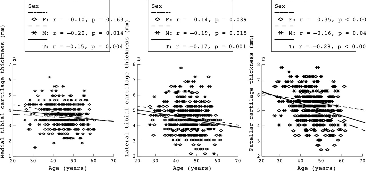
Correlation between age and knee cartilage thickness. There were significant negative associations between age and knee cartilage thickness at all three sites. T, total sample; F, female subjects; M, male subjects.
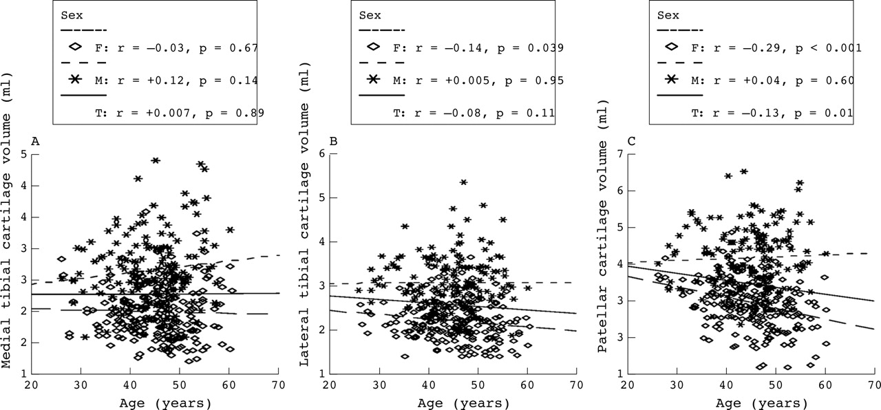
There were non-significant differences in knee cartilage volume between those aged over and under 45 years at all three sites (table 2⇑). Age was negatively associated with patellar cartilage volume (p<0.001) and lateral tibial cartilage volume (p = 0.039) in women, but did not correlate significantly with cartilage volume at other sites in women and at all sites in men (fig 3⇓). In multivariate analysis, age had no significant association with tibial cartilage volume but was negatively associated with patellar cartilage volume in the total sample and women. The association decreased and became non-significant in the total sample after further adjustment for ROA (table 3⇑).

Correlation between age and knee cartilage volume. There were significant negative correlations between age and patellar cartilage volume in the total population and lateral cartilage volume in women only. T, total sample; F, female subjects; M, male subjects.
Those aged >45 years had higher lateral tibial bone area but not patellar bone volume and medial tibial bone area than those aged <45 years (table 2⇑). Age had non-significant positive correlations with knee bone size in correlation analysis (p>0.05), with the exception of lateral bone area in men (p<0.01) (fig 4⇓). However, in multivariate analysis, age was significantly positively associated with medial and lateral tibial bone area and patellar bone volume in the total sample. These associations decreased in magnitude but remained significant for medial (p = 0.039) and lateral (p<0.001) tibial bone area after further adjustment for ROA score (table 3⇑). When men and women were analysed separately, age was significantly associated with lateral tibial bone area in men, and with patellar bone volume and lateral tibial bone area in women. The association between age and medial tibial bone area in women was of borderline significance (p = 0.07), and all the above associations decreased in magnitude after adjustment for ROA.

Correlation between age and knee bone size. There were no significant positive associations between age and knee bone size except that at the lateral tibial site in men. T, total sample; F, female subjects; M, male subjects.
DISCUSSION
This cross sectional study documents associations between age and knee cartilage defects, thickness, volume, and bone size in a convenience sample. In particular, the severity and prevalence of knee cartilage defects and bone size increased, whereas knee cartilage thickness decreased with increasing age at all compartments or sites. While patellar cartilage volume in women decreased with increasing age, tibial cartilage volume remained unchanged, suggesting knee cartilage thinning and defects and bone enlargement are the main processes that occur with aging.
Knee cartilage defects assessed by MRI are highly comparable to arthroscopic13–,15 and histological26 findings, and have been significantly associated with knee ROA27,28 and pain.29 However, the association between knee cartilage defects and age has not previously been reported. In this study we reported that age was positively associated with knee cartilage defect severity scores and prevalence in all compartments, and these associations were stronger in women and at the patellar site. The prevalence of any knee cartilage defect was 54% in subjects after the average age of 45 years, whereas the prevalence was 31% in subjects before the age of 45 years, suggesting that knee cartilage defects are quite common in older subjects, even in those without ROA.
There is limited information on cartilage loss with age. A necropsy study has previously reported that patellar cartilage from women aged over 50 years showed progressive thinning with increasing age, which was less obvious in men.30 Radiographic studies have shown significant negative correlations between increasing age and joint space in both men and women,9,10 whereas inconsistent associations are reported between joint space narrowing and age.9,12 The results from MRI studies have been inconsistent. Karvonen et al reported that cartilage thickness decreased significantly at the femoral condylar, but not at tibial and patellar sites,31 whereas Sargon et al reported that knee joint space declined linearly with increasing age.22 In an early study, Eckstein et al suggested that knee cartilage volume and mean cartilage thickness were not associated with age, while patellar maximal cartilage thickness was inversely associated with age.21 A subsequent larger study from Eckstein’s group reported a substantial decrease in thickness of cartilage in the knee at all sites, although this did not reach statistical significance at the tibia.23 This is consistent with our results. It is uncertain from the German study whether asymptomatic ROA was present as radiographs were not performed. Cicuttini et al reported that total and medial tibial cartilage volumes, which were standardised for bone size (leading to an indirect measure of cartilage thickness), were inversely associated with age in men consistent with our results, but there was no significant association between age and lateral cartilage volume.20 These inconsistencies are probably due to variations in study samples—generally small sample size and differing age and disease groups.
In the current study we found that age was not significantly associated with medial and lateral tibial cartilage volume either in men or women. This may reflect the coexistence of cartilage hypertrophy and cartilage thinning in early OA. However, tibial cartilage thickness decreased significantly with age, and these decreases were most obvious in men. In contrast, both patellar cartilage volume and thickness decreased with age, and these decreases were most obvious in women. These differences in volume changes at patellar and tibial sites may be real or may reflect chance variation, and these results will need to be replicated in other populations.
Previous studies have suggested that knee joint surface area is not associated with age in 27 healthy men,21 whereas the total cross sectional area of the tibial bone, assessed by peripheral quantitative computed tomography, became progressively greater with age in 512 men and 693 women.32 ROA was not assessed in this study so it is uncertain whether this was mediated by osteophytes. In the current sample, we found that age was positively associated with patellar bone volume as well as lateral and medial tibial bone area after adjustment for sex, height, weight, and offspring or control status. The reasons for the age related increase in knee bone size are unknown, as most studies suggest that insulin-like growth factor I and transforming growth factor β in bone decrease with age.33,34 The need to maintain adequate bone mechanical competence in the face of declining bone density may be one reason for the increase in bone cross sectional area in aging men.32 These associations may be of direct relevance to OA when combined with our previous reports that knee bone size is increased in subjects with early ROA19 and in the offspring of subjects at higher risk of OA.35
The age related increase in knee bone size is a possible explanation for age related knee cartilage alterations. In the present study the associations between age and knee cartilage defect scores in tibiofemoral compartments decreased in magnitude after adjustment for tibial bone area, but the associations between age, cartilage thickness, and patellar cartilage volume changed little after adjustment for bone size, suggesting that age related increases in bone area are linked to tibiofemoral cartilage defects, but not cartilage thickness or volume.
The relevance of these structural changes to OA remains uncertain. The associations between age and knee cartilage defect scores in tibiofemoral compartments became non-significant after adjustment for ROA. The associations between age and cartilage thickness, patellar cartilage volume, and knee bone size decreased in magnitude after adjustment for ROA, suggesting that age related alteration in all these factors is of direct relevance to tibiofemoral ROA. This was a cross sectional study and we cannot comment on causal directions. Thus longitudinal data will be required to determine which of these factors either singly or in combination is of most relevance to OA. In addition, we cannot comment on patellofemoral ROA, as these views are not available.
This study has a number of other potential limitations. Firstly, the study was primarily designed to look at genetic mechanisms of knee OA and used a matched design. The matching was broken for the current study, but adjustment for case-control status did not alter the results. Indeed, while there was a reduction in power, the results otherwise did not differ if examined in offspring and controls separately. Although the sample is a convenience sample, Miettinen states that for these associations to be generalisable to other populations three key criteria need to be met in the selection, sample size, and adequate distribution of study factors, all of which are met by this study.36 Secondly, measurement error may influence results. However, scoring of knee cartilage defects, volume, thickness, and bone size measurement was highly reproducible, suggesting this is unlikely. Thirdly, ROA assessment allows us to examine whether the changes are of relevance to ROA. However, given the relative rarity and mild severity of ROA in our sample, these results may not be generalisable to those with more severe OA. Fourthly, the oldest subject in this sample was 61 years, and thus we cannot comment on knee structural change after this age. Lastly, we are unable to comment on meniscal pathology at the current time.
In conclusion, this cross sectional study suggests that the most consistent knee structural changes with increasing age are an increase in cartilage defect severity and prevalence, cartilage thinning, and an increase in bone size, with inconsistent change in cartilage volume. Longitudinal studies will be required to determine which of these changes are primary and confirm their relevance to knee OA.
Acknowledgments
A special thanks to the subjects and orthopaedic surgeons who made this study possible. The role of Sr C Boon in coordinating the study is gratefully acknowledged. We would like to thank Martin Rush who performed the MRI scans, Kevin Morris for technical support, and Professor Dave Hosmer for statistical advice.
Supported by the National Health and Medical Research Council of Australia, Masonic Centenary Research Foundation.