Article Text

Abstract
Background: Crohn’s disease is associated with an increased number of macrophages in ileal and colonic mucosa. Data on macrophages in gut mucosa of patients with spondyloarthritis (SpA) are scarce.
Objective: To investigate macrophages and other antigen presenting cells in gut mucosa from patients with SpA and Crohn’s disease, given the relationship between both entities.
Methods: Biopsy specimens from patients with SpA, Crohn’s disease, ulcerative colitis, and from controls were immunohistochemically stained with different markers for macrophages and dendritic cells. Slides were scored semiquantitatively on a four point scale.
Results: SpA and Crohn’s disease were associated with large numbers of CD68+ macrophages. Colon mucosa of both patients with SpA and Crohn’s disease, but not ulcerative colitis, showed increased numbers of macrophages expressing the scavenger receptor CD163.
Conclusions: Macrophages expressing the scavenger receptor CD163 are increased in colonic mucosa in SpA and in Crohn’s disease, highlighting the relationship between these entities. The increased number of CD163+ macrophages in colon mucosa of patients with SpA suggests this is another argument for a role of macrophage scavenger receptors in this group of diseases.
- IBD, inflammatory bowel disease
- SRCR, scavenger receptor cysteine-rich
- SpA, spondyloarthritis
- TNFα, tumour necrosis factor α
- spondyloarthritis
- inflammatory bowel disease
- macrophages
- scavenger receptors
Statistics from Altmetric.com
- IBD, inflammatory bowel disease
- SRCR, scavenger receptor cysteine-rich
- SpA, spondyloarthritis
- TNFα, tumour necrosis factor α
The Th1 cytokine tumour necrosis factor α (TNFα) has a pivotal role in Crohn’s disease, clearly illustrated by the effect of anti-TNFα drugs.1 Blocking TNFα also improves the articular manifestations in spondyloarthritis (SpA).2 Together with the abundant presence of TNFα in sacroiliac joint biopsy specimens from patients with ankylosing spondylitis,3 this suggests a major role for TNFα in SpA as well.
TNFα is primarily produced by lymphocytes, monocytes, and macrophages.4 Besides their role in production of TNFα, macrophages play a different part in mucosal immunity, including antigen presentation and enhancement of immunoglobulin production. An increased number of mucosal macrophages in Crohn’s disease have been reported previously,5 whereas data for SpA are scarce. Studying macrophages in gut mucosa from patients with SpA, however, seems worthwhile because subclinical inflammatory gut lesions, which can evolve to clinically overt Crohn’s disease, are found in 25–75% of these patients.6
This study aimed at analysing macrophages and other antigen presenting cells in gut mucosa from patients with SpA and Crohn’s disease. The results were compared with those obtained in ulcerative colitis and controls. Special attention was paid to the macrophage scavenger receptor CD163, because CD163+ macrophages are increased in synovium from patients with SpA compared with patients with rheumatoid arthritis.7
METHODS
Patients with SpA
Ileal and colonic biopsy specimens were obtained from 21 patients with SpA fulfilling the European Spondylarthropathy Study Group criteria.8 This group included patients with ankylosing spondylitis (n = 16), psoriatic arthritis (n = 2), reactive arthritis (n = 1), juvenile SpA (n = 1), and undifferentiated SpA (n = 1). Their median age was 36.5 years (range 26–74). Six patients were taking sulfasalazine and 11 were receiving non-steroidal anti-inflammatory drugs. One of the patients was treated with corticosteroids. The remaining patients had no drug treatment.
As all tissue from these patients was collected during the past 4 years, there was no longer follow up period. Over that time, one of them developed a clinical Crohn’s-like colitis, confirmed histopathologically.
Patients with inflammatory bowel disease
Mucosal biopsy samples from resection specimens were obtained from patients with Crohn’s ileitis (n = 10), Crohn’s ileocolitis (n = 4), Crohn’s colitis (n = 6), and ulcerative colitis (n = 11). The diagnosis was independently established by endoscopic, radiological, and histological criteria. Samples were taken from areas with endoscopic or macroscopic involvement. The median age of the patients with Crohn’s disease was 31 years (range 13–67), and of those with ulcerative colitis 37 years (range 29–61). Twenty five patients with inflammatory bowel disease (IBD) were treated with corticosteroids or other immunosuppressive drugs; four patients were taking only sulfasalazine. The remaining two patients had no drug treatment.
Controls
Control ileal and colonic tissue was obtained from 13 patients who underwent a segmental colectomy for colorectal carcinoma, or a colonoscopy for irritable bowel syndrome or follow up of polyps. In cases of carcinoma, samples were taken at least 10 cm away from the tumour. The median age of these patients was 56 years (range 27–72).
This study was approved by the local ethical committee.
Immunohistochemistry
Frozen sections of ileal and colonic mucosa from all patients and controls were fixed in acetone and incubated with antibodies against CD68 and CD163. Moreover, we performed stainings for CD14, CD80, CD83, CD86, and HLA-DR on ileal and colonic mucosa from all controls and all patients with ulcerative colitis, Crohn’s ileitis, and Crohn’s ileocolitis, and from 10 patients with SpA and five with Crohn’s colitis. Table 1 lists the antibodies used.
Antibodies used in this study
Immunostaining was visualised using new fuchsin (for CD68) or 3-amino-9-ethylcarbazole (for the other markers), both obtained from DAKO Corporation (Carpintera, USA). Parallel sections were incubated with irrelevant isotype and concentration matched antibodies as negative control.
Evaluation of immunohistochemical staining
The number of positive cells in the lamina propria was analysed using magnification ×100 and coded by two independent observers who did not know the diagnosis and clinical data. A grading scale from 0 to 3 was applied, ranging from absent to numerous stained cells. As some histological markers are more abundant than others in gut tissue, the scoring system was calibrated for each marker separately by examining a representative number of samples. Thus, the sensitivity of the scoring system was different for each marker, allowing comparison of a specific marker between the different patient groups, but not comparison of different markers in one group. The analysis included all areas of the biopsy specimens, and a global score was given for each measure.
The scores obtained by both observers were concordant in more than 90% of cases. When the scores were discordant, differing by a maximum of one point, the mean of the two scores was used.
Statistical analysis
Patient groups were compared with controls; p values were calculated with the Mann-Whitney U test. A value of p<0.05 was considered significant.
Because the data were not compared between multiple groups and because the analysis of each marker was considered as an individual comparison between two groups, Bonferroni corrections for multiple comparisons were not applied in this exploratory study.
RESULTS
Increased number of CD163+ macrophages in both Crohn’s colitis and colon mucosa from patients with SpA
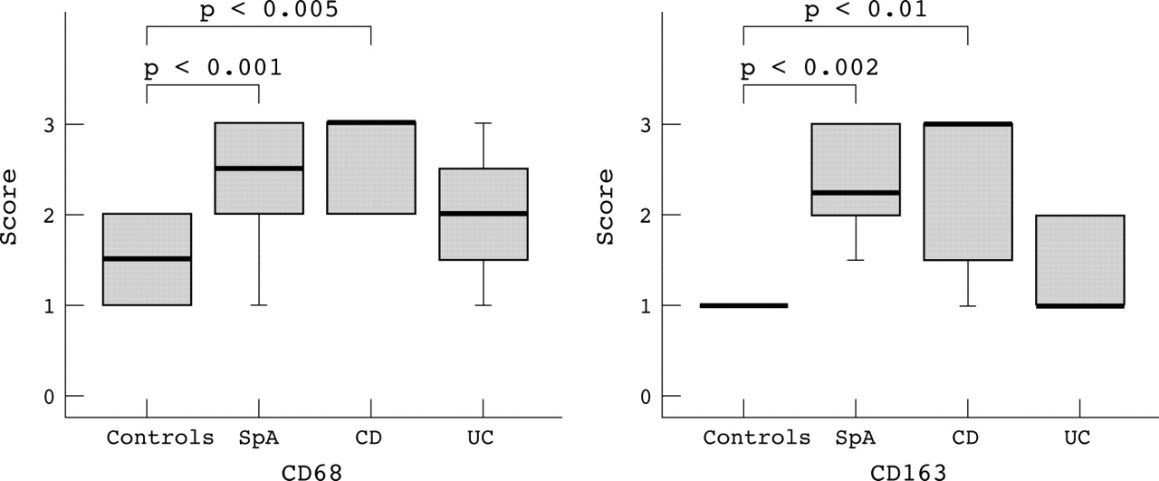
Control ileum and colon showed a subepithelial band of CD68+ macrophages. In patients with inflammatory disease multitudinous CD68+ cells were found in the ulcer bases; they were especially numerous in transmural inflammation in Crohn’s disease. CD68+ cells were increased in ileal (p<0.02) and colonic (p<0.01) Crohn’s disease and also in colon from patients with SpA (p<0.005).
In controls, CD163+ macrophages were also localised in the superficial lamina propria of the colon (fig 1A) and in ileal villous tips, but they were less numerous than CD68+ cells. Scarce CD163+ cells were found in the deeper layers of the lamina propria. Crohn’s colitis showed increased numbers of CD163+ cells throughout the mucosa (p<0.01) (fig 1B). In colon from patients with SpA, increased numbers of CD163+ cells were also detected (p<0.002) (fig 1C). This contrasts with the normal findings in ulcerative colitis (fig 1D). Figure 2 presents the results for the scores for CD68 and CD163 in colon.
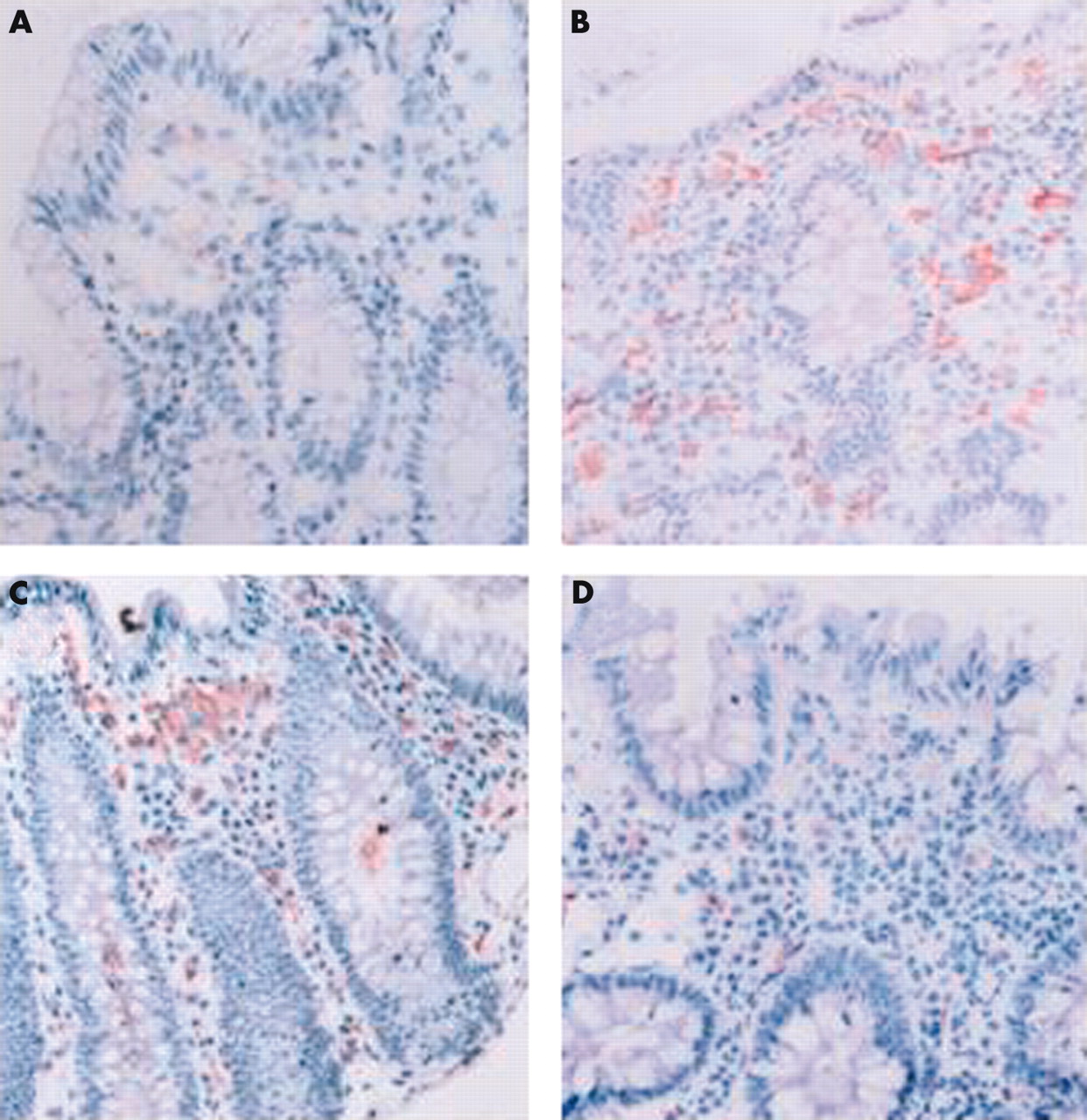
Immunohistochemical staining for CD163 (peroxidase method). (A) Macrophages weakly positive for CD163 in the superficial lamina propria of control colon. (B) Increase in the number of CD163+ cells in Crohn’s colitis. (C) Non-inflamed colonic mucosa from a patient with SpA showing a similar increase in the number of activated macrophages. (D) No increase in the number of CD163+ cells in ulcerative colitis.
Box and whisker plots representing the semiquantitative scores for CD68 and CD163 in the colonic lamina propria of patients with spondyloarthritis (SpA), Crohn’s disease (CD), and ulcerative colitis (UC) compared with controls. p Values were calculated with the Mann-Whitney U test.
The number of dendritic cells and HLA-DR positive inflammatory cells is unaltered in the mucosa of patients with IBD or SpA
CD83+ cells were present in T cell zones of lymphoid follicles in both normal and inflamed bowel mucosa. Although outside the follicles fewer CD83+ cells were found, clusters of positive cells were present in IBD but not in controls and patients with SpA.
CD80+ cells were scarce. CD86+ cells occurred scattered throughout the lamina propria and could be found in the dome area overlying lymphoid follicles; in follicles, however, they were scarce or absent. There was no increase in patient groups.
In all cases, HLA-DR+ inflammatory cells were found in the lamina propria. No significant differences between patients and controls were found.
DISCUSSION
We demonstrated an increased number of CD163+ macrophages in colon from patients with SpA and from those with Crohn’s disease, but not ulcerative colitis. The number of CD163+ cells was not related to the patient’s age or drug treatment (data not shown).
The transmembrane protein CD163 is a member of the scavenger receptor cysteine-rich (SRCR) superfamily. The SRCR proteins play a part in the development of the immune system and in the regulation of the immune response. Although the precise function of CD163 is unknown, a role in the regulation of the immune response of macrophages is supposed. Comparison of CD163− and CD163+ swine monocytes/macrophages showed that the latter are more mature.9 CD163+ cells induce a greater allogeneic response on T lymphocytes. Lipopolysaccharide stimulated CD163− and CD163+ macrophages both produce the proinflammatory cytokines interleukin 1 and TNFα, but anti-inflammatory interleukin 10 is only detected on the former population.9
An increase of CD163+ macrophages in colon mucosa in SpA as well as Crohn’s disease raises the possibility that these cells may induce a similar dysregulation of the cytokine balance in the colon in both disorders. Previously, we described also for T cells common characteristics in the cytokine profile.10,11 These observations, together with similar alterations in adhesion molecule expression,12,13 are in line with the concept that some patients with SpA have subclinical Crohn’s disease.
The CD163 protein scavenges haemoglobin by mediating endocytosis of haptoglobin-haemoglobin complexes.14 Compounds containing haem are potentially a valuable source of iron for invading micro-organisms; although bacteria have evolved several strategies to overcome iron shortage, increased scavenging of haptoglobin-haemoglobin complexes might alter the composition of the bacterial gut flora.
In conclusion, this study indicates that CD163+ macrophages are increased in colonic mucosa in SpA as well as in Crohn’s disease, highlighting the relationship between these entities. Because the macrophage receptor with collagenous structure is also susceptible to modulation in SpA,15 we suggest this is another argument for a role of macrophage scavenger receptors in this group of diseases. However, CD163 is the first scavenger receptor shown to be altered in colon mucosa from patients with SpA.
Acknowledgments
This work was supported by concerted action grant GOA96001 of Ghent University, FWO-Vlaanderen grant 3.0028.95, and Ghent University Research Foundation grant 00/DOC/017.