Article Text

Abstract
OBJECTIVE Previously an upregulation of E-cadherin and its associated molecules α-catenin, β-catenin and plakoglobin has been demonstrated in clinically overt inflammatory bowel disease (IBD). The aim of this study was to investigate the expression of the E-cadherin/catenin complex in subclinically inflamed bowel mucosa from spondyloarthropathy (SpA) patients.
METHODS Ileal and colonic biopsy specimens from 19 SpA patients with subclinical inflammatory gut lesions and from seven controls were stained with monoclonal antibodies against E-cadherin, β-catenin and plakoglobin and a polyclonal antibody against α-catenin. E-cadherin mRNA was detected using a riboprobe. Inflammation was histologically classified into acute, chronic active and chronic quiescent forms.
RESULTS In acute and chronic active bowel inflammation of SpA patients, upregulation of the E-cadherin/catenin glycoprotein complex could be observed. Chronic lesions in a quiescent state did not show such an upregulation. Furthermore, chronic inflammation was associated with an increase in E-cadherin mRNA.
CONCLUSIONS As some of the SpA patients with subclinical gut inflammation develop IBD, upregulation of the E-cadherin/catenin complex in inflamed bowel mucosa from SpA patients may point to early cellular changes in the development of IBD. However, at present it cannot be excluded that increased E-cadherin/catenin complex expression is a bystander phenomenon of active inflammation.
- spondyloarthropathy
- inflammatory bowel disease
- E-cadherin
- catenins
Statistics from Altmetric.com
In epithelial cells, homotypic homophilic intercellular adhesion is mediated by E-cadherin.1 This molecule is a transmembrane glycoprotein, mainly localised to the zonula adherens junctions of all normal epithelia.2 For normal function of E-cadherin, members of the catenin family (α-catenin, β-catenin and plakoglobin) are required.3 ,4 Both β-catenin and plakoglobin (γ-catenin) bind directly to E-cadherin; α-catenin links the bound β-catenin or plakoglobin to the actin cytoskeleton, thereby forming complexes of either E-cadherin/β-catenin/α-catenin or of E-cadherin/ plakoglobin/α-catenin.5-8
Association between down-regulation of E-cadherin in cancers, high invasion rates and metastasis has been described.9-11Changes in the expression of E-cadherin associated catenins, in particular β-catenin, the vertebrate homologue of theDrosophila segment polarity genearmadillo, also frequently occur in early dysplastic lesions of the colon.7 ,12
Previously we demonstrated an upregulation of E-cadherin and its associated catenins in clinically overt inflammatory bowel disease (IBD) (P Demetter, submitted data). This up-regulation was detectable in acute as well as in chronic active gut inflammation. Dynamic changes in the expression of this complex might be important for the maintenance of normal epithelial integrity under inflammatory conditions.
A prospective endoscopic and histological study of the terminal ileum and colorectum in 211 spondyloarthropathy (SpA) patients showed subclinical inflammatory gut lesions in 61%.13 Acute inflammation resembling acute infectious enterocolitis and a chronic type of ileocolitis were described.14 One third of the biopsy specimens featured chronic inflammation resembling Crohn's disease. After two to nine years, clinical and histological evidence of chronic IBD, particularly Crohn's disease, was found in 7% of patients,15 ,16 suggesting early lesions of Crohn's disease in SpA patients.
The aim of this study was to investigate whether subclinical inflammatory gut lesions of SpA are also associated with an upregulation of the E-cadherin/catenin complex. This could indicate whether or not E-cadherin/catenin complex upregulation is an early event in the development of IBD.
Methods
TISSUE SPECIMENS
Twenty seven formalin fixed, paraffin wax embedded specimens from 19 SpA patients featuring subclinically inflamed bowel mucosa were retrieved from the archival material of the Histopathology Department of the University Hospital in Gent. Blocks from seven patients with ileal lymphoid hyperplasia or irritable bowel syndrome were used as non-inflamed controls. All patients with SpA fulfilled the criteria of the European Spondyloarthropathy Study Group17: 10 of them suffered from ankylosing spondylitis, four from undifferentiated SpA, three from reactive arthritis and two from juvenile SpA. The disease group included 14 ileal and 13 colonic samples; the control group consisted of seven ileal and six colonic samples. Tables 1 and 2summarise the patient data. The patients were mostly treated with non-steroidal anti-inflammatory drugs, in some cases combined with salicylazosulphapyridine. None of them took systemic corticosteroids.
Clinical data of the investigated patients
Clinical characteristics of spondyloarthropathy patients included in this study
HISTOLOGICAL EVALUATION
Haematoxylin and eosin stained slides of all specimens were divided into acute, chronic active or chronic quiescent inflammation. Criteria used were described previously.14 ,18 In summary, acute inflammation is defined by the presence of neutrophils and/or eosinophils in the crypt and villus epithelium without alterations in the mucosal structure. In chronic active inflammation there is, in addition to architectural alterations, an active granulocytic infiltrate in the epithelium accompanied by an increased mononuclear cell infiltrate in the lamina propria. Chronic quiescent inflammation is characterised by structural changes only.
IMMUNOHISTOCHEMISTRY
Monoclonal antibodies against E-cadherin, β-catenin and plakoglobin (clones 36, 14 and 15, respectively) were obtained from Transduction Laboratories (Lexington, USA), and a polyclonal anti-α-catenin antibody (C-19) from Santa Cruz Biotechnology (Santa Cruz, USA). For unmasking of antigens, tissue sections (5 μm) were immersed in 0.1 M citrate buffer and heated in a microwave oven before applying the anti-E-cadherin, anti-α-caterin and anti-plakoglobin antibodies. For the detection of β-catenin no pretreatment was applied. Immunoreactivity was visualised using New Fuchsin or 3-amino-9-ethylcarbazole (AEC), both obtained from Dako Corporation (Carpintera, USA). For the demonstration of E-cadherin, α-catenin and plakoglobin a peroxidase method was used, whereas for the detection of β-catenin an alkaline phosphatase reaction was applied.
Negative controls consisted of duplicate sections stained simultaneously, in which the primary antibody was replaced by an isotype specific, irrelevant antibody.
EVALUATION OF IMMUNOHISTOCHEMICAL STAINING INTENSITY
After examining the control cases, the SpA gut biopsy specimens were studied by two observers (PD and CAC). As in some cases inflammatory changes were rather striking, real blind reading was not attempted. Differences in the expression of the studied molecules were indicated only if both observers agreed. In focal inflammation the inflamed areas were compared with non-inflamed mucosa in the same biopsy sample.
IN SITU HYBRIDISATION
The expression of E-cadherin mRNA was examined by in situ hybridisation for the following situations: acute ileitis (n=1), acute colitis (n=2), chronic active ileitis (n=3), chronic quiescent ileitis (n=1), chronic quiescent colitis (n=1), normal ileum (n=3) and normal colon (n=3). cRNA probes were generated in vitro according to the method described by Logel et al. 19 The transcription primers span a 250 bp region of E-cadherin cDNA (purchased from Gibco BRL, Gent, Belgium). The E-cadherin specific sense primer was modified 5' terminally with a T7 promoter consensus sequence, 5'-CCAAGCTTCTAATACGACT C ACTATAGGGAGATCACTGACACCAA CGATAATCC-3'. The E-cadherin specific antisense primer was modified 5' terminally with a T3 promoter consensus sequence, 5'-CAGAGATGCAATTAACCCTCACAAA GGGAGAGTTGGCAGTGTCTCTCCAAA TC-3'. This primerset was used in a PCR reaction on plasmid pMS13 containing E-cadherin cDNA.20The PCR product was generated with AmpliTaq Gold (Perkin Elmer, Branchburg, USA) in a Programmable Thermal Controller-100 (MJ Research, Watertown, USA) and then purified with an RNAse free Qiagen gel extraction kit (Qiagen GMBH, Hilden, Germany). Sense and antisense cRNA were transcribed and labelled with FITC-UTP in an RNA labelling kit (Boehringer Mannheim, Mannheim, Germany).
Paraffin wax sections of the tissues were hybridised with antisense RNA or sense RNA for 18 hours at 55°C. After hybridisation they were incubated with an anti-FITC antibody (BioGenex, San Ramon, USA) at room temperature for 20 minutes. Colour reaction was developed using New Fuchsin.
Results
IMMUNOHISTOCHEMISTRY
In normal ileum, positivity for E-cadherin and associated catenins presented as a distinct basolateral staining of the cells in the villi, especially in villous tips. Weak cytoplasmic staining was found in some instances. In crypts, staining was most prominent in the crypt base cells whereas other crypt cells expressed lower E-cadherin levels. For the associated catenins a similar pattern was found (fig1a).

E-cadherin/catenin immunoreactivity in normal and subclinically inflamed bowel mucosa. (a): Weak α-catenin expression in non-inflamed ileum. (b): Acute ileitis showing strong expression of α-catenin in all cells of the villi. (c): Focal upregulation of E-cadherin in villus epithelial cells in chronic active ileitis. (d): β-catenin immunoreactivity in villus and crypt cells in chronic active ileitis. (a) and (c) Bar = 40 μm; (b) and (d) bar = 25 μm.
In comparison with ileum, the colonic epithelial cells showed stronger expression of the E-cadherin/catenin complex. As in ileum, crypt cells stained weaker than superficial epithelial cells.
In acute and in chronic active subclinical gut inflammation of SpA patients we found focal upregulations of the molecules of the E-cadherin/catenin complex. This focality was linked to the intensity of inflammation, with stronger immunoreactivity in cells in more inflamed areas. Especially cells adjacent to epithelial breaches were strongly positive. In crypts and in superficial epithelium, invasion with inflammatory cells, in particular neutrophils but also lymphocytes, was associated with more intense staining (fig 1b-d). In chronic inflamed mucosa in a quiescent phase the expression of the E-cadherin/catenin complex resembled the findings in control biopsy specimens. In this condition focal changes in expression were only rarely seen. Results are presented in table 3.
Upregulation of E-cadherin/catenin complex in the different groups
IN SITU HYBRIDISATION
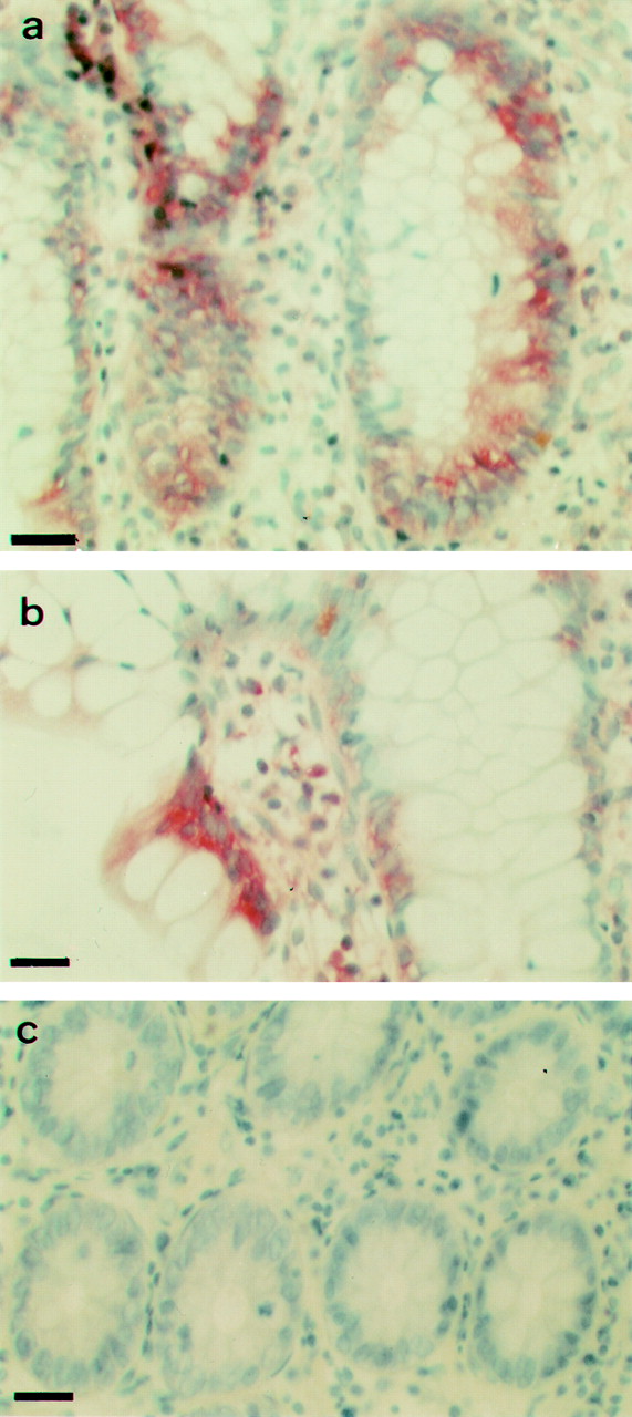
The labelling with the E-cadherin antisense mRNA probe showed strong mRNA positivity in both chronic active and chronic quiescent inflammation (fig 2a). Focal inflammation was accompanied by strong mRNA positivity in the adjacent area (fig 2b). In acute inflammation, however, the detectable expression was weak to negative, as in normal colon and ileum (fig 2c).

E-cadherin mRNA in situ hybridisation. (a): Strong positivity in chronic quiescent ileitis. (b): Focal upregulation in chronic quiescent ileitis. (c): Absence of detectable expression in normal colon. (a), (b) and (c) Bar = 25 μm.
Regardless of the inflammatory state, the E-cadherin mRNA signal was found in a pattern compatible with that of the protein expression, with strongest positivity in crypt bases and superficial epithelial cells.
Discussion
In this study we demonstrate an increased expression of the proteins of the E-cadherin/catenin complex in acute and chronic active subclinical gut inflammation in SpA patients. In chronic quiescent inflammation this upregulation was rare to absent. These findings are also seen in full blown IBD.21
Previously we found another similarity between IBD and SpA bowel mucosa. Flow cytometric analysis of T cells expanded from colonic biopsy specimens was conducted from patients with SpA featuring normal gut histology, patients with Crohn's disease and ulcerative colitis, and controls. On these T cells, the expression of the αE/β7 integrin, of which the αE subunit is also known as CD103, was increased in IBD and non-inflamed bowel mucosa from SpA patients but not in controls.22
As E-cadherin is not only involved in homophilic epithelial cell-cell adhesion but is also a ligand for the αE/β7 integrin on intra-epithelial T cells,23 these two changes in expression could be attributable to a hitherto unknown common environmental or genetic factor. Upregulation of E-cadherin might follow the changed expression of the αE/β7 integrin, as this molecule is already upregulated in histologically normal bowel mucosa from SpA patients.22 However, the expression of the E-cadherin/catenin complex in the latter situation remains to be examined. Interestingly, about half of the SpA patients included in this study did not show any macroscopically detectable ileocolonoscopic abnormalities, possibly pointing to upregulation of the E-cadherin/catenin complex as an early cellular change in the development of IBD. Seventeen of 19 patients included in this study were followed up by our group during 10 years or more after initial biopsy taking. Three of them developed full blown Crohn's disease during this period: patients 2, 15 and 16 (tables 2 and 3). Even in patient 2, ileocolonoscopy at time of subclinical histological gut inflammation was completely normal.
Effects on the expression of the E-cadherin/catenin complex may be moderated by the effects of trefoil peptides,24 called “trefoil” because of their distinctive cysteine-rich three leafed primary stucture.25 ,26 Trefoil peptides have been shown to be overexpressed in regions of the gastrointestinal tract particularly susceptible to mucosal damage, and are of potential functional importance in different inflammatory conditions in the gastrointestinal tract.25 ,27 One of these molecules, intestinal trefoil factor 3, may modulate epithelial cell adhesion and survival by affecting the complexes between E-cadherin, β-catenin and associated proteins.24 Moreover, the cooperative interaction of trefoil peptides with mucin glycoproteins, secreted by goblet cells, protects gastrointestinal mucosa from a variety of insults.28 However, the sequential expression of spasmolytic polypeptide, intestinal trefoil factor, epidermal growth factor and transforming growth factor α mRNAs in experimental ulceration in rat stomach makes clear that epithelial protection and restitution is a complex process.29 Anyway, the pronounced but temporally different patterns of mRNA induction after ulceration suggest that the trefoil peptides may fulfil immediate roles in the protection and restitution of epithelium.
Another molecule possibly involved in the upregulation of the E-cadherin/catenin complex is interleukin 12 (IL12), expressed and released by lamina propria mononuclear cells from patients with Crohn's disease.30 It has been shown that upregulation of IL12 production is a critical event in the initiation of bowel inflammatory lesions in Crohn's disease,31 which can evolve out of the subclinical gut lesions in SpA patients.15 ,16 As IL12 upregulates E-cadherin expression,32 it may be involved in the alterations we described.
In chronic quiescent inflammation, we detected an increase in E-cadherin mRNA without upregulation of the protein. This might be caused by the action of the matrix metalloproteinase stromelysin-1, which is upregulated in chronic bowel inflammation.33 ,34Stromelysin-1 possibly affects tissue repair at multiple stages.33 ,34 However, it may cleave E-cadherin and make it disappear from cell-cell contacts.35
In conclusion, we found an upregulation of the E-cadherin/catenin complex in subclinical gut inflammation in SpA patients. The meaning, however, is not fully understood. As part of these patients evolve into clinically overt inflammatory bowel disease, our finding can suggest on the one hand that this upregulation may be of major importance in the pathogenesis of Crohn's disease or SpA, or both. On the other hand, hitherto it cannot be excluded that the upregulation is secondary to active, albeit subclinical, inflammatory bowel disease. If the binding between E-cadherin and the αE/β7 integrin could be identified as crucial, therapeutic blocking approaches might be developed in the future to treat intestinal inflammation and SpA.
Acknowledgments
The authors wish to thank Dr Philip De Bruyne for preparation of the riboprobes. Dr Baeten is an FWO-Vlaanderen Research Assistant.
References
Footnotes
Funding: this work was supported by a concerted action grand GOA96001 of the University of Gent (Belgium), and an FWO-Vlaanderen grant 3.0028.95.
Linked Articles
- Correction