Article Text

Abstract
Objective To define the relationship between autoantigen citrullination and different peptidylarginine deiminase (PAD) enzymes in rheumatoid arthritis (RA).
Methods Citrullinated autoantigens were identified by immunoblotting control and ionomycin-activated human primary neutrophil lysate with RA sera. Autoantigen identity and citrullination sites were defined by mass spectrometry. PAD isoenzyme expression in human neutrophils was determined by immunoblotting. PAD substrate specificity was addressed in HL-60 cell lysates co-incubated with human recombinant PAD2, PAD3 and PAD4.
Results Although prominent protein citrullination is observed in ionomycin-activated neutrophils, RA sera only recognised a limited number of these citrullinated molecules. Among these, the authors identified that β and γ-actins are citrullinated on at least 10 arginine residues, generating a novel 47 kDa species that is frequently recognised by RA autoantibodies. Interestingly, the authors showed that the PAD enzymes expressed in human neutrophils (ie, PAD2, PAD3 and PAD4) have unique substrate specificities, independent of their subcellular distribution. Thus, only PAD2 was able to citrullinate native β/γ-actin, while histone H3 was only citrullinated by PAD4.
Conclusion These studies identified β and γ-actins as novel citrullinated autoantigens in RA, allowing enzyme specificity against intracellular substrates to be addressed. The studies provide evidence that PAD enzymes have the intrinsic capacity to select unique protein targets. The authors propose that unique PAD specificity may play a role in autoantigen selection in RA.
Statistics from Altmetric.com
Accumulating evidence suggests that enzymatic deimination (or citrullination) of proteins, a process catalysed by the peptidylarginine deiminase (PAD) enzymes,1 plays an essential role in rheumatoid arthritis (RA) pathogenesis.2 However, although advances have been made to support the pathogenic role of antigen citrullination in RA, important questions remain about the nature of the protein targets towards which anticitrullinated protein antibodies (ACPA) are directed, and about the interaction between these autoantigen substrates and the PAD enzymes.
Among the five PAD enzymes encoded by humans (PAD1–PAD4 and PAD6), PAD2 and PAD4 have gained attention as potential candidates that may drive the citrullination of self-antigens in RA. Both enzymes have been found in rheumatoid synovial tissue and fluid,3,–,5 and PAD4 polymorphisms are genetically associated with RA development.6 Besides their distinct tissue distribution (PAD2 is widely expressed while PAD4 is preferentially expressed by haematopoietic cells),1 the major difference between these enzymes is that PAD2 has a cytoplasmic distribution while PAD4 mainly resides in the cell nucleus.7 8 These observations raise the possibility that different PAD target different cellular substrates, defined by both the cell type and the subcellular localisation of the substrate. Therefore, distinct autoantibodies recognising such citrullinated substrates might indicate the activity of specific PAD in intracellular protein citrullination.
In RA, little attention has been paid to defining whether the selection of citrullinated autoantigens is driven by unique PAD enzymes. This can be explained partly because current evidence suggests that protein citrullination may occur extracellularly.5 In this case, substrate selection by the PAD would not be limited by their subcellular localisation. Under such circumstances, target specificity among PAD would be mechanistically irrelevant. Despite this, it is intriguing that except for PAD49 10 and the far upstream element-binding proteins,11 which are nuclear, the majority of cellular targets identified as potential citrullinated autoantigens in RA have extranuclear distribution (eg, filaggrin;12 13 vimentin;14 15 α-enolase, elongation factor 1a and adenyl cyclase-associated protein 1;16 F-actin capping protein α 1 subunit, asporin, cathepsin D, β-actin, histamine receptor, protein disulfide-isomerase, ER60 precursor and mitochondrial aldehyde dehydrogenase;17 collagen types I18 and II;19 eukaryotic translation initiation factor 4G1;20 aldolase, phosphoglycerate kinase 1, calreticulin and HSP60).11 The skewing of potential autoantigens to the extranuclear compartment raises questions about the role of PAD4 if autoantigen citrullination occurs intracellularly and about the random selection of targets if citrullination occurs extracellularly. It is possible that such substrate specificity reflects the activity of unique PADs either intracellularly (ie, cytoplasmic PADs) or extracellularly. These studies provide evidence to support that the PAD enzymes have the intrinsic capacity to select unique protein targets.
Methods
Subjects
Sera from 39 patients with established RA were obtained from a convenience institutional review board approved cohort followed at the Johns Hopkins Arthritis Center. Sera from 15 healthy adults were used as a comparison group.
Human PAD2–4 cloning and recombinant protein purification
Cloning and expression of recombinant (r)PAD4 was described elsewhere.10 Total RNA purified from ATRA-differentiated HL-60 cells (to clone PAD2) or human bone marrow (Cambrex, East Rutherford, NJ, USA) (to clone PAD3) was reverse-transcribed to generate complementary DNA. cDNA were further cloned into pET28a (Novagen, Gibbstown, NJ, USA) for prokaryotic expression to generate an N-terminal His6-tagged fusion protein that was purified using Ni-NTA agarose (Qiagen, Valencia, CA, USA) as described by the manufacturer.
Neutrophil isolation and activation
After IRB approval and informed consent, neutrophils were isolated from healthy donors as described10 and resuspended in Hank's balanced salt solution without calcium/magnesium. Calcium chloride was added to reach 2 mM and neutrophils were further incubated in the absence or presence of 1 μM ionomycin for 4 h at 37oC. After incubation, the cells were lysed and boiled in sodium dodecylsulphate (SDS) sample buffer for further analysis by immunoblotting. Alternatively, the cells were lysed with Isoelectric focusing (IEF) buffer (8 M urea, 2 M thiourea, 4% 3-[(3-Cholamidopropyl)-dimethylammonio]-1-propanesulfonate (CHAPS), 1% Dithiothreitol (DTT)).
Two-dimensional electrophoresis and mass spectrometry analysis
Samples in IEF buffer containing 0.2% Bio-lyte 3/10 ampholyte (BioRad, Hercules, CA, USA), 1.5% 2-hydroxyethyl disulfide and 0.05% bromophenol blue were resolved using 3-10 IPG strips (Bio-Rad) and a BioRad IEF cell system. The IPG strips were then incubated in equilibration buffer (6 M urea, 30% w/v glycerol, 2% SDS, 0.05 M tris pH 8.8) containing 1% DTT, followed by incubation in equilibration buffer containing 2.5% Iodoacetamide (IAA). For the second dimension, the samples were resolved by electrophoresis on 8% SDS-polyacrylamide gels and transferred into nitrocellulose membrane. The protein pattern was visualised by Ponceau-S before immunoblotting using sera from RA patients. Once the antigen of interest was mapped, the spot was sliced from a two-dimensional gel stained with GelCode blue (Thermo Scientific, Waltham, MA, USA) and analysed by mass spectrometry to define the protein identity and to identify citrullination sites (Proteomics Core/Mass Spectrometry Facility, Johns Hopkins School of Medicine).
In-vitro citrullination assays
Using siliconised tubes (Sigma, St. Louis, MO, USA), 1 μM human recombinant β-actin, γ-actin (GenWay, San Diego, CA, USA), or 700 nM purified actin from human platelets (Cytoskeleton, Denver, CO, USA) plus 700 nM human recombinant histone H3.1 (New England Biolabs, Ipswich, MA, USA) were incubated alone or co-incubated with 700 nM rabbit PAD2 (Sigma), human rPAD2, rPAD3 or rPAD4 in buffer A (100 mM Tris pH 7.6, 5 mM DTT, 10 mM calcium chloride). After 0–60 min at 37°C, reactions were stopped by adding SDS sample buffer and boiling. Non-citrullinated and citrullinated recombinant β-actin were also used for mass spectrometry analysis to identify citrullination sites, and to screen for anticitrullinated β-actin antibodies by immunoblotting using control and RA sera.
Cell lysates from PAD-negative undifferentiated HL-60 cells (3×10⁶ cells/ml) were generated in buffer B (20 mM Tris pH 7.6, 1% NP40 and protease inhibitors), sonicated, cleared by centrifugation, and further incubated alone or co-incubated with 700 nM human rPAD2, rPAD3 or rPAD4 in the presence of 5 mM DTT and 10 mM calcium chloride. After 60 min at 37°C, reactions were stopped by adding SDS sample buffer and boiling. Protein citrullination was determined by anti-modified citrulline (AMC) immunoblotting, according to the manufacturer's recommendations (Millipore, Billerica, MA, USA).
Results
RA autoantibodies recognise a limited number of citrullinated antigens in activated primary neutrophils
To understand better the independent role of the PAD enzymes in autoantigen citrullination in cells expressing multiple PAD, we initially focused on the study of human neutrophils. This cell type represents one of the most abundant inflammatory cells in the rheumatoid joint and has been widely used as a model for the study of protein citrullination. The cells constitutively express PAD4,7 21 and protein citrullination can be induced upon cell activation with different stimuli.22 In initial studies, we demonstrated that neutrophils express PAD2 and PAD3 in addition to PAD4 protein (figure 1A), making them a suitable system to study autoantigen citrullination by multiple PAD. To identify the patterns of citrullinated autoantigens generated in activated neutrophils, neutrophils were activated with ionomycin, and lysates of control and ionomycin-activated cells were analysed for protein citrullination (figure 1B) and recognition by RA sera (figure 1C). While protein citrullination was absent in control neutrophils, ionomycin treatment induced massive citrullination, modifying molecules across the entire range of molecular weights detected by SDS polyacrylamide gel electrophoresis. Two different patterns of reactivity with RA sera were noted: molecules that were only detected in activated neutrophils (the focus of this study), and antigens found in non-stimulated neutrophils, which either remained unchanged or disappeared upon cell activation. Interestingly, despite the large number of citrullinated proteins found in activated neutrophils (figure 1B), sera from RA patients only detected a few of these molecules (figure 1C and data not shown), confirming that RA autoantibodies recognise only a very small subset of the proteins citrullinated during PAD activation. Moreover, except for a few antigens that were co-detected by different sera, patterns of autoantigen recognition among RA sera were quite distinct.

Peptidylarginine deiminase (PAD) expression in human primary neutrophils and autoantigen recognition by rheumatoid arthritis (RA) sera in control and ionomycin-activated neutrophils. (A) PAD2, 3 and 4 are expressed in human neutrophils. Samples from freshly isolated neutrophils were analysed by immunoblotting with antibodies against human PAD2, PAD3 and PAD4. (B, C) Primary human neutrophils in Hank's balanced salt solution containing 2 mM calcium chloride were incubated in the absence (−) or presence (+) of 1 μM ionomycin for 4 h at 37oC. Samples were analysed by electrophoresis on 13% sodium dodecylsulphate polyacrylamide gels and immunoblotted using an anti-modified citrulline (AMC) antibody (B) or anti-cyclic citrullinated peptide-positive sera from RA patients (C). Data from four representative sera are shown in (C). The unfilled arrows denote antigens detected in non-stimulated neutrophils, filled arrows mark antigens generated upon neutrophil activation and the asterisk denotes the 47 kDa species that was further analysed by mass spectrometry.
β/γ-Actins are citrullinated in ionomycin-activated neutrophils and targeted by RA autoantibodies
To define the identity of autoantigens recognised by autoantibodies in activated neutrophils, we used two-dimensional electrophoresis analysis and protein sequencing. We sought to identify an approximately 47 kDa species that was among the few autoantigens detected by multiple RA sera (ie, a similar band was recognised by ∼30% of RA patients' serum) (figure 1C and data not shown). We mapped the isoelectric point and molecular weights of the autoantigen by immunoblotting (figure 2B), and then identified, harvested and sequenced the corresponding protein spot from a companion gelcode blue-stained gel. The protein was identified as cytoplasmic actin, a molecule that was previously described as a citrullinated autoantigen among many others detected in a proteomic analysis of RA synovial tissue.17 An immunoblot using standard antibodies recognising the two major cytoplasmic actins β and γ-actin (which only differ by four amino acids at the amino terminus)23 24 showed the detection of higher molecular weight forms of these molecules only in ionomycin-activated neutrophils. Interestingly, these higher forms co-migrated exactly with the 47 kDa species detected by RA sera (figure 3A). To define further β and γ-actins as targets of citrullination, recombinant β or γ-actin was incubated in the absence or presence of rabbit PAD2, and protein citrullination was determined by AMC immunoblotting. Interestingly, in the presence of rabbit PAD2, β and γ-actins migrated as higher molecular weight species (figure 3B,C, upper panels), similar to endogenous actin in ionomycin-activated neutrophils. Moreover, we confirmed that the higher molecular weight species of β/γ-actin generated by rabbit PAD2 were indeed citrullinated (figure 3B, lower panel). Strikingly, when sera from RA patients were used to immunoblot β/γ-actin, only the citrullinated forms were detected (figure 3C, lower panel). Indeed, antibodies against purified citrullinated β-actin were identified by immunoblotting in 51% of RA sera (table 1), but only in one of 15 controls (data not shown). Among the RA patients, anticitrullinated actin antibodies tended to be more frequent in men and anticyclic citrullinated peptide-positive individuals, although these differences were not quite statistically significant in our sample.
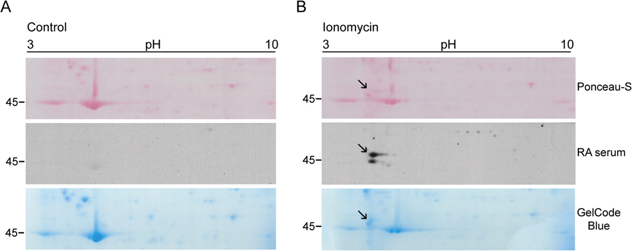
Two-dimensional mapping of the 47 kDa rheumatoid arthritis (RA) autoantigen generated in ionomycin-activated neutrophils. Primary human neutrophils in Hank's balanced salt solution containing 2 mM calcium chloride were incubated in the absence (A) or presence (B) of 1 μM ionomycin for 4 h at 37oC. Samples were resolved in the first dimension using 3-10 IPG strips and then by electrophoresis on 8% sodium dodecylsulphate polyacrylamide gels. Proteins were visualised by Ponceau S staining (upper panel) before immunoblotting with RA patient sera (middle panel). Data from one representative serum is shown. Once the antigen was mapped, the spot was sliced from a GelCode blue-stained gel (lower panel) and used for protein sequencing. Note that the 47 kDa species detected by RA sera (marked with the black arrow) is absent in control cells.

β and γ-Actin are citrullinated autoantigens in rheumatoid arthritis (RA). (A) β and γ-actin are modified in ionomycin-activated neutrophils and the novel species co-migrates with the 47 kDa RA autoantigen. Samples from control (lanes 1, 3 and 5) and ionomycin-activated neutrophils (lanes 2, 4 and 6) were analysed by immunoblotting using monoclonal antibodies against human β and γ-actin (Sigma) or RA serum. The filled arrow denotes the 47 kDa species detected by anti-actin antibodies and RA sera. (B, C) β and γ-actin are citrullinated and recognised by RA autoantibodies. Recombinant human β-actin (lanes 1 and 2) or γ-actin (lanes 3 and 4) was incubated in the absence (lanes 1 and 3) or presence (lanes 2 and 4) of rabbit peptidylarginine deiminase 2 (PAD2). After 1 h at 37oC, samples were divided in two and analysed by electrophoresis on 8% sodium dodecylsulphate polyacrylamide gels. After electrophoresis, proteins were visualised by Ponceau S staining (upper panels) before anti-modified citrulline (AMC) immunoblotting (B, lower panel) or immunoblotting with RA sera (C, lower panel). Data from one representative serum containing anticitrullinated actin antibodies are shown in (C).
Characteristics of 39 RA patients according to anticitrullinated β-actin antibodies
PAD2, PAD3 and PAD4 have distinct macromolecular substrate specificity
It is presently unknown whether the citrullination of intracellular substrates occurs intra or extracellularly in vivo in RA. In addition, if citrullination occurs extracellularly, it is not known whether all PAD modify all substrates similarly. To gain insight into potential substrate specificity among PADs, we directly addressed the citrullination activity of human rPAD2, rPAD3 and rPAD4 against cellular substrates generated from HL-60 cell lysates that were sonicated and cleared by centrifugation. Using this approach, the PAD enzymes would have similar access to cellular substrates independent of the subcellular localisation of the substrate or the enzyme. Interestingly, when HL-60 cell lysates were incubated in the absence or presence of human rPAD2, rPAD3 or rPAD4, different patterns of protein citrullination were generated (figure 4A). These differences were particularly striking when specific substrates were analysed (figure 4B). Therefore, only rPAD2 was able to citrullinate β/γ-actin, as evidenced by a characteristic change in its molecular weight, while histone H3 was only citrullinated by rPAD4. Furthermore, PAD3 (a cytoplasmic enzyme like PAD2) was unable to citrullinate actin, as well as histone H3. Further analysis using purified components confirmed the substrate preferences of these enzymes (figure 4C). Therefore, although rPAD2 and rPAD4 were able to modify purified human platelet actin (composed of both β and γ-actin) and recombinant histone H3, the citrullination of actin and histone H3 was more efficient by rPAD2 and rPAD4, respectively. Moreover, rPAD3 was the least efficient in citrullinating these substrates. In regard to histone H3, it is interesting that differences between PAD2, PAD3 and PAD4 were more evident when H3 citrullination was determined using the AMC method (which detects all modified arginines), compared with the detection of citrullines specifically at residues 2, 8 and 17. Therefore, it appears that in addition to substrate preference, PAD have different access to arginines within the same substrate.

Peptidylarginine deiminase (PAD) 2, 3 and 4 have distinct substrate specificities. (A, B) Sonicated HL-60 cell lysates were incubated in the absence (lane 1) or presence of human rPAD2, rPAD3 or rPAD4 (lanes 2–4, respectively) for 1 h at 37oC. After terminating the reactions, samples were analysed by electrophoresis on 13% sodium dodecylsulphate (SDS) polyacrylamide gels and immunoblotted with anti-modified citrulline (AMC) antibodies (A) or antibodies against human β-actin, γ-actin (Sigma) and citrullinated histone H3 (citrulline 2 + 8 + 17) (Abcam, Cambridge, MA,USA) (B). (C) A mixture of purified human actin plus recombinant histone H3 was incubated in the absence (lane 1) or presence of human rPAD2 (lanes 2–5), rPAD3 (lanes 6–9) or rPAD4 (lanes 10–13) for 5–60 min at 37oC. After terminating the reactions, samples were analysed in duplicated (actin) or triplicated (histone H3) by electrophoresis on 13% SDS polyacrylamide gels. After electrophoresis, proteins were visualised by immunoblotting with antibodies against AMC, citrullinated histone H3 (citrulline 2 + 8 + 17) (Cit-H3), human β/γ-actin and histone H3 (Abcam).
Citrullination sites in cytoplasmic actin form clusters in the three-dimensional structure
In order to define the distribution of peptidylcitrullines in actin, citrullination sites were identified using two different approaches: endogenous citrullinated actin from activated neutrophils was isolated from a two-dimensional gel and subjected to protein sequencing (figure 5A) and recombinant β-actin was citrullinated in vitro with human rPAD2 and analysed by mass spectrometry (figure 5B). As the amino acids surrounding all arginine residues are identical in the cytoplasmic actins, we did not include γ-actin for this analysis. Peptides identified using the two approaches covered 69% of the actin sequence (which included 72.2% of the arginine content), leaving the citrullination status of five arginines (ie, Arg-95, 116, 147, 290 and 335) undetermined. Five citrullination sites overlapped between endogenous and recombinant citrullinated actin (ie, positions 183, 196, 206, 312 and 372) and two divergences were found (ie, Cit-177 and 210, respectively). Peptides containing Cit-62, 354 and 356 were only isolated from citrullinated recombinant β-actin. Three arginines were confirmed not to be modified by PAD2 (Arg-28, 37 and 39). Although we found no consensus sequences that may explain the targeting of distinct arginines by PAD2, it is noteworthy that non-modified arginines are flanked by proline residues at positions −1 or +1, supporting previous observations that this sequence (ie, Pro-Arg or Arg-Pro) disfavours arginine deimination by PAD.25 26 The three-dimensional structure of β-actin has been resolved and consists of two domains (called the large and small domains) with a cleft containing the bound nucleotide (usually ATP; figure 5C).27 Interestingly, the large domain of β-actin contains the majority of the citrulline residues (ie, eight of the 10 peptidylarginines) identified in actin. Although these residues are far apart in the primary sequence, they form clusters in the three-dimensional protein structure. One cluster is located inside the actin cleft and includes residues 62, 183, 206 and 210. The second cluster is located in the surface of the large domain (residues 196, 254 and 256) opposite to the cleft region.

Cirullination sites in actin. (A–C) Amino acid sequence (γ-actin in A and β-actin in B) and tertiary structure (C) of actin. The structure of actin consists of two domains called the large and small domains (shown in blue and orange, respectively), with a cleft containing the bound nucleotide (marked as ATP in C). The amino acid sequence covered by mass spectrometry analysis of endogenous (A) and recombinant (B) citrullinated actin is shown in red, the arginines that are not citrullinated are shown in green and the potential arginine targets for citrullination are shown in yellow. The tertiary structure of actin (C) reveals clustering of citrullination sites around the cleft and the surface of the large domain opposite to the cleft region. The model shown in (C) was generated from the Molecular Modelling DataBase (NCBI) using the software Cn3D according to coordinates generated by Schutt et al.27
Discussion
Citrullinated autoantigens have emerged as key targets of the immune response in RA. Understanding the mechanisms that generate these antigens may identify unique pathways that regulate antigen drive in this disease, which might be relevant to the development of novel therapies. In this study, we identified several unique features of protein citrullination of potential relevance to RA pathogenesis: (1) although a large number of proteins are citrullinated in activated neutrophils, ACPA only recognise a limited number of these molecules; (2) cytoplasmic actins represent a novel target for citrullination and a novel autoantigen in RA; (3) primary human neutrophils express three different PAD isoenzymes (ie, PAD2, PAD3 and PAD4); and (4) the citrullination activity of each PAD appears to be specific and directed preferentially against distinct substrates, independent of their cellular localisation.
Accumulating evidence from this and other studies strongly supports the notion that although citrullination plays a critical role in antigen recognition by ACPA, the modification per se is not the only determinant that confers antibody binding.2 Although the structural context in which citrulline occurs may be shared among autoantigens, the striking observation that different RA sera have unique patterns of reactivity (see figure 1) strongly supports the hypothesis that different ACPA recognise unique sequences.2 28 Whether these unique patterns of antigen recognition among RA patients reflect the citrullination of distinct autoantigens in different microenvironments is not yet known.
Citrullination sites have been confirmed in only a few RA autoantigens generated in vitro or isolated from cells or tissues.10 16 29 Ten potential citrullination sites were identified in peptides generated from endogenous and recombinant citrullinated actin. Among these sites, two distribution patterns of peptidylcitrullines appear relevant. In the linear structure of actin, citrulline residues are found in pairs separated by two to five amino acid residues, which resemble the peptidylcitrulline distribution found in short peptides used for ACPA detection.12 19 30 31 However, in the tertiary structure of the molecule, peptidylcitrullines formed clusters, in which citrulline residues far from each other in primary sequence converge to form citrulline-enriched regions within the molecule. Interestingly, a similar pattern of peptidylcitrulline distribution is observed in the tertiary structure of the autoantigen PAD4.10 As the number and/or the conformation of citrulline residues appear to play an important role in peptide recognition by ACPA,2 12 30 it is possible that clusters of citrullines may allow more efficient antibody binding to citrullinated molecules. Considering that citrullinated autoantigens in RA are unlikely to be unified by common immunogenic sequences (as ACPA do not crossreact among citrullinated autoantigens), but are probably unified by common structural features, it would be important to address whether the clustering of peptidylcitrullines within the tertiary structure of molecules plays a role in autoantigen selection.
The sites and circumstances of citrullination of autoantigens in vivo in RA remain unclear. While fibrinogen is citrullinated extracellularly, probably by PADs that have leaked out of damaged cells,4 32 it is unknown where and under what circumstances the citrullination of intracellular autoantigens occurs. The studies in this paper highlight the striking variation in the specificity of ACPA autoantibodies in different RA patients, and raise important questions about the mechanisms underlying such variation. Importantly, these studies question the assumptions that all PAD share similar specificities and that the proposed extracellular presence of PADs in RA obliterate potentially important subcellular barriers. In spite of the cytoplasmic co-localisation of actins, PAD2 and PAD3, it was surprising that these enzymes generated distinct patterns of citrullinated proteins. Moreover, the finding that in cell lysates, actin and histone H3 are only citrullinated by PAD2 and PAD4, respectively, strongly supports the existence of substrate specificity among PAD. In this regard, studies using purified substrates demonstrate that although purified actin or histone H3 can be citrullinated by PAD2, PAD3 and/or PAD4, the enzymes have a clear substrate preference, which becomes more evident when the enzymes are exposed to a larger number of substrates (eg, the full cellular content). In addition, these data strongly support that target selection by PAD is independent of any other cellular component, and therefore is an intrinsic property of the enzyme. Interestingly, previous observations by Nakayama-Hamada et al33 demonstrated that human rPAD2 citrullinates purified fibrinogen and filaggrin several orders of magnitude more efficiently than human rPAD4, supporting the notion that PAD enzymes have either unique and/or preferential activity against macromolecular substrates.
Based on differences in the sequence alignment between PAD2, PAD3 and PAD4,34 there are two major regions in PAD that may participate in substrate selection, the N-terminal domain and the active site cleft. With regard to the active site cleft, it is interesting to note that Arg-374 in PAD4 is not conserved in PAD2 or PAD3 (which contain Gly at position 374). This Arg is directly involved in the recognition of histone N-terminal peptides35 and is absolutely required for H3 citrullination and for PAD4 enzyme activity.10 35 Interestingly, recent studies have shown that PAD3 has poor reactivity against small synthetic benzoylated arginine derivatives, known PAD4 substrates.36 The mutation of PAD3 Gly-374 to Arg was unable to restore the ability of PAD3 to citrullinate these molecules, suggesting that PAD3 and perhaps PAD2 have very different substrate specificities driven by other amino acid contacts outside the catalytic site. The N-terminal domain is not well conserved among the PAD enzymes and therefore may contribute to substrate specificity, potentially through extended macromolecular interactions outside the active site (ie, through exosites). Defining these additional interactions may have important implications in the designing of inhibitors against specific PAD.
The discovery that different PAD isoforms have distinct substrate specificities has significant implications for RA. It is possible that only one or a few PADs may account for autoantigen citrullination in vivo. In this regard, future therapies targeting PAD enzymes might therefore be validly focused on PAD-selective inhibitors, instead of pan-PAD inhibitors, which may have higher toxicity by affecting PADs with no relevance to RA. Further studies that elucidate PAD activation, specificity and functional consequences as they relate to specific ACPA antigens are of high priority.
Acknowledgments
The authors would like to thank Xiaoming Zhu for her valuable technical assistance and Dr Joan Bathon for providing valuable RA serum samples. FA is a Lowe Family Scholar in the Johns Hopkins Bayview Center for Innovative Medicine and is supported by a Dana Foundation Scholars Program in Human Immunology, the Donald B. and Dorothy L. Stabler Foundation and NIH grant P30 AR053503. AR is supported by NIH grant R37 DE-12354 and ACR-REF Within our Reach grant. ED is supported by NIH grant T32 AR048522. JTG is supported by NIH grant 1K23AR054112-01.
References
Footnotes
-
Competing interests None.
-
Patient consent Obtained.
-
Ethics approval This study was conducted with the approval of the Johns Hopkins Institutional Review Board.
-
Provenance and peer review Not commissioned; externally peer reviewed.