Article Text

Abstract
Objectives To analyse the impact of tumour necrosis factor inhibitors (TNFis) on spinal radiographic progression in ankylosing spondylitis (AS).
Methods Patients with AS in the Swiss Clinical Quality Management cohort with up to 10 years of follow-up and radiographic assessments every 2 years were included. Radiographs were scored by two readers according to the modified Stoke Ankylosing Spondylitis Spine Score (mSASSS) with known chronology. The relationship between TNFi use before a 2-year radiographic interval and progression within the interval was investigated using binomial generalised estimating equation models with adjustment for potential confounding and multiple imputation of missing values. Ankylosing Spondylitis Disease Activity Score (ASDAS) was regarded as mediating the effect of TNFi on progression and added to the model in a sensitivity analysis.
Results A total of 432 patients with AS contributed to data for 616 radiographic intervals. Radiographic progression was defined as an increase in ≥2 mSASSS units in 2 years. Mean (SD) mSASSS increase was 0.9 (2.6) units in 2 years. Prior use of TNFi reduced the odds of progression by 50% (OR 0.50, 95% CI 0.28 to 0.88) in the multivariable analysis. While no direct effect of TNFi on progression was present in an analysis including time-varying ASDAS (OR 0.61, 95% CI 0.34 to 1.08), the indirect effect, via a reduction in ASDAS, was statistically significant (OR 0.75, 95% CI 0.59 to 0.97).
Conclusion TNFis are associated with a reduction of spinal radiographic progression in patients with AS. This effect seems mediated through the inhibiting effect of TNFi on disease activity.
- ankylosing spondylitis
- anti-tnf
- epidemiology
This is an Open Access article distributed in accordance with the Creative Commons Attribution Non Commercial (CC BY-NC 4.0) license, which permits others to distribute, remix, adapt, build upon this work non-commercially, and license their derivative works on different terms, provided the original work is properly cited and the use is non-commercial. See: http://creativecommons.org/licenses/by-nc/4.0/
Statistics from Altmetric.com
Introduction
The introduction of tumour necrosis factor inhibitors (TNFis) nearly two decades ago has considerably improved the treatment of ankylosing spondylitis (AS) in patients with insufficient response to conventional treatment by reducing symptoms and signs of the disease, together with reduced inflammatory activity in the sacroiliac joints and the spine.1 Besides inflammation, spinal damage caused by new bone formation contributes to impairment of spinal mobility and function in AS.2 Therefore, retarding spinal radiographic progression in addition to improving symptoms should remain an important treatment goal.3 While an association between disease activity and future spinal radiographic progression has been demonstrated,4 5 the deductive reasoning that lowering inflammation by TNFi might inhibit radiographic damage remains elusive. Three open-label extensions of randomised controlled trials of TNFi in AS over 2 years failed to demonstrate inhibition of radiographic progression in comparison with a historical cohort of patients not treated with biologicals.6–8 However, TNFi use was associated with a lower odds of spinal radiographic progression in an observational study.9 Methodological shortcomings of this latter publication and requirements for prospective cohort analyses to elucidate this controversial issue have been amply discussed.10 We hereby present a longitudinal analysis of up to 10 years of follow-up, with 2-year clinical and radiographic intervals, with the aim of investigating the relationship between treatment with TNFi, subsequent course of disease activity and spinal radiographic progression.
Methods
Study population
We used data from the ongoing Swiss Clinical Quality Management (SCQM) cohort of patients with a clinical diagnosis of axial spondyloarthritis (axSpA).11 Clinical assessments, following the recommendations of ASAS,12 were performed at annual visits. Radiographs of the cervical and lumbar spine were recommended every 2 years. Patients were included in the present study if they fulfilled the modified New York criteria for AS13 with central reading of the pelvic radiographs and if they had at least two sets of spinal radiographs with an interval of 2 years±1 year. Sensitivity analyses were performed in patients with interval duration between radiograph sets of 2 years±6 months. The study was approved by the Ethics Committee of the Canton of Zurich (KEK-ZH-Nr. 2014–0439). Written informed consent was obtained from all patients.
Assessment of radiographic progression
All available radiographs per patient were scored at the same time according to the modified Stoke Ankylosing Spondylitis Spine Score14 (mSASSS) by two trained readers (XB and MdH) independently, blinded to all other data, but with known chronology. Averaged scores per vertebral corner (VC) were used. An independent adjudicator (AC) scored all of the radiographs from patients with an absolute difference in mSASSS status scores between the primary readers of ≥5 units in at least one radiograph set. Only scores of radiographs with ≤3 missing VCs per cervical or lumbar segment were used. Individual missing VCs were imputed using an adaptation algorithm (see online supplementary materials). Radiographic progression was defined as an increase in mSASSS of at least 2 units over an interval of 2 years, based on calculations of the smallest detectable change (SDC).15 In addition, we assessed the proportion of patients with formation of at least one new syndesmophyte over a period of 2 years.16
Supplementary file 1
Statistical analysis
Reliability between the two readers for mSASSS was explored by Bland-Altman plot on 2-year progression intervals of mSASSS and by calculating the SDC for 2-year progression scores and the intraclass correlation coefficient (ICC; type 2, k) on mSASSS. The relationship between TNFi treatment and radiographic progression over time was investigated using binomial generalised estimating equations (GEE) to account for repeated measurements within a patient. An ‘exchangeable’ correlation structure was chosen (see online supplementary materials). The duration of radiographic intervals was added as a covariate to the model to account for differences in interval lengths. Progression of ≥2 mSASSS units in 2 years was modelled by using the binomial family and the logistic link function. To account for missing values, the GEE was fitted using multiple imputation of missing covariate data (see online supplementary materials).
The GEE analyses were adjusted for baseline radiographic damage16 17 (mSASSS or the presence of syndesmophytes18 in different models), sex,19 symptom duration, human leucocyte antigen B27 (HLA-B27) status,19 smoking status,17 20 21 the number of physical activity sessions per week,22 presence of peripheral arthritis,23 body mass index categories and treatment with non-steroidal anti-inflammatory drugs (NSAIDs)24 25 recorded as a dichotomous variable (yes/no) at every visit. To address the issue of confounding by indication, a model was fitted that was additionally adjusted for the Ankylosing Spondylitis Disease Activity Score (ASDAS) value before start of TNFi (ASDAS at inclusion for non-TNFi patients).
Based on available data on the potential impact of TNFi on progression to date,6–9 26 27 different longitudinal models were run that varied with regards to the variable representing TNFi treatment: use of TNFi prior to the radiographic interval as yes/no, as number of years of continuous use of TNFi, or alternatively, as ≤4 years versus >4 years of TNFi use,9 26 27 treatment with TNFi during the 2 year radiographic interval as yes/no or as duration of use of <50% versus ≥50% of the radiographic interval.
Disease activity variables (Bath Ankylosing Spondylitis Disease Activity Index (BASDAI) and C reactive protein (CRP) or ASDAS) after start of TNFi were regarded as potential intermediate variables mediating the effect of TNFi on radiographic progression and were therefore not included in the main statistical models. To investigate the potential mediating effect of disease activity on the impact of TNFi (independent variable) on radiographic progression (dependent variable), we estimated the indirect effect and tested it with the Sobel test with second-order estimator of the SE, as described by Hayes.28 The direct effect of TNFi on radiographic progression was tested by introducing disease activity variables (BASDAI, CRP or ASDAS) at start of each radiographic interval in the main models.
Results
A total of 432 patients with AS presented with at least one 2-year radiographic interval during the observation period in SCQM. Mean (SD) time between radiographs was 2.1 (0.4) years. Interobserver reliability was ‘good’ (ICC 0.85). The SDC of progression in a 2-year radiographic interval was 1.89 mSASSS units, which is below the threshold of 2 mSASSS units defining progression. A Bland-Altman plot is shown in the online supplementary figure S1. Adjudication was performed in 130 patients. Baseline disease characteristics are shown in table 1.
Baseline characteristics at first radiograph
Adjusted longitudinal analyses
Mean (SD) radiographic progression was 0.9 (2.3) mSASSS units in 2 years. Any TNFi use before a radiographic interval (yes vs no) was used as main TNFi variable in the adjusted analyses (figure 1A). Prior TNFi treatment versus no TNFi treatment was associated in multivariable analyses of 616 radiographic intervals in 432 patients with a reduction of the odds for radiographic progression during the next 2-year interval by 50% (OR 0.50, 95% CI 0.28 to 0.88, p=0.02). Baseline mSASSS (OR 1.06, 95% CI 1.04 to 1.09, p<0.001) and male sex (OR 2.16, 95% CI 1.09 to 4.30, p=0.03) were associated with an increase in radiographic damage after 2 years. The impact of prior TNFi use in reducing radiographic progression during the next 2-year radiographic interval was confirmed in an adjusted model with progression defined as the appearance of at least one new syndesmophyte in 2 years (OR 0.55, 95% CI 0.33 to 0.94) (figure 1B). Treatment with NSAIDs at baseline, smoking, HLA-B27, peripheral arthritis, overweight, obesity and physical exercise were not found to be associated with an effect on radiographic progression in both models. The estimated impact of prior TNFi use was not affected by the additional adjustment for disease activity measures (ASDAS) before treatment start, performed to address the issue of confounding by indication (table 2).

Multivariable analysis of 616 radiographic intervals from 432 patients after multiple imputation of missing covariate data for the identification of factors associated with (A) radiographic progression defined as an increase of ≥2 mSASSS units per 2 years and (B) radiographic progression defined as the formation of at least one new syndesmophyte per 2 years. Analysis performed in 616 radiographic intervals from 432 patients after multiple imputation of missing covariate data. BMI, body mass index; HLA-B27, human leucocyte antigen B27; mSASSS, modified Stoke Ankylosing Spondylitis Spine Score; NSAIDs, non-steroidal anti-inflammatory drugs; Ref, reference; TNFi, tumour necrosis factor inhibitor. *mSASSS at start of each 2-year radiographic interval in A and presence of syndesmophytes at start of each 2-year radiographic interval (yes vs no) in B.
Impact of pretreatment Ankylosing Spondylitis Disease Activity Score (ASDAS) on spinal radiographic progression
The magnitude of the effect of all variables on progression was also confirmed in the subset of patients with radiographic interval duration of 2 years±6 months and in a complete case analysis of 403 radiographic intervals from 301 patients (see online supplementary tables S1 and S2, respectively).
A beneficial effect of TNFi treatment before a radiographic interval on progression was also confirmed in adjusted models with alternative variable choices for TNFi use, as summarised in table 3 and presented in full in the online supplementary tables S3 and S4. These data also suggest that a longer duration of TNFi treatment is associated with a stronger protective effect, since each additional year of continuous TNFi therapy before a radiographic interval was associated with a reduced risk of progression (model 2 in table 3). Moreover, >4 years of treatment before the radiographic interval resulted in a lower estimate of progression than ≤4 years of TNFi use (model 3 in table 3). In contrast to prior TNFi use, TNFi treatment during a 2-year radiographic interval (assessed either as ‘yes/no’ or as ‘duration of TNFi treatment during the interval (≤50% vs >50%)’) was not associated with a reduction of progression in the respective interval (models 4 and 5 in table 3 and online supplementary tables S5 and S6).
Impact of alternative variable choices for TNFi use on spinal radiographic progression from different multivariable models*
Impact of reduction of disease activity by TNFi on radiographic progression
TNFi treatment before a 2-year radiographic interval was associated with a reduced disease activity at the start of that interval, as assessed by the ASDAS: −0.96 units, 95% CI −1.15 to −0.77, p<0.001. The association of disease activity measures at baseline of a 2-year radiographic interval with progression in the respective interval was analysed in separate GEE models with baseline mSASSS as dependent variable (models 6–8 in table 4). A higher ASDAS increased the probability of radiographic progression (OR 1.39, 95% CI 1.06 to 1.81). When adding ASDAS as a covariate to the multivariable model displayed in figure 1A, it was significantly associated with radiographic progression (table 4, model 9). The TNFi variable in the model without ASDAS captures the total TNFi effect on progression. In a model with ASDAS, the TNFi variable coefficient estimated the direct effect of TNFi on radiographic progression, which was not significant (OR 0.61, 95% CI 0.34 to 1.08, p=0.09). The estimated indirect effect of TNFi on progression via a reduction in ASDAS was on the contrary statistically significant (OR 0.75, 95% CI 0.59 to 0.97, p=0.01).
Impact of time-varying Ankylosing Spondylitis Disease Activity sScore (ASDAS) on spinal radiographic progression
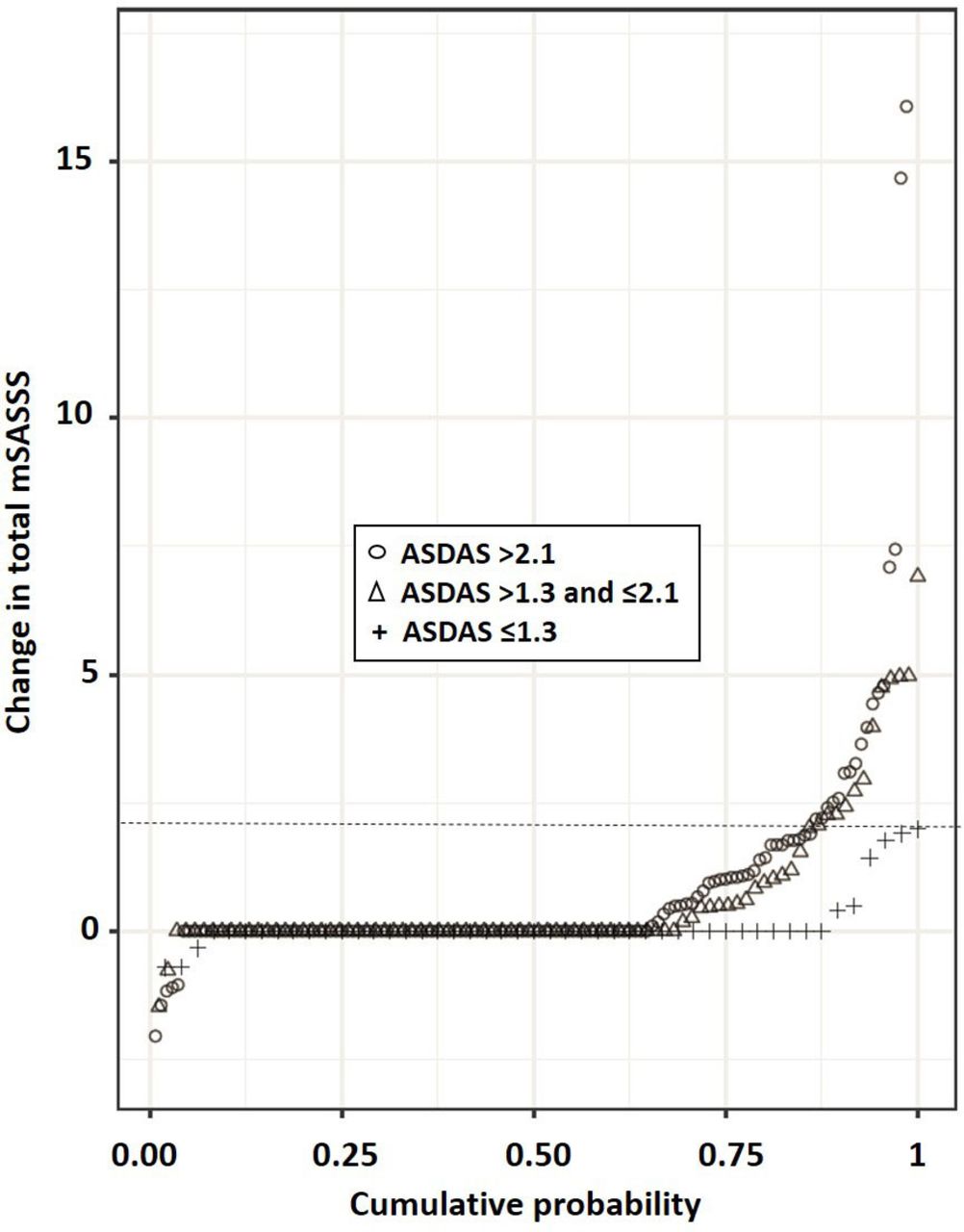
When analysing the unadjusted radiographic progression in 2-year intervals following the use of TNFi, responders to TNFi (defined by an ASDAS ≤2.1 at start of the radiographic interval) had a significantly lower mean spinal progression than non-responders to TNFi (defined by ASDAS >2.1), with a mean mSASSS progression of 0.31 versus 1.45 units (Kruskal-Wallis test p=0.02). Radiographic progression was entirely inhibited in TNFi patients reaching an inactive disease status (ASDAS ≤1.3) at the start of the next 2-year radiographic interval (mean spinal progression in these patients was 0.01 mSASSS units vs 0.52 mSASSS units in patients with ASDAS between 1.3 and 2.1, Kruskal-Wallis test p=0.01). Cumulative probability plots of spinal progression after stratification by ASDAS status at the start of the next radiographic interval are presented for patients treated with TNFi in figure 2. Stratification by baseline CRP status (elevated CRP vs normal CRP) segregated less well between radiographic progressors and non-progressors in this population than stratification by the ASDAS (see online supplementary figure S2).
Discussion
The current longitudinal analysis of a large observational national cohort, consisting of both patients treated with TNFi and untreated patients, supports the notion of an effect of TNFi treatment on inhibition of radiographic spinal progression in patients with AS. As residual confounding cannot be completely ruled out in an observational context, causality could not be proven, although it remains the most plausible explanation for our findings. Long-term randomised controlled trials to definitively clarify this issue will never be performed for ethical reasons.

Cumulative probability plot of 2-year progression in the modified Stoke Ankylosing Spine Score (mSASSS), illustrating the change in mSASSS values from baseline of each individual radiographic interval to 2 years in patients already treated with TNFi at start of the respective interval, stratified by the ASDAS cut-off level reached at the beginning of each radiographic interval: ASDAS >2.1 (n=111, 136 radiographic intervals), ASDAS >1.3 and ≤2.1 (n=68, 85 radiographic intervals) and ASDAS ≤1.3 (inactive disease status; n=40, 48 radiographic intervals). Radiographic progression was defined as an increase in mSASSS of ≥2 in 2 years (dotted line). ASDAS, Ankylosing Spondylitis Disease Activity Score.
We demonstrate an association between TNFi use and reduced risk of spinal structural damage, both in terms of mSASSS and new syndesmophyte formation. The odds of radiographic progression were nearly halved over the next 2 years in patients having started TNFi treatment before this 2-year interval. The pattern of correlations demonstrated in our mediational analyses is consistent with the hypothesis that the impact of TNFi on spinal radiographic progression is mediated by its decreasing effect on disease activity (ASDAS, BASDAI or CRP). Only a trend for a direct effect of TNFi on reduction of spinal progression could be found. ASDAS outperformed BASDAI and CRP alone for the association of disease activity with radiographic progression, confirming previous analyses.4
We present important clues concerning the period of time needed before the inhibitory effects can be objectified: around 2 years of continuous TNFi use, as there was no impact of TNFi treatment during a 2-year radiographic interval, while there was an effect if the treatment was started before this interval. Our study therefore reconciles conflicting results of previous investigations. Treatment with TNFi over 2 years in three open-label extensions of randomised control trials in AS failed to demonstrate an inhibition of radiographic progression during this period.6–8 The principal explanation for this seems to be that inflammation needs to be suppressed for at least 2 years in order to demonstrate an inhibition of radiographic progression. In another study with fewer patients with AS, less radiographic progression was only demonstrated after 4 years of follow-up.26 This was also demonstrated in two observational studies.9 27 We confirm the finding that longer periods of treatment with TNFi translate into a stronger inhibition of progression in two different models: one accounting for the number of years of continuous TNFi therapy and the second using a cut-off of 4 years of treatment.
Radiographic spinal progression was nearly entirely inhibited in the following 2-year radiographic interval in patients with AS reaching an inactive disease status (ASDAS ≤1.3)29on treatment with TNFi. While a treat-to-target approach has been recommended, the content of the target was not mentioned.30 Our study suggests that ASDAS ≤1.3 might be an adequate target, if the goal of treatment is inhibition of further spinal radiographic damage in addition to control of signs and symptoms, provided that the target seems realistic based on the clinical context.
The issue of whether NSAIDs alone may have an impact on structural damage remains controversial. Continuous versus on demand use of celecoxib has been shown to inhibit progression in a randomised controlled trial, particularly in the subgroup of patients with elevated CRP levels.24 31 However, in a more recent study with similar design, diclofenac had no impact on progression.32 Conflicting results have also been found in observational studies.9 25 In our study, use of NSAIDs was not associated with an independent effect on progression. We acknowledge, however, that data collection in SCQM does not allow for calculation of the recommended NSAIDs index.33 We confirm in our study that baseline structural damage and male sex are the major drivers of radiographic progression.16–19
We have not addressed the issue of whether any particular TNFi agent might have a greater impact on spinal radiographic progression than another, due to insufficient patient numbers. The fact that the impact of TNFi on progression seems to be mediated by a decrease in disease activity would argue against major differences between individual TNFi. It also remains to be demonstrated whether new biological agents and in particular anti-interleukin-17 drugs34 also have an impact on radiographic progression.
Strengths of our investigation include the prospective study design, standardised regular clinical and radiographic assessments at 2-year intervals allowing for the implementation of a longitudinal analysis and statistical methods (GEE analyses) that take into account potential confounders and the within-patient correlation of structural damage. We acknowledge the fact that our analyses were based on only one radiographic interval in around 2/3 of patients. The scoring of radiographs with knowledge of chronology might be seen as an additional limitation, but it has been shown to be more sensitive to change than reading with paired time order,35 and readers were blinded to clinical data, including treatment.
In addition to the possibility of residual confounding, which has already been highlighted, observational studies might be prone to other risks of bias. We have addressed the issue of confounding by indication by performing sensitivity analyses with adjustment for disease activity measures and other covariates before the start of treatment. Involvement of a multitude of rheumatologists throughout Switzerland in this real-life cohort might be the source of inconsistent data collection and measurement errors. We have previously demonstrated that adherence to ASAS treatment recommendations and response rates to TNFi were similar in private practices and academic centres.36 Classification as AS was established by central reading of the pelvis radiographs. We have used multiple imputation techniques for missing values and have provided complete case analyses to allow for the evaluation of the robustness of our investigation.
In conclusion, our data suggest that TNFi therapy in AS has a clinically relevant inhibitory effect on spinal radiographic progression if treatment is continued for at least 2 years and that this effect is mediated by a decrease in disease activity.
Acknowledgments
We would like to thank all patients and their rheumatologists for participation, the members of the SCQM axSpA scientific board for contribution to pelvis X-ray scoring and helpful discussions and the entire SCQM staff for data management and support. A list of rheumatology private practices and hospitals that are contributing to the SCQM registries can be found on: http://www.scqm.ch/institutions
References
Footnotes
CM and AS contributed equally.
Handling editor Tore K Kvien
Contributors AC, AS and CM designed the study. All investigators substantially contributed to the acquisition, analysis or interpretation of data. AC wrote the article, and all coauthors revised the manuscript critically for important intellectual content. CM and AS were responsible for the implementation of the statistical analyses. AC had full access to all of the data in the study and takes responsibility for the integrity of the data and the accuracy of the data analysis. All authors agreed on the final content of the submitted manuscript.
Funding The SCQM Foundation is supported by the Swiss Society of Rheumatology and by AbbVie, Bristol-Myers-Squibb, Celgene, Janssen-Cilag, Merck Sharp & Dohme, Novartis, Pfizer, Roche and UCB and has received project-based financial supports from the Arco Foundation, Switzerland, as well as from the Swiss Balgrist Society, Switzerland. This study was supported by the Stiftung für Rheumaforschung and a research grant from the investigator initiated studies program of Merck Sharp & Dohme. The study sponsors had no role in the study design or in the collection, analysis or interpretation of the data, the writing of the manuscript or the decision to submit the manuscript for publication. Publication of this article was not contingent upon approval by the study sponsors.
Competing interests JB has received consulting fees from Merck Sharp & Dohme, Pfizer and Roche. AC has received consulting and/or speaking fees from AbbVie, Celgene, Eli Lilly, Janssen-Cilag, Merck Sharp & Dohme, Novartis, Pfizer and UCB. MJN has received consulting and/or speaking fees from Abbvie, Novartis and Pfizer. DvdH has received consulting fees from Abbvie, Amgen, Astellas, AstraZeneca, BMS, Boeringer Ingelheim, Celgene, Daiichi, Eli-Lilly, Galapagos, Gilead, Janssen, Merck, Novartis, Pfizer, Regeneron, Roche, Sanofi and UCB and is director of Imaging Rheumatology BV. UW has received speaking fees from AbbVie. AS, GT, LMW, MC, MdH, PE, PZ, RBML, RK, RM and XB
declare no conflict of interest. No non-financial conflicts of interest exist for any of the authors.
Patient consent Obtained.
Ethics approval Ethics Committee of the Canton of Zurich.
Provenance and peer review Not commissioned; externally peer reviewed.
Data sharing statement All data supporting our findings are shown in the article.