Article Text

Abstract
Objective To develop and validate new classification criteria for adult and juvenile idiopathic inflammatory myopathies (IIM) and their major subgroups.
Methods Candidate variables were assembled from published criteria and expert opinion using consensus methodology. Data were collected from 47 rheumatology, dermatology, neurology and paediatric clinics worldwide. Several statistical methods were used to derive the classification criteria.
Results Based on data from 976 IIM patients (74% adults; 26% children) and 624 non-IIM patients with mimicking conditions (82% adults; 18% children), new criteria were derived. Each item is assigned a weighted score. The total score corresponds to a probability of having IIM. Subclassification is performed using a classification tree. A probability cut-off of 55%, corresponding to a score of 5.5 (6.7 with muscle biopsy) ‘probable IIM’, had best sensitivity/specificity (87%/82% without biopsies, 93%/88% with biopsies) and is recommended as a minimum to classify a patient as having IIM. A probability of ≥90%, corresponding to a score of ≥7.5 (≥8.7 with muscle biopsy), corresponds to ‘definite IIM’. A probability of <50%, corresponding to a score of <5.3 (<6.5 with muscle biopsy), rules out IIM, leaving a probability of ≥50 to <55% as ‘possible IIM’.
Conclusions The European League Against Rheumatism/American College of Rheumatology (EULAR/ACR) classification criteria for IIM have been endorsed by international rheumatology, dermatology, neurology and paediatric groups. They employ easily accessible and operationally defined elements, and have been partially validated. They allow classification of ‘definite’, ‘probable’ and ‘possible’ IIM, in addition to the major subgroups of IIM, including juvenile IIM. They generally perform better than existing criteria.
- dermatomyositis
- polymyositis
- autoimmune diseases
Statistics from Altmetric.com
Introduction
Idiopathic inflammatory myopathies (IIM), collectively known as myositis, are heterogeneous disorders characterised by muscle weakness and muscle inflammation.1 The most common subgroups in adults are dermatomyositis (DM), polymyositis (PM) and inclusion body myositis (IBM),2 and in children, juvenile DM (JDM).
This criteria set has been approved by the European League Against Rheumatism (EULAR) Executive Committee and the American College of Rheumatology (ACR) Board of Directors as Provisional. This signifies that the criteria set has been quantitatively validated using patient data, but it has not undergone full validation based on an independent dataset, using both cases and controls. This validation step is still needed before the criteria are fully validated.
The International Myositis Assessment and Clinical Studies (IMACS) Group has developed consensus on outcome measures and definitions of improvement to be used in clinical trials for myositis.3 4 A prerequisite for clinical trials and other clinical studies is the inclusion of well-defined patient groups. A wide variety of diagnostic or classification criteria for myositis are used,2 5–16 but are generally derived empirically and not validated. The criteria of Bohan and Peter7 8 are most widely used, but have limitations. Because they do not clearly specify how to exclude other forms of myopathy, they may misclassify IBM patients as PM,13 17–19 and muscular dystrophies with inflammation as myositis, and each criterion is not defined explicitly. New discoveries in the last decade, such as myositis-specific autoantibodies that are associated with distinct clinical phenotypes,2 20–22 may provide opportunities to improve the precision of classification, but have not been tested adequately.11 23
The aim of this project was to develop classification criteria for adult and juvenile IIM. The specific goal was to define the minimum essential, easily available clinical and laboratory features to (1) distinguish IIM from mimicking conditions with high sensitivity and specificity, and (2) distinguish the major subgroups of IIM.
Methods
Study design
The International Myositis Classification Criteria Project (IMCCP), an international collaboration with experts from adult and paediatric rheumatology, neurology, dermatology, epidemiology and biostatistics, was established in 2004 and followed at our best the European League Against Rheumatism (EULAR) and American College of Rheumatology (ACR) recommendations for development of classification criteria from that time or published soon thereafter.24 25 A steering committee (online supplementary 1) and a larger working committee with experts in IIM were formed (see online supplementary appendix).
Using the nominal group technique, experts in IIM from the steering committee and the working committee26–29 designed the study and validation experiments, assembled and defined candidate criteria from published myositis criteria2 5–16 and other characteristics of myositis, and determined and assembled the IIM subgroup diagnoses and comparator conditions that were studied. A pilot study to assess the practicality of capturing the items showed a fair agreement of data availability from IIM and non-IIM cases (online supplementary 2). Input was obtained from myositis experts, by email to the IMACS network and requesting comments on the items, to maximise face and content validity.24 25 The steering committee revised the list of variables based on the comments and further suggestions from the IMACS network and 93 variables (online supplementary 3) were selected by the steering committee for study in cases and comparators. A glossary and definitions were developed according to an ACR glossary30 31 (online supplementary 4). Data were abstracted from patients’ records and entered into a web-based database.
Inclusion criteria for cases and comparators were (1) diagnosis for at least 6 months prior to study inclusion; (2) physician certainty of diagnosis—either known IIM or, as comparators, known non-IIM cases where myositis was considered in the initial differential diagnosis; and (3) patients with the most recent and complete data were prioritised to acquire the most complete data in a consistent manner. A maximum of 40 cases and an equal number of comparators were collected from each centre.
The study was approved by the ethics committees at each site.
Data analysis and candidate criteria selection
The association of each variable with the diagnosis (IIM, non-IIM) was assessed by ORs and tested with Fisher’s exact test. The treating physician diagnosis was considered the gold standard for analysis. Three classification techniques were explored: (1) a sum-of-items model in which a patient was classified as a case if the patient had a specified number of items from a set of items, (2) a probability-score model and (3) a classification tree. The ensuing candidate criteria were examined with respect to statistical performance and clinical relevance. Due to the observed superior discriminating performance of the probability-score model, the other models were set aside.
Criteria development
The probability-score model summed score points associated with the signs and symptoms present. The score points were obtained as coefficients of a logistic regression model used to combine multiple variables for predicting IIM. The statistical significance of the resulting increase in the goodness-of-fit of the model was assessed using the Wald test. The improvement in predictive ability was measured by the increment in specificity and sensitivity and summarised by the area under the receiver operating characteristic curve (AUC).
Paediatric experts are using fewer muscle biopsies for classification of JDM in clinical practice than adult rheumatologists. Thus, a second model not including biopsy variables was developed. Assessment of statistical performance for each score/probability cut-off value provided the basis for a recommendation of a cut-off value for IIM classification by the steering committee. The proposed cut-offs were then defined as possible, probable and definite IIM. To facilitate use of the new criteria, a web-based calculator for the probability-score model was developed.
The new classification criteria were compared with previous IIM criteria. Their statistical performance, and number of patients per IIM subdiagnosis classified as IIM by the different criteria sets, were calculated.
To distinguish subgroups of patients classified with IIM according to the new criteria, a classification tree was developed. The tree was based on the variables in the new classification criteria, statistical analyses, as described in a separate methodology paper and on expert opinion.
Validation
The new criteria were internally cross-validated. Samples of equal size to the original sample were drawn from the entire population at random with replacement, so-called ‘bootstrap’ samples.32 The bootstrap sample represented the training sample, and the remaining subjects not contained in the bootstrap sample constituted the validation sample. The probability score was applied to each bootstrap training sample separately and then used to predict IIM in the validation sample. The procedure was repeated in over 200 bootstrap samples, and the average AUC was calculated.
The performance of the new criteria for IIM including the subgroups was tested for sensitivity in two independent cohorts, the Euromyositis Register (https://euromyositis.eu/) and the Juvenile Dermatomyositis Cohort Biomarker Study and Repository (JDRG) (UK and Ireland) (https://www.juveniledermatomyositis.org.uk/).
The program Stata V.13 (StataCorp) was used for data management and statistical analyses. The statistical program R (R Core Team (2014). R: a language and environment for statistical computing. R Foundation for Statistical Computing, Vienna, Austria. URL http://www.R-project.org/) was used for some analyses.
A report detailing the methodology will be submitted as a separate publication (manuscript submitted).
Results
Study population
Data from 976 IIM patients (74.5% adults; 25.5% children) (table 1) were collected between 2008 and 2011 from 23 European, 17 North American, 1 South American and 6 Asian sites, representing IIM subgroups of JDM (n=248), PM (n=245), DM (n=239), IBM (n=176), amyopathic DM (ADM) (n=44), hypomyopathic DM (n=12), immune-mediated necrotising myopathy (IMNM) (n=11) and juvenile PM (n=1). A total of 624 comparators (81.6% adults; 18.4% children) (table 1) representing a broad spectrum of conditions that can mimic IIM were included, comprising systemic inflammatory diseases (36.5%), muscle dystrophies (16.0%), drug-associated or toxin-associated myopathies (7.9%), motor neuron diseases/neuropathies (7.7%), metabolic myopathies (6.9%), myalgias (4.5%), dermatological diseases (3.7%), endocrine myopathies (3.7%), infectious myopathies (4.5%), mitochondrial myopathies (2.4%), neuromuscular diseases (2.6%), other myopathies (1.9%), immune-mediated skin conditions (0.5%) as well as other diagnoses (1.3%) (online supplementary 5 and 6).
Demographic data of the International Myositis Classification Criteria Project cohort
Candidate criteria selection and criteria development
Based on statistical models, 16 variables from six categories best distinguished IIM cases from comparators (table 2), and each variable was assigned a weight (score) based on its influence to discriminate IIM from non-IIM. A total score was computed by adding score points corresponding to each criterion being present. The score can be converted into a probability of IIM (figure 1A,B) by:

Probability of having idiopathic inflammatory myopathies (IIM) based on the EULAR/ACR classification criteria for IIM. Each score obtained from the classification criteria corresponds to a probability of having the disease, without muscle biopsy data (A) or with muscle biopsy data (B). Each score and probability of disease display a unique set of sensitivity (blue line) and specificity (red line) measurements for the classification criteria not including muscle biopsy data (C) or including muscle biopsy data (D). The most optimal point of accuracy should be stated in publications and be appropriate to the intended purpose, with the recommendation of using a minimum of 55% probability (score of 5.5 without biopsies; 6.7 with biopsies) for classifying a case as IIM (‘probable IIM’) (dotted line). ‘Definite IIM’ corresponds to a probability of at least 90% (score of ≥ 7.5 without biopsies; ≥ 8.7 with biopsies). ACR, American College of Rheumatology; EULAR, European League Against Rheumatism.
The European League Against Rheumatism/American College of Rheumatology (EULAR/ACR) classification criteria for adult and juvenile idiopathic inflammatory myopathies (IIMs)
Probability of IIM including muscle biopsy=1/[1+exponential (5.33–score)]
or,
Probability of IIM without muscle biopsy=1/[1+exponential (6.49–score)]
or by using the online web calculator (www.imm.ki.se/biostatistics/calculators/iim).
Sensitivity and specificity for varying probability cut-offs are shown in figure 1C,D.
Cut-points for classification
The best balance between sensitivity and specificity was found for a probability of 55%–60% for the criteria not including muscle biopsy data, and 55%–75% when including muscle biopsies, or a total aggregated score of score of ≥5.5 and ≤5.7 (≥6.7 and ≤7.6 if biopsy is available). The IMCCP proposes that a patient may be classified as IIM if the probability exceeds a predetermined cut-off of at least 55% (corresponding to a score of ≥5.5, or ≥6.7 if biopsies are included) based on maximisation of statistical performance and best balance between sensitivity and specificity. The level of probability ≥55% and <90% was defined as ‘probable IIM’. The steering committee recommends, based on expert opinion, that ‘definite IIM’ should equal a probability of ≥90%, corresponding to having total aggregate score of ≥7.5 without muscle biopsy and ≥8.7 with muscle biopsy.
Patients falling in the probability range ≥50% and <55% will be classified as ‘possible IIM’. For a patient to be classified as a non-IIM patient, the probability would have to be <50% (score of < 5.3 without biopsies; < 6.5 with biopsies).
As suggested by paediatric experts and dermatologists, for patients with pathognomonic skin rashes of DM or JDM, classification criteria were developed, which did not include muscle biopsy data (table 2). However, where no skin rash is present, a muscle biopsy is required for classification, as determined by a consensus of expert opinion within the IMCCP steering and working committees. Both sets apply equally well to adult IIM patients and to juvenile patients with DM and should be used when IIM is suspected and no better explanation for the symptoms exists, as agreed on by expert opinion. Definitions for the criteria items are presented in table 2.
Identification of subgroups
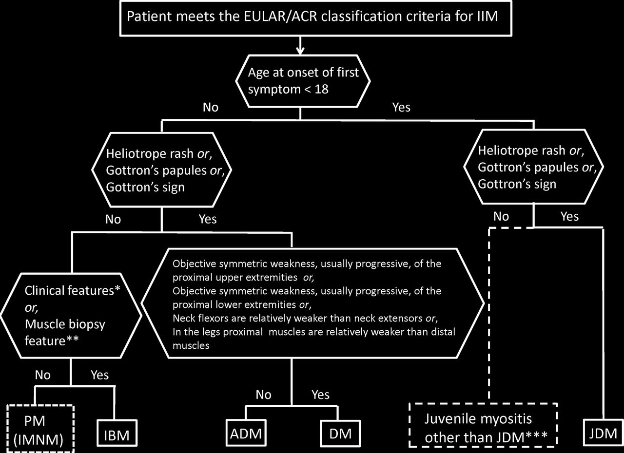
A patient classified with IIM by the EULAR/ACR classification criteria (probability of IIM ≥55%) can be further subclassified with a classification tree (figure 2). Age at onset of first symptom (≥18 years of age) distinguishes adult from juvenile IIM. Thereafter, clinical findings and muscle biopsy features subclassify adult IIM patients into PM, IBM, ADM or DM. Based on our dataset, juvenile patients with skin rash can be classified into JDM. Three subgroups cannot be further separated using our criteria because of small sample sizes: juvenile PM, IMNM and hypomyopathic DM.
Classification tree for subgroups of IIM. A patient must first meet the EULAR/ACR classification criteria for IIM (probability of IIM ≥55%). The patient can then be subclassified using the classification tree. The subgroup of PM patients includes patients with IMNM. For IBM classification, one of the following, *finger flexor weakness and response to treatment: not improved, or **muscle biopsy: rimmed vacuoles, is required for classification. ***Juvenile myositis other than JDM was developed based on expert opinion. IMNM and hypomyopathic DM were too few to allow subclassification. ACR, American College of Rheumatology; ADM, amyopathic dermatomyositis; DM, dermatomyositis; EULAR, European League Against Rheumatism; IBM, inclusion body myositis; IIM, idiopathic inflammatory myopathies; IMNM, immune-mediated necrotising myopathy; JDM, juvenile dermatomyositis; PM, polymyositis.
Among patients with IIM by the EULAR/ACR classification criteria (probability of IIM ≥55%), and with sufficient data to allow subclassification (n=703), the number of cases in the subgroups as defined according to the classification tree was enumerated (table 3). The agreement between the classification tree subgroups and the physician-diagnosed subgroups in the dataset was high (92.6% agreement, kappa=0.90, p<0.00001). The agreement proportions, with a probability of 55%, were 1.00 for JDM, 0.89 for DM, 0.94 for ADM, 0.92 for IBM and 0.93 for PM. Raising the probability cut-off of IIM to 90% yielded 94.9% agreement, kappa=0.93, p<0.00001. With a probability cut-off of 90%, the agreement proportions were 1.00 for JDM, 0.96 for DM, 0.95 for ADM, 0.93 for IBM and 0.88 for PM.
Comparison of physician-diagnosed IIM subgroups with IIM subgroups defined according to the classification tree among patients meeting the EULAR/ACR classification criteria for IIM
Performance of EULAR/ACR criteria compared with published criteria
Performance of the EULAR/ACR criteria was compared with published criteria for IIM7 8 10 11 14 15 using the IMCCP dataset (table 4). The new criteria including muscle biopsy features displayed high sensitivity (93%) and specificity (88%). There was slightly lower performance without biopsy variables (sensitivity and specificity 87% and 82%, respectively). Among the assessed criteria, the Targoff criteria11 showed the highest sensitivity (93%) and specificity (89%). Other criteria had either high sensitivity and low specificity (Bohan and Peter7 8 and Tanimoto criteria10), or low sensitivity and high specificity (Dalakas and Hohlfeld14 and ENMC criteria15).
Performance of the EULAR/ACR classification criteria for IIM and existing classification and diagnostic criteria for IIM
We studied how different criteria could classify patients with diverse IIM subdiagnoses in the IMCCP dataset (table 4). The EULAR/ACR classification criteria correctly classified most patients with all IIM subdiagnoses. When biopsy data were used, the performance improved for IBM (94% with biopsy data vs 58% without biopsy data) and PM (86% with biopsy data vs 79% without biopsy data). The Bohan and Peter,7 8 Tanimoto10 and Targoff11 criteria correctly classified all IIM subdiagnoses except ADM, a diagnosis not included in those criteria. The Dalakas and Hohlfeld criteria14 could not classify any subdiagnoses. The ENMC criteria15 correctly classified DM and JDM cases but no other subdiagnoses.
A comparison between the EULAR/ACR classification criteria (55% probability cut-off) and the Bohan and Peter criteria7 8 showed 89% agreement (kappa=0.71, p<0.00001) without including muscle biopsy data, and 93% agreement (kappa=0.73, p<0.00001) using muscle biopsy findings. Comparison between the newly developed criteria and the Targoff criteria11 demonstrated that the agreement was 89% (kappa=0.74, p<0.00001) and 93% (kappa=0.82, p<0.00001) without or with inclusion of muscle biopsy data, respectively.
Validation
Internal validation
Using the criteria without muscle biopsy data, 733 observations were used, resulting in AUC=0.942 and cross-validated area=0.933. Using the criteria with muscle biopsy data, 507 observations were included, resulting in AUC=0.962 and cross-validated area=0.942.
External validation for sensitivity
Data from 592 cases (PM=281, DM=256, IBM=33, JDM=18 and ADM=4) in the Euromyositis register were used where clinical, laboratory and muscle biopsy data were available (Karolinska University Hospital, Stockholm, Sweden; Prague Hospital, Prague, Czech Republic; Oslo University Hospital, Oslo, Norway) (online supplementary 7). When there was sufficient information available, the EULAR/ACR classification criteria confirmed IIM diagnosis using a 55% probability cut-off for classification of IIM with no misclassification, yielding 100% sensitivity. Using the criteria without muscle biopsies, 489 (83%) patients were classified as IIM, and 103 (17%) patients could not be classified due to missing data. For the criteria with biopsies, 204 (34%) were classified as IIM and 388 (66%) could not be classified due to missing muscle biopsy data in the register. Results for the IBM and PM subgroups improved when biopsy data were included: 97% of IBM cases could be classified compared with 73% when biopsy data were not included. For PM, 80% and 76%, respectively, could be classified. Raising the IIM classification cut-off from 55% to 90% decreased the total number of cases that could be classified to only 63% (not including muscle biopsies) or 28% (including muscle biopsies) due to absence of some muscle biopsy variables in the Euromyositis registry database.
The Juvenile Dermatomyositis Biomarker Study and Repository (UK and Ireland)
The JDRG register included 332 juvenile IIM cases in the study (definite JDM=292, probable JDM=20, definite juvenile PM=4, probable juvenile PM=2, focal myositis=6 and other IIM=8) (online supplementary 8). Muscle biopsy data were not available for all, thus the EULAR/ACR classification criteria without muscle biopsy data were used to test sensitivity in this dataset. Three hundred and seven (92%) cases could be classified using the 55% cut-off and no case was misclassified, yielding 100% sensitivity. The remaining 25 cases (8%) could not be classified due to missing data. Raising the cut-off stepwise to 60%, 70%, 80% or 90% yielded classification of 92%, 88%, 87% or 64% cases, respectively, where classification was possible.
Web calculator
A web calculator was developed (www.imm.ki.se/biostatistics/calculators/iim) as an aid to use the EULAR/ACR classification criteria. A probability range of classification can be obtained, providing the minimum and maximum probability. In addition to the probabilities acquired, the aggregated scores will be displayed. Whenever sufficient data are entered, the subclassification will be displayed.
Discussion
Classification criteria are essential for inclusion of comparable patients in studies. No validated classification criteria for IIM currently exist. The EULAR/ACR classification criteria for IIM offer advantages that previous criteria lack. They are data driven, exhibit high sensitivity and specificity, and use a limited number of accessible, defined clinical and laboratory variables. Internal validation and testing in external cohorts confirmed excellent performance. Importantly, the new criteria capture the most frequent IIM subgroups and can be used for both adults and children for research studies and clinical trials.
The new EULAR/ACR classification criteria provide a score with a corresponding probability of having IIM. This provides investigators flexibility in inclusion criteria for different types of studies, for example, clinical trials requiring high specificity would warrant a high probability of IIM in the inclusion criteria, whereas epidemiological studies requiring high sensitivity would need inclusion criteria with lower probability of IIM.
The new criteria are based on data from children and adults with different ethnicities from centres in Europe, America and Asia, and use symptoms, signs and other measures that are routinely assessed. A limitation is still that a majority of the patients were Caucasian, and even though we included data from 298 patients from Asia, we cannot exclude that there can be differences in manifestations between different ethnic groups, hence we still need to validate the criteria in Asian and African populations. Importantly, in patients with a typical DM skin rash, the criteria can be used without muscle biopsy data. For JDM, 97% of patients were correctly classified using the new criteria without muscle biopsy data. The new criteria also offer practical advantages in the number of variables needed to be tested. If a sufficient probability is reached, there is no requirement to test all items. Each criterion is well defined, lessening the opportunities for ad hoc interpretation. The skin rash typical of DM contributed with high weights in the probability score. Skin biopsy is recommended in the absence of muscle symptoms.33 34 The EULAR/ACR classification criteria are the first myositis criteria to be validated and tested for sensitivity in other cohorts and revealed no misclassification.
Compared with most previous criteria, the new criteria are superior in sensitivity, specificity and classification accuracy. Classification criteria should have high sensitivity and specificity. The EULAR/ACR criteria demonstrated sensitivity and specificity of 87% and 82%, respectively, with even higher accuracy when muscle biopsies were included, 93% and 88%, respectively. Correctly classified patients were 86% and 91%, respectively, with and without inclusion of biopsies, and the criteria performed equally well for adult and juvenile cases. The Targoff criteria11 also showed good statistical properties, but were not able to capture all subgroups of IIM as ADM patients were not included. Furthermore, the variables were not clearly defined in the Targoff criteria, and testing of more variables is required, including electromyography, which is not always easily accessible and may be painful for patients. Importantly, the EULAR/ACR criteria can be applied to patients with myositis with overlap diagnoses, such as mixed connective tissue disease or systemic lupus erythematosus with myositis, since these patients were included among IIM cases.
There are limitations of the study; no controls or comparators were included in the external validation cohort since the IMCCP study was designed before those recommendations from ACR/EULAR were in place, requiring future validation. A validation study using comparators is underway, but we encourage additional validation studies in different populations. Another limitation largely unavoidable in observational data is the high frequency of missing data in the derivation dataset and validation samples, reflecting differences in practice patterns in evaluating patients. Nevertheless, 80% of cases and comparators had muscle biopsy data available, whereas MRI data and electromyography were only available for 38% and 29% of cases, respectively, reflecting their limited usage in clinical diagnosis. However, MRI data and electromyography examination are still important for diagnostic purposes of IIM. Patients studied had to have their disease for at least 6 months, which did not allow us to study new-onset patients. Importantly, these criteria are proposed as classification criteria in research and in clinical trials, not as diagnostic criteria.35 There is also some possibility that the cut-points established for probable and definite myositis will need adjustment when tested with new populations of patients.
It took almost 10 years to assemble sufficient numbers of patients with these rare diseases, and three subgroups did not have enough subjects to study adequately. During this period, a new IIM subgroup became recognised, IMNM,36 of which only a few cases were included into the study. IMNM cases could thus not be distinguished from PM in the subclassification tree. Another subgroup with few cases was juvenile PM, making a data-derived distinction from JDM impossible. However, paediatric rheumatology experts in the IMCCP recommended that the adult subclassification of IIM could be used for juvenile PM by extrapolation (figure 2). IBM cases were identified in the subclassification tree by the clinical features of finger flexor weakness and no response to treatment, or by the presence of rimmed vacuoles in muscle biopsies.37
Another limitation was the low frequency of myositis-specific autoantibodies documented. Five myositis-specific autoantibodies were included: anti-Jo-1, anti-Mi-2, anti-SRP, anti-PL7 and anti-PL12 antibodies, and all were strongly associated with IIM. However, only anti-Jo-1 autoantibody had a significant number of observations (n=1062) to permit analyses and inclusion in the classification criteria. A future update of the EULAR/ACR classification criteria should include the more recently identified myositis-specific autoantibodies,21 22 in addition to more patients with IMNM, ADM, hypomyopathic DM and juvenile cases other than JDM.
Recommendations
Patients with pathognomonic skin rashes (heliotrope rash, Gottron’s papules and/or Gottron’s sign) of JDM or DM are accurately classified with the EULAR/ACR classification criteria without including muscle biopsy data. For patients without these skin manifestations, muscle biopsy is recommended. For DM patients without muscle involvement, a skin biopsy is recommended.
The EULAR/ACR classification criteria provide a score and a corresponding probability of having IIM. Each probability displays a unique sensitivity and specificity. The best balance between sensitivity and specificity can be found for a probability of 55%–60% (total aggregated score of ≥5.5 and ≤5.7) for the criteria not including muscle biopsy data, and 55%–75% (total aggregated score ≥6.7 and ≤7.6) when including muscle biopsies. These cases are designated ‘probable IIM’. The recommended cut-off needed for classifying a patient as IIM is ≥55%.
‘Definite IIM’ corresponds to a probability of ≥90% or a total aggregate score of 7.5 or more without muscle biopsy and 8.7 with muscle biopsy, and is recommended in studies where a high specificity is required.
A patient is termed ‘possible IIM’ if the probability is ≥50% and <55% (a minimum score of 5.3 without biopsies and 6.5 with biopsies).
For clarity and transparency, both the descriptive term (‘possible’, ‘probable’ or ‘definite’) and the probability and the aggregated score should be reported in studies.
Conclusions
New classification criteria for IIM and the major IIM subgroups have been developed. These data-driven criteria have a good feasibility, high sensitivity and specificity, have been partly validated in external cohorts and are superior to previous criteria in capturing different subgroups of IIM. Revision of the criteria in the future will be important when additional validated myositis autoantibody tests, imaging and other tests are available in more IIM cases and comparator cases without IIM.
Acknowledgments
We thank Elin Forslund for assistance with data registration. We thank Dr Andrew Mammen and Dr Mike Ward for critical reading of the manuscript. We are grateful for contribution of clinical data from investigators and for participants contributing with valuable input at IMCCP meetings.
References
Footnotes
This article is published simultaneously in Arthritis & Rheumatology.
FWM and LGR contributed equally,
AT and MB contributed equally.
Handling editor Tore K Kvein
Contributors All authors were involved in drafting the article or revising it critically for important intellectual content and approved the final version to be published. All authors had full access to all of the data in the study and take responsibility for the integrity of the data and the accuracy of the data analysis. Study conception and design: IEL, AT, MB, VPW, CP, MdV, LA, AAA, RJB, MHL, JAS, KD, BMF, HK, PAL, BAL, FWM, LGR. Acquisition of data: IEL, AT, MB, VPW, CP, MdV, LA, AAA, RJB, MHL, JAS, RA, SA, HC, RGC, KD, MMD, BMF, IG-DLT, PG, TH, JDK, HK, PAL, BAL, YL, CVO, MO, AMR, LR-S, HS, AS-O, YWS, JV, SRY, FWM, LGR, The International Myositis Classification Criteria Consortium, working committee members. Analysis and interpretation of data: IEL, AT, MB, VPW, CP, MdV, LA, AAA, RJB, MHL, JAS, RA, BMF, IG-DLT, PG, HK, PAL, BAL, YL, FWM, LGR.
Funding Financial support came from the European League Against Rheumatism (EULAR), American College of Rheumatology (ACR), The Myositis Association (TMA) and in part by the Intramural Research Program of the NIH, National Institute of Environmental Health Sciences and the European Science Foundation for the Euromyositis Register, the Swedish Research Council K2014-52X-14045-14-3 and through the regional agreement on medical training and clinical research (ALF) between Stockholm County Council and Karolinska Institutet. However, the project also received support (not financial support/funding) from different associations: the American Academy of Neurology (AAN), the Childhood Arthritis and Rheumatology Research Alliance (CARRA, CARRA Inc is funded by NIH-NIAMS), Friends of CARRA, and the Arthritis Foundation, the European Neuromuscular Centre (ENMC), the International Myositis Assessment and Clinical Studies Group (IMACS), the Muscle Study Group (MSG), the Rheumatologic Dermatology Society (RDS), the Pediatric RheumatologyEuropean Society (PReS) network for JDM and the Pediatric Rheumatology International Trials Organization (PRINTO).
Disclaimer The views expressed in this article are those of the authors and do not necessarily reflect the position or policy of the Department of Veterans Affairs or the United States government, or the NHS, the National Institute for Health Research or the Department of Health (UK).
Competing interests JAS has received research grants from Takeda and Savient and consultant fees from Savient, Takeda, Regeneron, Merz, Iroko, Bioiberica, Crealta and Allergan. JAS serves as the principal investigator for an investigator-initiated study funded by Horizon pharmaceuticals through a grant to DINORA, Inc., a 501 (c)(3) entity. JAS is a member of the executive committee of OMERACT, an organisation that develops outcome measures in rheumatology and receives arms-length funding from 36 companies; a member of the American College of Rheumatology’s (ACR) Annual Meeting Planning Committee (AMPC); Chair of the ACR Meet-the-Professor, Workshop and Study Group Subcommittee; and a member of the Veterans Affairs Rheumatology Field Advisory Committee. HC and RGC’s work in myositis is partly funded by grants from Arthritis Research UK (18474) and the Medical Research Council (MR/N003322/1). JV’s work in myositis is supported by Project (Ministry of Health, Czech Republic) for conceptual development of research organization 00023728.
Ethics approval Ethical committees at each participating centre.
Provenance and peer review Not commissioned; externally peer reviewed.
Collaborators The collaborators are included in the appendix.