Article Text

Abstract
Objectives To determine whether ultrasound can identify anti-cyclic citrullinated peptide (anti-CCP) antibody-positive patients without clinical synovitis (CS) who progress to inflammatory arthritis (IA).
Methods In a prospective study, anti-CCP-positive patients without CS underwent ultrasound imaging of 32 joints (wrists, metacarpophalangeal joints, proximal interphalangeal joints and metatarsophalangeal joints (MTPs)) and were monitored for the development of IA. Associations between baseline ultrasound findings (grey scale (GS), power Doppler (PD) and erosions) and (1) progression to IA and (2) development of CS within an individual joint were measured.
Results Consecutive anti-CCP-positive patients (n=136; mean age 51 years, 100 women) were followed up for median of 18.3 months (range 0.1–79.6). At baseline 96% had GS, 30% had PD and 21% had one or more erosions. IA developed in 57 patients (42%) after median of 8.6 months (range 0.1–52.4). Ultrasound abnormalities (GS ≥2, PD ≥1 or erosion ≥1) were found in 86% at baseline compared with 67% of non-progressors (χ2=6.3, p=0.012). Progression to IA was significantly higher in those with ultrasound findings in any joint (excluding MTPs for GS) (GS ≥2: 55% vs 24%, HR (95% CI) 2.3 (1.0 to 4.9), p=0.038; PD ≥2: 75% vs 32%, 3.7 (2.0 to 6.9), p<0.001 and erosion ≥1: 71% vs 34%, 2.9 (1.7 to 5.1), p<0.001). Furthermore, progression occurred earlier with PD ≥2 (median 7.1 vs 52.4 months) and erosion ≥1 (15.4 vs 46.5). At the individual joint level, the trend for progression to CS was more significant for GS and PD (GS ≥2: 26% vs 3%, 9.4 (5.1 to 17.5), p<0.001; PD ≥2: 55% vs 4%, 31.3 (15.6 to 62.9), p<0.001).
Conclusion Ultrasound features of joint inflammation may be detected in anti-CCP-positive patients without CS. Ultrasound findings predict progression (and rate of progression) to IA, with the risk of progression highest in those with PD signal.
Trial registration number NCT02012764; Results.
- Ant-CCP
- Ultrasonography
- Synovitis
- Early Rheumatoid Arthritis
Statistics from Altmetric.com
Introduction
Increasing emphasis is being placed on the early diagnosis patients with inflammatory arthritis (IA).1 In the very early stages of the disease, however, making the diagnosis may be particularly challenging as patients often present with non-specific musculoskeletal (MSK) symptoms. Additional biomarkers may be required to identify these patients.
Studies have shown that serological markers including rheumatoid factor (RF) and the more specific anti-cyclic citrullinated protein (anti-CCP) antibodies may be present years before clinically apparent disease.2 MSK ultrasound has also been shown to be more sensitive than clinical examination in identifying subclinical synovitis,3 and more sensitive than radiography in detecting erosions.4 Grey-scale (GS) change can delineate synovial thickening and power Doppler (PD) is particularly useful in synovial tissue, where velocity of blood flow is low, to detect hyperaemia.5
Ultrasound has been shown to be of value in determining disease persistence in patients presenting with early IA.6 In patients without clinical synovitis (CS), the role of ultrasound is still not clear. In one study of anti-CCP-positive patients with MSK symptoms, preliminary analyses found the presence of ultrasound PD in the wrist and hands together with clinical, serological and genetic markers to be of value to determine progression to IA.7 However, in another study of patients with RF±anti-CCP-positive arthralgia, ultrasound was found to be predictive of the development of IA at a joint level, but not at a patient level.8
The aims of this study were to determine whether GS, PD and erosions on ultrasound:
identified the patients who progressed to IA;
predicted the timing of progression to IA and
within a joint predicted progression to CS in that joint.
Methods
The patient cohort has previously been described.7 In brief, patients aged >18 years with MSK symptoms with a positive anti-CCP2 test were invited to participate in the study. Patients were followed up in a dedicated research clinic at Chapel Allerton Hospital, Leeds, UK, and attended for three monthly assessments for the first year then annually or until a clinical diagnosis of IA (defined by at least one tender and swollen joint) was made. Patients were seen sooner if necessary. Patients with a history of IA diagnosed by a rheumatologist, clinically detected IA at baseline and those on disease-modifying anti-rheumatic drugs (DMARDs) were excluded.
Ultrasound examination was performed on all patients by a single rheumatologist (JLN) experienced in MSK ultrasound, who was blinded to the clinical examination for the majority of scans. Patients recruited to the study had scans of the wrists, metacarpophalangeal joints (MCPs), proximal interphalangeal joints (PIPs) and metatarsophalangeal joints (MTPs) bilaterally. In addition, other joints could be scanned if symptomatic. The scans were performed at baseline, 6 and 12 months, then annually and at withdrawal to confirm an IA diagnosis.
As these patients had no CS, a control group without IA of the same age range with no or stable joint symptoms was scanned to ensure that there was some measure of what could be expected in otherwise healthy individuals.
Ultrasound scans were initially performed using a Philips (ATL HDI 5000) machine with 5–12 and 8–15 MHz transducers. The machine was later changed to a General Electric S7 machine and a 6–15 MHz transducer was used. PD was assessed using a pulse repetition frequency (PRF) set between 700 and 1000 MHz, medium wall filter and gain adjusted until the background noise was suppressed. Doppler frequency for the Philips (ATL HDI 5000) was 6 MHz and for the General Electric S7 was 10 MHz.
The Outcome Measures in Rheumatoid Arthritis Clinical Trials (OMERACT) definitions were used to define synovitis and erosions.9 The ultrasound scans of the wrist included the radiocarpal, intercarpal and ulnocarpal joint compartments. The highest score of these compartments was used for the wrist score. GS and PD were scored using a semi-quantitative method from 0 to 3, using the European League against Rheumatism (EULAR)-OMERACT scoring system.10 The longitudinal dorsal midline scans were used to score each joint. Transverse scans were performed to confirm pathology. Presence of erosion(s) was also recorded for each joint if seen and confirmed on longitudinal and transverse scans. Intra-reader reliability for the assessor performing the ultrasound scanning has been reported to be excellent, with 100% agreement between two repeated assessments of the presence of PD signal in 33 joints.11
Statistical analysis
All analyses were conducted in Stata (V.13.1). Spearman's rank correlation and Wilcoxon rank-sum tests were used for identifying associations between clinical characteristics and total ultrasound scores summated across the joints scanned. Cox proportional hazards regression was used for assessing the associations at the patient level between baseline ultrasound findings and the timing of progression to IA. The additional value of scanning other affected joints was assessed, as was the value of repeated scanning 6 months after baseline. At the joint level, clustered Cox models (taking into account clustering of joints within patients) were used for investigating whether the presence of GS, PD or erosion(s) in a joint (wrist, MCP, PIP or MTP) at baseline was predictive of that joint later developing CS. Individual joints that had not progressed when the patient developed CS were censored in the analysis. We also assessed the association between the semi-quantitative scores in the joint and the odds of the joint developing swelling.
Model performance was assessed using Harrell's C, a measure of concordance which expresses the proportion of randomly selected pairs of survival times that are ‘correctly’ ordered by the model predictions.
Because there was a change in the ultrasound machine during the course of the study, sensitivity analyses were performed. We first restricted the dataset to the patients scanned immediately before and after the change to investigate whether there was a difference in the ultrasound scores; this approach was chosen to eliminate the possibility that operator learning curve could have produced any observed differences. We then repeated our main analysis, excluding patients who had been scanned following the change in machine (see online supplementary tables S1 to S3 for details).
Results
Clinical findings
A total of 167 individuals were referred. Twenty-five were found to have CS at baseline and were excluded. One was lost to follow-up after the initial clinic visit and was excluded. A further five were excluded because they had received a steroid injection (n=1) or tacrolimus (n=1) prior to the baseline assessment, or were already receiving DMARD therapy at baseline (n=3). The clinical characteristics of the 136 patients included are described in table 1. Fifty-seven (41.9%) were diagnosed with an IA after a median follow-up of 18.3 months (range 0.1–79.6 months). At the point of developing IA, CS was detected mainly in the wrists and the second and third MCP, PIP and MTP joints (see online supplementary figure S1). Of the patients who developed an IA, 49 patients (86.0%) fulfilled the 2010 American College of Rheumatology EULAR rheumatoid arthritis (RA) classification criteria12 and 8 patients (14%) developed undifferentiated IA (ie, IA not fulfilling these criteria). None developed psoriatic arthritis, gout or another IA. Forty-nine patients (86%) had ultrasound scans on development of CS—synovitis was confirmed in all. The remaining 79 patients who did not develop IA had been followed up for a median of 28.1 months (range 4.7–79.6).
Baseline characteristics of anti-CCP-positive individuals with non-specific musculoskeletal symptoms and no clinical inflammatory arthritis at baseline (n=136)
Ultrasound findings
Controls
Forty-eight controls had scans of the wrists, MCPs, PIPs and MTPs. Median (range) GS across all joints was 4 (0–20). In most of the different joint types, GS change was uncommon and mainly low grade. However, GS grades above 1 were frequently observed in the first to fourth MTP joints; 41.7% of controls had at least one MTP joint scoring >1 for GS. Only one control had PD signal, a score of 1 in a single first MTP joint. None had erosions in any of the joints in the hands or wrist, but two each had a small erosion in one of their fifth MTP joints without accompanying synovitis.
Patients
Baseline ultrasound findings are summarised in table 2. Almost all patients had evidence of GS in at least one joint (95.6%) and the majority (67.7%) scored ≥2 in at least one joint (table 2). The majority of patients (67.2%) did not have any PD present; maximum PD score in any joint was 1 in 18.2% and 2 in 14.6% (table 2). None had a joint scoring 3 for PD. Ultrasound erosions were seen in 28 patients (20.6%)—5.9% (8/136) had erosions in the MCPs and 16.2% (22/136) had erosions in the MTPs. Of these, 68% (19/28) had associated PD; of the remainder, all had some associated GS change.
Descriptive data summarising ultrasound PD findings in wrists, MCPs, PIPs and MTPs (maximum score observed bilaterally) at point of presentation in a cohort of 136 anti-CCP-positive individuals with non-specific musculoskeletal symptoms

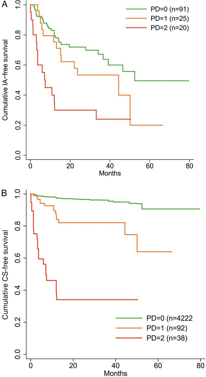
Survival plot of anti-CCP positive patients showing (A) cumulative inflammatory arthritis (IA)-free survival according to PD score observed at baseline in any joint (MCP, PIP, wrist, MTP) at the patient level and (B) clinical synovitis (CS)-free survival according to PD score observed at baseline at the individual joint level. Anti-CCP, anti-cyclic citrullinated peptide; MCP, metacarpophalangeal joint; MTP, metatarsophalangeal joint; PIP, proximal interphalangeal joint of the finger; PD, power Doppler.
There were no substantive associations with either total GS or total PD scores and clinical findings. The number of joints with erosions was higher in patients with high-positive RF and/or anti-CCP (three times the upper limit of normal),12 despite the majority having no erosions (the 95th and 99th percentiles in this group were 2 and 2, respectively, compared with 0 and 1 in those with low positive RF and anti-CCP; p=0.033).
Ultrasound findings and progression to IA at the patient level
Of the patients who progressed, 86% had at least one ultrasound abnormality (GS ≥2/PD ≥1/erosion ≥1) present at baseline compared with 67% of non-progressors (χ2=6.3, p=0.012).
Patients with a baseline GS score of 1 or 2 in any MCP, PIP, wrist or MTP joint were descriptively more likely to progress to IA than those without GS (table 3). However, only six patients had no evidence of GS, so power was limited. When MTPs were excluded, patients who scored 2 or more in the remaining joints at baseline were at higher risk of progression to IA than those with no GS synovitis in those joints (mean time to IA 36.8 months vs 57.1; HR=2.3, p=0.038). (The median is usually the preferred summary for survival time, but due to the small proportion of patients with GS=0 progressing to IA, the median could not be calculated for this group.) Patients with a baseline PD score of ≥2 were at higher risk of progression than those with no PD (median 7.1 months vs 52.4 months; HR=3.7, p<0.001; table 3 and figure 1A). Patients with an erosion present in at least one joint were also at higher risk of progression (median 7.5 months vs 50.1 months; HR=2.9, p<0.001; table 3).
Patient-level risk of progression to IA according to ultrasound findings at baseline

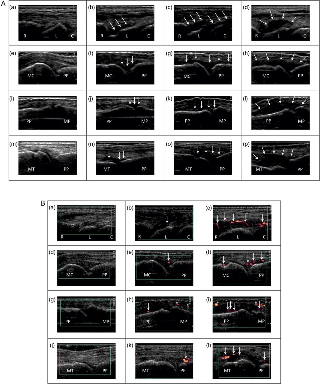
(A) Spectrum of ultrasound grey scale on the dorsal aspects of the wrists ((a) to (d)), MCPs ((e) to (h)), PIPs ((i) to (l)) and MTPs ((m) to (p)) of anti-CCP-positive patients without clinical synovitis: grade 0 ((a), (e), (i) and (m)), grade 1 ((b), (f), (j) and (n)), grade 2 ((c), (g), (k) and (o)) and grade 3 ((d), (h), (l) and (p)). (B) Spectrum of ultrasound power Doppler on the dorsal aspects of the wrists ((a) to (c)), MCPs ((d) to (f)), PIPs ((g) to (i)) and MTPs ((j) to (l)) of anti-CCP-positive patients without clinical synovitis: grade 0 ((a), (d), (g) and (j)), grade 1 ((b), (e), (h) and (k)) and grade 2 ((c), (f), (i) and (l)). Anti-CCP, anti-cyclic citrullinated peptide; C, capitate; L, lunate; MC, metacarpal; MCPs, metacarpophalangeal joints; MP, middle phalanx; MT, metatarsal; MTPs, metatarsophalangeal joints; PIPs, proximal interphalangeal joints; PP, proximal phalanx; R, radius; *, extracapsular blood vessel.
Maximum and total scores of PD were also significantly associated with progression to IA (details in online supplementary section).
In patients who had a repeat scan at 6 months, if patients were categorised according to whether ultrasound synovitis had remained absent, resolved, appeared or persisted compared with baseline, then although for some groups sample size was small, the descriptive data suggested that this might help further categorise patients (see online supplementary table S4).
Ultrasound findings and progression to CS at the joint level
Thirty-two joints were scanned for each individual (10 MCPs, 10 PIPs, two wrists, 10 MTPs), yielding a total of 4352 joints. GS score was 1 in 621 joints (14.3% of those scanned), 2 in 349 (8.0%) and 3 in 29 (0.7%). PD score was 1 in 92 joints (2.1%) and 2 in 38 (0.9%) (figures 2A and 2B). Erosions were found in 38 joints (0.9%).
The proportion of joints found to be clinically swollen in patients who progressed to IA, according to joint type, is summarised in the online supplementary figure S1. Proportions of each joint type that progressed to CS, according to baseline ultrasound findings are summarised in online supplementary table S5. Descriptively, of the joints scanned, a slightly greater proportion of those with higher GS score progressed to CS when all joints were included; this trend appeared to be more pronounced when MTPs were excluded. The same general pattern was found for presence of erosions. Progression to CS was rare in joints scoring 0 for PD (3.5% progressed) compared with joints scoring 1 (15.2%), while a substantial proportion of the joints scoring 2 progressed (55.3%).
Clustered Cox regression indicated that the presence of GS in at least one joint at baseline (any score >0) was associated with a 2.5-fold increase in the risk of the joint developing CS (HR=2.5 (95% CI 1.61 to 3.8), p<0.001). When MTPs were excluded, the HR increased to 4.6 (95% CI 3.0 to 7.2; p<0.001). Using semi-quantitative scoring, joints scoring 1 (HR=1.9, p=0.010) or ≥2 (HR=3.3, p=0.311) were more likely to progress than joints without GS (table 4). When MTPs were excluded, the HRs increased (score 1: HR=3.3, p<0.001; score ≥2: HR=9.4, p<0.001).
Joint-level risk of progression to CS according to ultrasound findings at baseline
The presence of PD in a joint at baseline (any score >0) was associated with 10-fold increase in risk of the joint developing CS (HR=10.3 (5.9, 18.2), p<0.001). Using semi-quantitative scoring, a score of 1 increased the risk of CS fivefold relative to a score of 0 (HR=5.2, p<0.001), while a score of 2 was associated with very large increase in risk (HR=31.3, p<0.001; table 4) and more rapid progression (figure 1B). It should be noted, however, that the CI around the HR for PD=2 was wide (15.6 to 62.9), as a consequence of the small number of joints scoring 2 (n=38).
The presence of erosions was associated with a 2.8-fold increase in risk. Although this was a substantive effect, power was low because few joints had erosions present (n=38), and the effect was not significant at the 5% level (p=0.076).
There was potential value of scanning additional joints, for example, the knees and shoulders if symptomatic (see online supplementary section for details).
Discussion
Ultrasound is increasingly being used as an extension of clinical practice.13 The advantages of ultrasound include the ability to image both bone and soft tissue structures without using ionising radiation, allowing for repeat testing. Ultrasound is also relatively rapid to perform and allows dynamic assessment of multiple joints. Although user-dependant, rheumatologists are becoming skilled at performing ultrasound.14 ,15 While the role of ultrasound in determining progression of patients with undifferentiated arthritis to RA,6 and in assessing disease activity in patients with RA with or without DMARD therapy13 ,15–17 has been well documented, its role in patients at risk18 is less well described.
To our knowledge this is the first study to show the predictive value of ultrasound in anti-CCP-positive patients without CS for the development of IA. This was seen both at a patient level and at an individual joint level. In contrast, another study by van der Stadt et al also examined the use of ultrasound in patients with arthralgia in detecting IA. This study found ultrasound to be predictive of the development of CS at a joint level, however not IA at a patient level.8 Importantly this cohort also included patients who were anti-CCP-negative/RF-positive compared with the patients in the present study who were all anti-CCP positive. Also the ultrasound protocol was different to the present study in that painful or tender joints, as well as the adjacent and contralateral joints were scanned.
There are little published data on the prevalence of ultrasound findings in patients with MSK symptoms without CS and positive serology. Work by van der Stadt et al8 reported the presence of PD in 17% of anti-CCP-positive or RF-positive patients with arthralgia versus 30% of patients in our cohort.
The optimal set of joints to be scanned in patients with IA remains an area of debate.19 In this study, a core set of 32 joints (wrists, MCPs, PIPs and MTPs) was used as they are the most commonly affected joints in patients with IA. As these patients were likely to develop synovitis in only a few joints in the earliest stages, scanning fewer joints may have missed findings on ultrasound. For the majority of patients, this encompassed the joint areas in which CS developed. In a few patients, the large joints were the predominant symptomatic area. Ultrasound of the knees and shoulder were helpful in these. A pragmatic approach therefore may be to scan the wrists, hands±feet (MTPs) of all patients and other joints if symptomatic. Analysis of the ultrasound scans at 6 months in patients who had not progressed to IA at that stage also suggest value of repeat scanning, particularly if symptoms change.
Scoring was based on the OMERACT semi-quantitative scores from 0 to 3 for GS and PD. Higher scores for both GS and PD (particularly for PD) were associated with an increased risk of developing CS at a joint level. Whilst Doppler signal may be present in joints of healthy individuals,20 we used a PD cut-off score of 1 as positive as these patients tended to have relatively little synovitis at disease onset. GS was predictive of progression to CS in the hands. In the feet, however, GS was less discriminating between those who progressed and those who did not. While the absence or presence of erosions was used for analysis in this cohort, a proportion of healthy individuals may have erosions and erosion size may have been more discriminating.21
Other imaging modalities including MRI have demonstrated subclinical inflammation in patients with anti-CCP-positive arthralgia22 and anti-CCP-negative arthralgia with inflammatory symptoms.23 Subclinical arthritis has also been visualised on macrophage positron emission tomography (PET) in Anti-CCP antibody-positive arthralgia, with a subgroup of PET-positive patients developing features of an IA within 2 years of follow-up.24
Our study has its limitations. First, the ultrasonographer was not completely blinded to the clinical setting, although it is hard to see how this baseline examination could influence the outcome. Second, the ultrasound machine was changed to a newer one for the last 23 baseline scans. Excluding the patients scanned on the second machine, the results remained significant. We, therefore, do not think that the change in machines affected the study conclusions.
In this study, the focus was on the joints. In a recent study, however, MRI-detected tenosynovitis was noted to be a common finding in early arthritis.25 Further investigations in this regard may be warranted.
In summary, our study shows that ultrasound features of joint inflammation may be detected in anti-CCP-positive patients without CS and can predict progression to IA. Our findings suggest that ultrasound will be an important part of prognosis assessment and management of these patients.
Acknowledgments
The authors would like to thank all the participating general practitioners and health professionals and the UK Clinical Research Network teams for the referrals from primary care to the study. The authors would also like to acknowledge Ged Connolly-Thompson, Andrea Paterson and Jonathan Thompson for helping with data management.
References
Supplementary materials
Supplementary Data
This web only file has been produced by the BMJ Publishing Group from an electronic file supplied by the author(s) and has not been edited for content.
- Data supplement 1 - Online supplement
Footnotes
Handling editor Tore K Kvien
Contributors JLN performed the ultrasound scans and was responsible for the data collection and writing of the manuscript. LH was one of the study clinicians. EMAH was responsible for the statistical analysis. PE was the study lead and was responsible for the study design. Co-authors read and revised the manuscript.
Funding This study presents independent research supported by the National Institute for Health Research (NIHR) Leeds Clinical Research Facility. This study was supported by AbbVie who provided funding for the anti-CCP testing.
Competing interests None declared.
Ethics approval NHS Health Research Authority National Research Ethics Service Committee Yorkshire & The Humber – Leeds West.
Provenance and peer review Not commissioned; externally peer reviewed.