Article Text

Abstract
Objective Systemic lupus erythematosus (SLE) is a clinically heterogeneous disease with limited reliable diagnostic biomarkers. We investigated whether gene methylation could meet sensitivity and specificity criteria for a robust biomarker.
Methods IFI44L promoter methylation was examined using DNA samples from a discovery set including 377 patients with SLE, 358 healthy controls (HCs) and 353 patients with rheumatoid arthritis (RA). Two independent sets including 1144 patients with SLE, 1350 HCs, 429 patients with RA and 199 patients with primary Sjögren's syndrome (pSS) were used for validation.
Results Significant hypomethylation of two CpG sites within IFI44L promoter, Site1 (Chr1: 79 085 222) and Site2 (Chr1: 79 085 250; cg06872964), was identified in patients with SLE compared with HCs, patients with RA and patients with pSS. In a comparison between patients with SLE and HCs included in the first validation cohort, Site1 methylation had a sensitivity of 93.6% and a specificity of 96.8% at a cut-off methylation level of 75.5% and Site2 methylation had a sensitivity of 94.1% and a specificity of 98.2% at a cut-off methylation level of 25.5%. The IFI44L promoter methylation marker was also validated in an European-derived cohort. In addition, the methylation levels of Site1 and Site2 within IFI44L promoter were significantly lower in patients with SLE with renal damage than those without renal damage. Patients with SLE showed significantly increased methylation levels of Site1 and Site2 during remission compared with active stage.
Conclusions The methylation level of IFI44L promoter can distinguish patients with SLE from healthy persons and other autoimmune diseases, and is a highly sensitive and specific diagnostic marker for SLE.
- Systemic Lupus Erythematosus
- Autoimmune Diseases
- Gene Polymorphism
Statistics from Altmetric.com
Video abstract
Introduction
Systemic lupus erythematosus (SLE) is a chronic, remitting and relapsing, multisystem, autoimmune disease. The worldwide prevalence of SLE is approximately 20–150 cases per 100 000 individuals.1 SLE is much more prevalent in women, particularly during the childbearing years, and has a female-to-male ratio of 9:1.2 Autoantibodies play an important role in the pathogenesis of SLE, and the diverse clinical manifestations of the disease are associated with the deposition of antibody-containing immune complexes, leading to inflammation in the kidney, brain, skin and other organ systems.1 An important feature of SLE is the presence of autoantibodies, such as anti-nuclear antibodies (ANAs), anti-double strand DNA (dsDNA) antibody and anti-Smith (Anti-Sm) antibody, which have been used as conventional serological markers in patients with SLE.3 ,4 However, the currently available laboratory markers for SLE have significant limitations. ANA tests have a very high sensitivity (almost 100%) for a diagnosis of SLE but have a relatively low specificity (65%).5 Anti-dsDNA antibody is highly specific for SLE (94%). However, it is not particularly sensitive owing to the fact that it may be present transiently, and occurring in only 50–60% of patients with lupus at some point in the course of their disease.6 ,7 Anti-Sm is highly specific (99%), but has a low sensitivity (25–40%) for SLE.8 Due to the significant heterogeneity of the disease and the complex and rigorous process required to validate individual biomarkers, there is currently a very limited number of consensus biomarkers to aid in the diagnosis of SLE.9
Genetic factors likely contribute to the risk of developing SLE. Genome-wide association studies conducted throughout the past few years have identified more than 55 genetic loci associated with SLE risk such as IRF5, ITGAM, STAT4, MECP2 and many others.10 However, incomplete concordance in identical twins and the fact that most cases of SLE are sporadic rather than familial indicate the requirement for additional factors and mechanisms in the pathogenesis of SLE.1 ,11 ,12
In the last 10 years, epigenetic mechanisms have been increasingly recognised to play an important role in the pathogenesis of SLE. Our previous work has demonstrated reduced global genomic DNA methylation in SLE, and reduced DNA methylation levels in promoters of specific autoimmune related genes such as ITGAL,13 TNFSF7,14 PRF115 and CD40LG16 in SLE CD4+ T cells. Moreover, multiple genome-wide DNA methylation studies have been recently performed in SLE, leading to the identification of additional differentially methylated genes in this disease, most of which are hypomethylated in patients with SLE.11 ,17 ,18 Importantly, there is a robust hypomethylation of interferon (IFN)-regulated genes in SLE, independent of the cell type examined.19–21 Based on these observations, we decided to investigate if specific DNA methylation changes in the peripheral blood can be useful for the diagnosis of SLE.
Materials and methods
Study design
First, we screened for differentially methylated cytosines of cytosine-guanine dinucleotides (CpGs) in DNA samples from peripheral blood between patients with SLE and healthy controls (HCs). Second, we identified whether differential methylation in two CpG sites, located in the promoter region of IFI44L (IFN-induced protein 44-like), can be used to distinguish patients with SLE from HCs and patients with rheumatoid arthritis (RA). This was performed using a discovery cohort consisting of 377 patients with SLE, 358 HCs and 353 patients with RA using pyrosequencing. Next, we validated the diagnostic value of IFI44L promoter methylation in SLE using a larger cohort from China consisting of 529 patients with SLE, 569 HCs, 429 patients with RA and 199 patients with primary Sjögren's syndrome (pSS). The third independent cohort consisting of 615 patients with SLE and 781 HCs of European descent was used for additional validation. The disease characteristics of all patients with SLE are presented in the online supplementary table S1. We then compared the difference in IFI44L promoter methylation in patients with SLE with and without renal involvement. In addition, we compared the difference in IFI44L promoter methylation in 30 patients with SLE with paired blood samples that were obtained from the same patient in active disease stage and remission stage. We also investigated whether IFI44L hypomethylation might be triggered by a high type-1 IFN response.
Patients and controls
Peripheral blood samples were collected in EDTA-treated collection tubes. HCs had no history of cancer, cardiovascular diseases, autoimmune diseases or known infectious diseases. All study participants signed a written informed consent. All patients with SLE fulfilled the American College of Rheumatology (ACR) classification criteria for SLE and disease activity was assessed using the SLE Disease Activity Index (SLEDAI) (SLEDAI score >4 was considered active).22 All patients with RA satisfied the ACR Diagnostic Criteria for RA.23 All patients with pSS fulfilled the European classification criteria for pSS.24 SLE with renal damage was defined as clinical and laboratory manifestations that meet ACR criteria for renal involvement (persistent proteinuria >0.5 g per day or >3+ by dipstick, and/or cellular casts including red blood cells (RBCs), haemoglobin, granular, tubular, or mixed).22 ,25 In addition, the ACR criteria recommended that ‘active urinary sediment’ (>5 RBCs/high-power field (hpf), >5 white blood cells/hpf in the absence of infection) can be substituted for cellular casts.3 ,25 ,26 The baseline characteristics of all samples analysed in this study are presented in table 1.
Baseline characteristics of all subjects*
Isolation of genomic DNA, bisulfite treatment, DNA methylation profiling and pryosequencing for DNA methylation
Genomic DNA was extracted from peripheral blood using GeneJET Whole Blood Genomic DNA Purification Mini Kit (Thermo Fisher Scientific). Infinium Human Methylation 450 BeadChips (Illumina) were used to assay genome-wide DNA methylation. Bisulfite conversion of DNA samples was done using the EZ DNA Methylation Kit (Zymo Research). Differentially methylated CpGs were selected using an algorithm in IMA Bioconductor. In this study, we assessed the mean-difference β-value (Δβ) between the two sample groups for each CpG site. Specifically, we considered a probe as differentially methylated if the absolute Δβ was higher than 0.1 and the statistical test was significant (p value <0.05). The detailed description of data analysis is presented in the online supplementary text.
DNA methylation levels within the IFI44L promoter were measured by bisulfite pyrosequencing. Bisulfite-treated genomic DNA was used as template to amplify the target fragment of IFI44L promoter with the specific primer pair: forward, 5′-TGTGGATAGTGATAATTTGTTATAAAGTAA-3′; reverse, 5′-AACCTCATCCAATCTTAAAACACTTATA-3′ tagged with biotin at 5′ end. The PCR product was sequenced by pyrosequencing with the specific probe: 5′-AATGTTGTTATTTTATTTTAGATAG-3′. All samples from the discovery cohort were sequenced in The Second Xiangya Hospital of Central South University using PyroMark Q24 (Qiagen), and all samples from the validation cohorts were sequenced using PyroMark Q96 (Qiagen) in Shanghai Biotechnology Corporation or EpigenDx (Hopkinton).
Isolation of peripheral blood mononuclear cells (PBMCs) and stimulation with IFN-α and serum of patients with SLE
A total of 20 mL of venous peripheral blood was drawn from healthy subjects and patients with SLE and preserved in heparin. PBMCs were isolated by Ficoll-Hypaque density gradient centrifugation, and cultured in Roswell Park Memorial Institute (RPMI) 1640 medium (Life Technologies) with 10% fetal bovine serum (Life Technologies). IFN-α (PeproTech) was dissolved in phosphate buffered saline buffer. PBMCs were treated with IFN-α (1000 U/L) or 10% serum from patients with SLE. Cells were harvested at 12 h, 24 h and 48 h for DNA methylation analyses.
Statistical analysis
According to the normality test (Shapiro-Wilk test), the DNA methylation data in the discovery and validation cohorts are not normally distributed. Therefore, the Wilcoxon rank-sum test was used to compare the difference between two groups. One-way analysis of variance was used to analyse the data from IFN-α or SLE serum stimulation experiments. For paired data, the Wilcoxon signed-rank test was used. Two-sided p values <0.05 were considered to indicate statistical significance. The area under the receiver operating characteristic (ROC) curve (AUC) was calculated, and 95% confidence intervals (CIs) were reported. The Youden’s index was defined for all points of an ROC curve, and the maximum value of the index may be used as a criterion for selecting the optimum cut-off point. The positive predictive value (PPV) and negative predictive value (NPV) of the biomarkers were also calculated. Considering that factors such as sex, age and cell counts of peripheral blood have influence on DNA methylation results, we used binary logistic regression to avoid confounding bias from sex, age and cell counts. All statistical analyses were performed with SPSS V.17.0 software.
Results
Promoter methylation of IFI44L in peripheral blood of patients with SLE and HCs
We first performed a genome-wide DNA methylation study in blood samples from three patients with active SLE (SLEa), three patients with inactive SLE (SLEi) and three HCs using Infinium Human Methylation 450 BeadChip arrays. Applying the thresholds of Δβ >|0.10| and p value <0.05, we found 3719 CpG sites representing 2099 genes to be differentially methylated when comparing patients with SLEa with HCs and 983 CpG sites representing 539 genes to be differentially methylated when comparing patients with SLEi with HCs (see online supplementary tables S2 and S3). All of the differentially methylated CpGs in IFI44L were located within 1500 base-pairs upstream of the transcription start site and the 5′-untranslated region, including cg00855901, cg03607951, cg06872964, cg17980508, cg13304609 and cg05696877 (see online supplementary table S4), among which cg06872964 is one of very significant differentially methylated CpG sites comparing patients with SLE with HCs (ΔβSLEa vs HC=−0.31 and ΔβSLEi vs HC=−0.30) (see online supplementary figure S1). In addition, our previous studies have revealed significant hypomethylation of the IFI44L promoter in naive CD4+ T cells and neutrophils of patients with SLE compared with HCs.19 ,21 Therefore, we assessed the diagnostic value of IFI44L promoter methylation level in SLE using peripheral blood samples.
Discovery study
In the discovery cohort from China, we measured the DNA methylation within the IFI44L promoter (from 79 085 190 to 79 085 311 on Chromosome 1 (HG19)) in blood samples from 377 patients with SLE, 358 HCs and 353 patients with RA using pyrosequencing. The analysed DNA fragment contains two CpG sites, corresponding to a CpG site located at Chr1: 79 085 222 (hereafter referred to as Site1) and cg06872964 located at Chr1: 79 085 250 (hereafter referred to as Site2) (figure 1A). The methylation levels of Site1 and Site2 in SLE blood samples were markedly lower than in HC and RA samples, respectively (mean±SD; Site1: 44.75±18.79%, 84.89±7.61% and 76.83±14.35%; Site2: 11.81±7.93%, 38.65±7.21% and 31.32±11.67%; p<0.001 for all comparison; figure 1B, C and online supplementary table S5). The ROC curve analysis for SLE as compared with HC revealed that the methylation levels of Site1 and Site2 had very high AUC values of 0.968 (95% CI: 0.954 to 0.981) and 0.982 (95% CI: 0.972 to 0.992), respectively. The sensitivity and specificity for SLE of Site1 methylation level at a cut-off level of 74.5% were 91.5% and 95.3%, respectively, and the PPV and NPV were 95.30% and 91.18%, respectively (figure 1D). The sensitivity and specificity for SLE of Site2 methylation level at a cut-off level of 25.5% were 93.6% and 96.4%, respectively, and the PPV and NPV were 96.45% and 93.24%, respectively (figure 1E). In patients with SLE compared with patients with RA, the AUC value of Site1 was 0.900 (95% CI: 0.877 to 0.923, sensitivity: 84.1% and specificity: 85.8% at a cut-off level of 64.5%, PPV: 86.38%, NPV: 83.24%; figure 1F) and the AUC value of Site2 was 0.921 (95% CI: 0.901 to 0.941, sensitivity: 87.3% and specificity: 83.6% at a cut-off level of 19.5%, PPV: 85.01%, NPV: 85.76%; figure 1G).

DNA methylation level of the IFI44L promoter and its sensitivity and specificity in the discovery cohort (Chinese cohort 1). (A) shows the position of the interrogated sequence within the IFI44L promoter region in the human genome (HG19). (B) and (C) show that the two CpG sites within the IFI44L promoter are significantly hypomethylated in the blood of patients with systemic lupus erythematosus (SLE) compared with healthy controls (HCs) and patients with rheumatoid arthritis (RA) (p<0.001 for all comparisons). (D–G) show the receiver operating characteristic (ROC) curves of the DNA methylation levels at Site1 and Site2 in patients with SLE compared with HCs (D and E) and patients with RA (F and G), respectively.
Validation studies
We further validated the results in a larger independent cohort from China that included 529 patients with SLE, 569 HCs, 429 patients with RA and 199 patients with pSS. The methylation levels of Site1 and Site2 were significantly lower in SLE blood samples than in HC, RA and pSS samples, respectively (mean±SD; Site1: 39.26±18.31%, 86.86±5.44%, 78.72±13.82% and 66.96±15.73%; Site2: 10.74±8.38%, 45.59±13.08%, 45.13±16.69% and 28.56±11.74%; p<0.001 for all comparisons; figure 2A, B and online supplementary table S5). In ROC curve analysis for SLE as compared with HC, the methylation levels of Site1 and Site2 showed high AUC values of 0.986 (95% CI: 0.980 to 0.992) and 0.989 (95% CI: 0.985 to 0.994), respectively. The sensitivity and specificity of Site1 methylation level at a cut-off level of 75.5% were 93.6% and 96.8%, respectively, and the PPV and NPV were 96.49% and 94.03%, respectively (figure 2C). The sensitivity and specificity of Site2 methylation level at a cut-off level of 25.5% were 94.1% and 98.2%, respectively, and the PPV and NPV were 98.03% and 94.59%, respectively (figure 2D). Compared with RA, the AUCs for SLE were 0.936 (Site1: 95% CI: 0.921 to 0.952, sensitivity: 91.3%, specificity: 85.3%, cut-off <67.5%, PPV: 88.46%, NPV: 88.62%; figure 2E) and 0.972 (Site2: 95% CI: 0.962 to 0.982, sensitivity: 91.9%, specificity: 94.6%, cut-off <21.5%, PPV: 95.48%, NPV: 90.22%; figure 2F). Compared with pSS, the AUCs for SLE were 0.864 (Site1: 95% CI: 0.837 to 0.890, sensitivity: 72.6%, specificity: 87.9%, cut-off <46.5%, PPV: 94.12%, NPV: 54.52%; figure 2G) and 0.909 (Site2: 95% CI: 0.887 to 0.930, sensitivity: 87.3%, specificity: 78.9%, cut-off <18.5%, PPV: 91.67%, NPV: 69.78%; figure 2H).
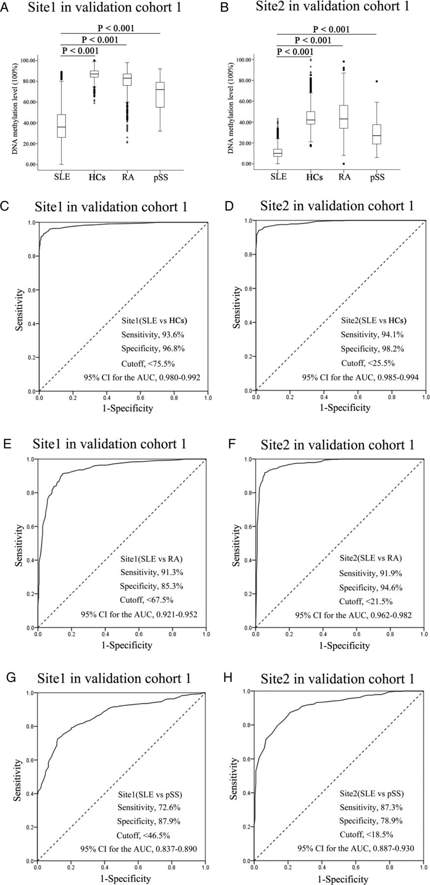
DNA methylation level of the IFI44L promoter and its sensitivity and specificity in the validation cohort 1 (Chinese cohort 2). (A) and (B) show the two CpG sites within the IFI44L promoter that are significantly hypomethylated in the blood of patients with systemic lupus erythematosus (SLE) compared with healthy controls (HCs), patients with rheumatoid arthritis (RA) and patients with primary Sjögren's syndrome (pSS) (p<0.001 for all comparisons). (C–H) show the receiver operating characteristic (ROC) curves of the methylation levels at Site1 and Site2 in patients with SLE compared with HCs (C and D), patients with RA (E and F) and patients with pSS (G and H), respectively.
The diagnostic value of IFI44L promoter methylation levels were also validated in an European-derived cohort consisting of 615 patients with SLE and 781 HCs. The methylation levels of Site1 and Site2 were significantly lower in SLE blood samples compared with HC samples (mean±SD; Site1: 57.04±23.42% and 85.88±11.08%; Site2: 16.97±13.60% and 41.10±14.76%; p<0.001 for both comparisons, figure 3A, B). The ROC curve analysis for SLE compared with HC showed the AUC value of Site1 was 0.872 (95% CI: 0.853 to 0.890) with sensitivity of 73.3% and specificity of 86.8% at a cut-off level of 76.9% (PPV: 81.29%, NPV: 80.50%) and the AUC value of Site2 was 0.879 (95% CI: 0.860 to 0.897) with sensitivity of 82.1% and specificity of 81.8% at a cut-off level of 30.3%, respectively (PPV: 78.05%, NPV: 85.20%) (figure 3C, D).

DNA methylation level of the IFI44L promoter and its sensitivity and specificity in the validation cohort 2 (European-derived cohort). (A) and (B) show two CpG sites within the IFI44L promoter that are significantly hypomethylated in blood samples from systemic lupus erythematosus (SLE) patients compared with healthy controls (HCs) (p<0.001 for both comparisons). (C) and (D) show the receiver operating characteristic (ROC) curves for the DNA methylation levels at Site1 and Site2 in patients with SLE compared with HCs.
Analysis of the association between IFI44L promoter methylation and clinical characteristics of SLE
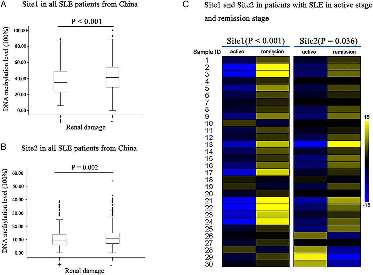
We divided all 906 Chinese patients with SLE into two groups: SLE with renal involvement (n=290) and SLE without renal involvement (n=616). We then determined if IFI44L methylation levels were different between the two SLE groups. We found that the methylation levels at Site1 and Site2 were significantly lower in patients with SLE with renal involvement compared with patients with SLE without renal involvement (mean±SD; Site1: 37.98±18.81% and 43.11±18.49%; p<0.001; figure 4A; Site2: 9.94±7.77% and 11.74±8.38%; p=0.002; figure 4B).

Analysis of the association between IFI44L promoter methylation level and renal involvement in systemic lupus erythematosus (SLE). (A) and (B) show that DNA methylation levels at Site1 and Site2 within the IFI44L promoter are significantly lower in patients with SLE with renal involvement compared with patients with SLE without renal involvement, respectively (p<0.001 and p=0.002). (C) show that DNA methylation levels of Site1 and Site2 within the IFI44L promoter are increased in 86.67% (26/30) and 66.67% (20/30) patients with SLE in clinical remission compared with patients in the active disease stage (p<0.001 and p=0.036, respectively).
Analysis of DNA methylation changes in the IFI44L promoter region was performed in 30 patients with SLE with paired blood samples obtained during active disease and remission. Among the 30 paired samples, 86.67% (26/30) and 66.67% (20/30) showed increased DNA methylation levels at Site1 and Site2 of the IFI44L promoter during remission compared with active disease (mean±SD; Site1: 48.10±18.45% and 35.57±17.52%; p<0.001; Site2: 14.77±9.33% and 10.40±9.08%; p=0.036; figure 4C).
IFI44L promoter methylation in PBMCs stimulated with IFN-α and serum of patients with SLE
IFI44L is a type-1 IFN induced gene. Previous studies have shown and repeatedly confirmed an increase in type-1 IFN levels in patients with SLE.27 To test the hypothesis that IFI44L hypomethylation is triggered by a high type-1 IFN response in patients with SLE, we measured IFI44L methylation levels in normal and SLE PBMCs with and without stimulation by IFN-α (1000 U/L) or 10% serum from patients with SLE. The pyrosequencing results showed that the methylation levels of Site1 and Site2 within the IFI44L promoter did not significantly change in PBMCs stimulated with IFN-α (figure 5A, B, online supplementary table S6 and figure S2A, B) or serum from patients with SLE (figure 5C, D, online supplementary table S6 and figure S2C, D) at 12 h, 24 h and 48 h in comparison with untreated controls.

DNA methylation level of the IFI44L promoter in peripheral blood mononuclear cells (PBMCs) of healthy persons treated with interferon α (IFN-α) or serum from patients with systemic lupus erythematosus (SLE). (A) and (B) show that there is no significant difference in DNA methylation levels of Site1 and Site2 within the IFI44L promoter between healthy PBMCs treated with IFN-α (1000 U/L) and untreated controls. (C) and (D) show that DNA methylation levels of Site1 and Site2 within the IFI44L promoter are not significantly different in healthy PBMCs treated with 10% serum from patients with SLE compared with untreated controls. All data represent the mean±SD of three independent experiments per group.
Discussion
The results of our study suggest that DNA methylation levels of IFI44L in the peripheral blood may be useful in the evaluation and diagnosis of SLE. The specificity and sensitivity of IFI44L promoter methylation in discriminating between SLE and HC were superior to those of currently available tests. Moreover, IFI44L promoter methylation levels can discriminate between SLE and other autoimmune diseases such as RA and pSS.
IFNs activate intracellular antimicrobial programmes and influence the development of innate and adaptive immune responses.28 Although the function of IFI44L is unknown, increased IFI44L expression is a component of the type-1 IFN response signature and also part of the cellular response to viral infections.29 An increased expression of type-1 IFN-regulated genes, termed IFN signature, has been well documented in the peripheral blood and tissues of patients with SLE.30 Interestingly, IFN-α therapy (given to patients with malignancy or hepatitis C) could induce autoimmunity.31 ,32 In this study, we suspected that DNA hypomethylation of two CpG sites within the IFI44L promoter might be due to the increased type-1 IFN in patients with SLE. However, we found that neither IFN-α nor serum derived from patients with SLE could downregulate DNA methylation level of IFI44L promoter in PBMCs from HCs and patients with SLE. This suggests that DNA hypomethylation of IFI44L observed in patients with SLE is not directly induced by type-1 IFN.
In this study, we found that the DNA methylation level of IFI44L promoter was significantly lower in patients with SLE with renal involvement than in patients with SLE without renal involvement, consistent with a recently reported more robust hypomethylation in IFN-regulated genes in patients with SLE with renal involvement.33 Although our results suggest that the DNA methylation level of IFI44L promoter may be associated with renal damage in patients with SLE, it does not seem to be a good biomarker for renal damage in SLE due to a low AUC value between SLE with renal damage and SLE without renal damage (AUC of Site1: 0.585, 95% CI: 0.544 to 0.625; AUC of Site2: 0.565, 95% CI: 0.525 to 0.605).
An excellent diagnostic biomarker should be highly sensitive and specific for a disease, and be feasible and reliably detected. DNA methylation changes are ideal for novel biomarker development in SLE. DNA methylation detection is performed using stable genomic DNA, as compared with RNA that is much more easily degradable.34 In addition, whole blood is easy to collect and pyrosequencing is reproducible and not operator-dependent.35 Indeed, it is more objective and already more standardised than serological tests such as ANA and other autoantibodies detections.
Acknowledgments
The authors thank Dr Xingli Li, Dr Hanqi Yin and Xin Huang for statistical consultation; and their collaborators who provided additional DNA samples for their studies, including Dr Enrique de Ramón Garrido, the Department of Internal Medicine, Hospital Universitario Carlos Haya, Málaga, Spain; Dr Norberto Ortego Centeno, Unidad de Enfermedades Autoinmunes Sistémicas, UGC Medicina Interna, Hospital Universitario San Cecilio, Granada, Spain; Dr Ma de los Angeles Aguirre, Servicio de Reumatología, Hospital Universitario Reina Sofía, Instituto Maimónides de Investigación Biomédica (IMIBIC), Córdoba, Spain; Dr Carlos Vasconcelos, Centro Hospitalar de Porto/Hospital San Antonio, Porto, Portugal and Dr Berta Martins da Silva, Laboratory of Imunogenetics and Autoimmunity and Neurosciences, Unidade Multidisciplinar Invest Biomed, Inst Ciencias Biomed Abel Salazar, Porto, Portugal.
References
Supplementary materials
Supplementary Data
This web only file has been produced by the BMJ Publishing Group from an electronic file supplied by the author(s) and has not been edited for content.
- Data supplement 1 - Online supplement
- Data supplement 2 - Online table S1
- Data supplement 3 - Online table S2
- Data supplement 4 - Online table S3
- Data supplement 5 - Online table S4
- Data supplement 6 - Online table S5
- Data supplement 7 - Online table S6
Footnotes
Handling editor Tore K Kvien
Contributors MZ, QL and AHS designed and performed the experiments, analysed the data and drafted the manuscript; YZ, BZ, MW, TJ, QT, YL, JJ, SL, YT, HW, PR, MdMAG, MJCP, ROC, CF-R, ER, RF, CC, MEA-R, ZX, JC, FL, GL, HZ, XL and YL performed sample collection and the experiments.
Funding This work was supported by the National Natural Science Foundation of China (No. 81220108017, No. 81430074, No. 81270024 and No. 81522038), the Hunan Provincial Natural Science Foundation of China (14JJ1009), the National Key Clinical Speciality Construction Project of National Health and Family Planning Commission of the People's Republic of China, and the National Institute of Allergy and Infectious Diseases, NIH under award number R01AI097134.
Competing interests None declared.
Ethics approval The ethical committee of the Second Xiangya Hospital of Central South University and the ethics committees and institutional review boards at all the authors’ institutions approved this study.
Provenance and peer review Not commissioned; externally peer reviewed.
Data sharing statement All data are available from the corresponding author upon request.