Article Text

Abstract
Background: The precise anatomical location of pathology associated with inflammatory back pain (IBP) in early spondyloarthropathy (SpA) remains unclear.
Objective: To use MRI to study the sacroiliac joint (SIJ) and lumbar spine (LS) and explore the relationship between sites and extent of inflammation and HLA-B27 status over 12 months.
Methods: 54 patients with IBP; median duration 24 weeks (54% HLA-B27 positive; median Bath Ankylosing Spondylitis Disease Activity Index (BASDAI) 5.65) and 22 control subjects (11 with mechanical back pain; 11 volunteers) were recruited and 63% (n = 34) were reassessed at 1 year. Fat saturation and T1-weighted MRI was performed with images being scored for active bone marrow oedema (BMO) lesions representative of inflammation.
Results: At baseline 46/54 (85%) patients had BMO (SIJs and LS) compared with 40% in the control group. The majority of affected patients had inflammation at the SIJ level (96% (n = 44); 23.5% (n = 12) LS) and 28.3% (n = 13) at both sites simultaneously. The SIJ activity score confirmed more severe inflammation (BMO grade 2 or 3: 52.2%) in the IBP group (controls = BMO grade 1: 100%; p<0.001). HLA-B27 was associated with both the severity (p = 0.009) and number of baseline SIJ lesions (p = 0.045) and with persistence (SIJ or LS) at 1 year (p = 0.02). 90% of reattenders fulfilled European Spondyloarthropathy Study Group criteria; 73.5% showed MRI inflammation despite clinical improvement (median BASDAI 5.65 to 3.05; p<0.009).
Conclusion: LS and SIJ involvement may occur simultaneously in very early SpA and may be differentiated from non-inflammatory back pain by the severity of MRI lesions. HLA-B27 is associated with both the severity of osteitis and its persistence.
Statistics from Altmetric.com
Inflammatory back pain (IBP), typically perceived in the sacroiliac region, is often the earliest harbinger for the development of axial spondyloarthropathy (SpA), including ankylosing spondylitis (AS).1 Radiographic confirmation of sacroiliitis is mandatory for the diagnosis of AS but diagnostic changes are not usually demonstrable until the disease has been present for several years.2 Magnetic resonance imaging (MRI) has emerged in the past decade as the preferred method for visualising the sacroiliac joint (SIJ).3 4 The MRI findings of sacroiliitis are now well established and bone marrow oedema (BMO), as determined by fat suppression methods, which is due to an osteitis is the most characteristic change.5 6
However, the relationship between IBP symptoms and BMO or osteitis has not been well defined. Previous MRI studies have looked at subjects with recent-onset IBP, although these cohorts had relatively long disease durations (>12 months)7 8 9 and some were preselected on the basis of HLA-B27.9 Furthermore, no controls were included and only the SIJ was studied as it has traditionally been assumed that IBP originates in the SIJ regions and only spreads upwards at a later stage. Therefore the diagnostic utility of MRI in early IBP is not clear.
The purpose of this study was to investigate the likeliest anatomical sites contributing to IBP of recent onset such as the SIJ and the lumbar spine (LS). Given the pivotal role of the HLA-B27 gene in the evolution of AS we were especially interested in looking at the relationship between HLA-B27 and the magnitude of osteitis and its link to surrogates of inflammation such as C-reactive protein (CRP). To undertake this we explored the evolution of SIJ and lumbar spinal lesions occurring in subjects with IBP using MRI at baseline and again at 1 year.
Patients and methods
Consecutive patients who attended the early arthritis and early inflammatory back pain clinics at a large teaching hospital between 1999 and 2001 were recruited after approval from the local ethics committee was obtained. All study subjects provided informed consent. Inclusion criteria included a clinical history of short duration of low IBP, normal or equivocal plain radiography of the LS and SIJs and a high clinical suspicion of a diagnosis of SpA. IBP was defined according to Calin’s criteria10 and considered as present if fulfilling at least four of the five following characteristics: insidious onset, age <40 years, associated with morning stiffness, improved by exercise and persistent for at least 3 months. Clinical assessments included a core set of validated outcome measures such as the visual analogue scores (VAS) for spinal pain during day and night, Bath Ankylosing Spondylitis Functional Index (BASFI)11 and Bath Ankylosing Spondylitis Disease Activity Index (BASDAI).12 CRP measurements and HLA-B27 typing were performed using standard techniques.
Magnetic resonance imaging
MRI of the SIJs and LS was performed in all patients (n = 54) using a Philips 1.5 T magnet, Gyroscan ASC NT (Philips Medical Systems, Best, the Netherlands). In addition, a control group comprising 22 subjects (11 patients with mechanical low back pain who did not fulfil Calin’s criteria for IBP and 11 healthy asymptomatic subjects with no present or past history of back pain or family history of spondylitis, inflammatory bowel disease or psoriasis) were also scanned. Subjects were examined in the supine position with a flat surface coil positioned under the sacrum. The following sequences were acquired: T1-weighted turbo spin echo (SE) and T2-weighted SPIR (spectral presaturation with inversion recovery, which represents a specific term for fat suppression) coronal oblique sequences plus T1 FFE SPIR post-gadolinium (Gd-DTPA) (vol 1.5 mm) of the sacroiliac joints and a T2 SPIR sagittal sequence of the lumbar spine. MR parameters were as previously described.13
MRI scoring
All images were reported for the presence of acute inflammatory changes using the activity score proposed within the Leeds Scoring System,13 14 which has been reported by OMERACT to be equivalent to other scoring systems for assessing inflammatory changes.15 In the SIJs, four quadrants (superior ilial, superior sacral, inferior ilial, inferior sacral) were assessed at both sides (right and left) in each patient for the presence of BMO in the subchondral region and the bone marrow cavity. BMO was defined as low signal intensity on T1 with enhancement after Gd administration, and/or high or intermediate bone marrow signal with irregular contour on a T2 SPIR image.16 17 Its presence or absence in the SIJ was recorded and severity was ranked on a semiquantitative scale (0, absent; grade 1, mild (<25%); grade 2, moderate (25–75%); grade 3, severe (>75%) of the quadrant affected). An overall score of inflammatory activity was calculated as the sum of the scores of BMO making the total maximal score for each abnormality 12 for each joint (24 for each patient).
In the LS, BMO lesions were defined as above and recorded as present or absent in the vertebral body and posterior elements, including spinous processes and facetal joints. Facet joint lesions were scored as affected when BMO was identified as covering the “perifacetal area” meaning the facet joint and pedicle. In addition, soft tissue lesions were identified as high signal on a T2-SPIR image in the paraspinal soft tissues and scored as being present or absent. A total count of lesions per spinal area for each subject was obtained.
Reliability of scoring system
To assess the reliability of the scoring system in early disease, training sessions were arranged between two experienced assessors (POC and DMG) who reviewed and scored MRI scans together to agree on definitions and discuss any discrepancies. After these sessions all baseline scans were scored independently and at different sittings by both scorers in order to calculate inter-reader and intrareader reliability values. The inter-reader reliability of this system for BMO findings in these early cohorts was (range) 0.67–0.85 for the SIJ and 0.80–0.93 for the LS. As this represents substantial agreement18 between the scorers only one reader scored the follow-up scans. For all scoring the assessors were unaware of the patients’ clinical characteristics and time sequence. Baseline and follow-up scans were scored paired at two different sittings in order to calculate intraobserver statistics. These agreements using ICC statistics varied between (range) 0.77 and 0.91 for individual quadrants in the SIJ and (range) between 0.74 and 1.00 for the LS regions.
The smallest detectable difference (SDD) was calculated as 1.96×(SDdifferences between repeat baseline scores). SDD ranged from 1 to 2 in individual SIJ quadrants, suggesting that scores of 1 are “probable” lesions whereas scores of ⩾2 are “definite” lesions. SDD did not exceed 1 in any of the LS regions. Where total lesion scores were produced, the SDDs were 4 and 2 for the SIJs and LS, respectively. To allow for a meaningful difference to assess longitudinal change within patients, the smallest detectable change (SDC) was then calculated (1.96×(SDdifferences between repeat change scores))/√2×√k where k is the number of measurements to be made for each patient in the main study (here k = 1). This agreement was very good at the spine (SDC = 1 for all individual sites, and for total score) and moderate for the SIJ (SDC range 1–2 for individual sites, total score SDC = 4). The reliability for assignment of grades of inflammation was substantial: κ = 0.73.
Patients in the inflammatory group were then invited to reattend for repeat MRI and clinical assessment at 12 months.
Statistical analysis
Normally distributed interval-level data were summarised using mean (SD). Non-normally distributed and ordinal variables were summarised using median (interquartile range). Nominal variables were presented as No (%). Differences between subgroups were assessed using Mann–Whitney U tests and χ2 tests with Wilcoxon signed-rank tests being applied to assess changes over time within the cohort. Means of the change of scores from their baseline values were obtained for the reattenders at follow-up. All analyses were carried out using SPSS, version 15.0.
Results
Fifty-four patients with IBP (62% male, mean age 30 years, 54% HLA-B27 positive, median disease duration from onset of back pain 24 weeks (range 2–260), median BASDAI (IQR) 5.65 (3.28–7.53)); 11 patients with mechanical back pain (63% male, mean age 34 years) and 11 healthy controls (54% male, mean age 35 years) were recruited. In the IBP group 82% of the patients were receiving non-steroidal anti-inflammatory drugs (NSAIDs) at the time of study entry with no patients receiving disease-modifying antirheumatic drugs (DMARDs) or steroid treatment at this stage.
Patients in the IBP group were invited for review and repeat imaging at a mean follow-up of 12.5 months (range 12–16). Twenty patients failed to reattend, one owing to pregnancy and the rest were either unable to be contacted or declined repeat scanning. Of the reattenders (63%; n = 34; 55.8% were HLA-B27 positive), 71% (n = 24) reported continuing symptoms of IBP. At follow-up, 56% (n = 19) were receiving NSAIDs and 17.6% (n = 6) were receiving DMARDs (sulfasalazine). All clinical outcomes at this point including the CRP showed an improvement in comparison with baseline (tables 1 and 2).
Demographics and clinical characteristics of the study patients who attended at both end points
Baseline characteristics of the patients who attended for follow-up and patients who defaulted and were not re-evaluated
Magnetic resonance imaging
Baseline MRI data were available in 76 subjects (54 in the IBP group and 22 in the control group). A positive scan, defined by at least one BMO lesion in at least one region in either the SIJ (score >0 = grade 1 or above) or LS was seen in 85% of patients (n = 46) in the IBP cohort with no lesions seen in the rest (n = 8). The majority of patients (n = 31) had lesions at the SIJ only with a minority (n = 2) having lesions solely at the LS and not the SIJ. A total of 13 patients (28.3%) had lesions occurring simultaneously at both sites (SIJ and LS),
Most of the affected patients (96%, n = 44/46) had lesions in any of the SIJ regions with the majority having bilateral involvement (61%, n = 27). Of the affected patients, 24 patients (55%) had a score ⩾2 in at least one SIJ region (table 3). The mean number of lesions per patient was 3.2 (0–8) with the right lower ilial region being most frequently affected (46.2% of patients, n = 25) and with more patients scoring grade 3 (severe) on this region. However, there was no significant difference between the summated activity scores in the ilial and sacral regions (median (IQR) ilial 1.00 (0.00–4.25), sacral 1.00 (0.00–3.25)).
Number (%) of subjects with moderate/severe bone marrow oedema lesions in the sacroiliac joint (SIJ; score⩾2) and/or lesions in the lumbar spine (>0) according to whether they had inflammatory back pain, mechanical back pain, or no pain symptoms
On the LS (n = 51 scans available), BMO lesions (total n = 31) were seen in 23.5% of patients (n = 12) with the vertebral body being most frequently affected (19.6%, 10 patients with a total of 18 lesions) followed by the spinous processes (7.8%, four patients with seven lesions), facetal joints (3.9%, two patients with four lesions) (fig 1) and soft tissue regions (interspinous ligament: 3.9%, two patients with two lesions).
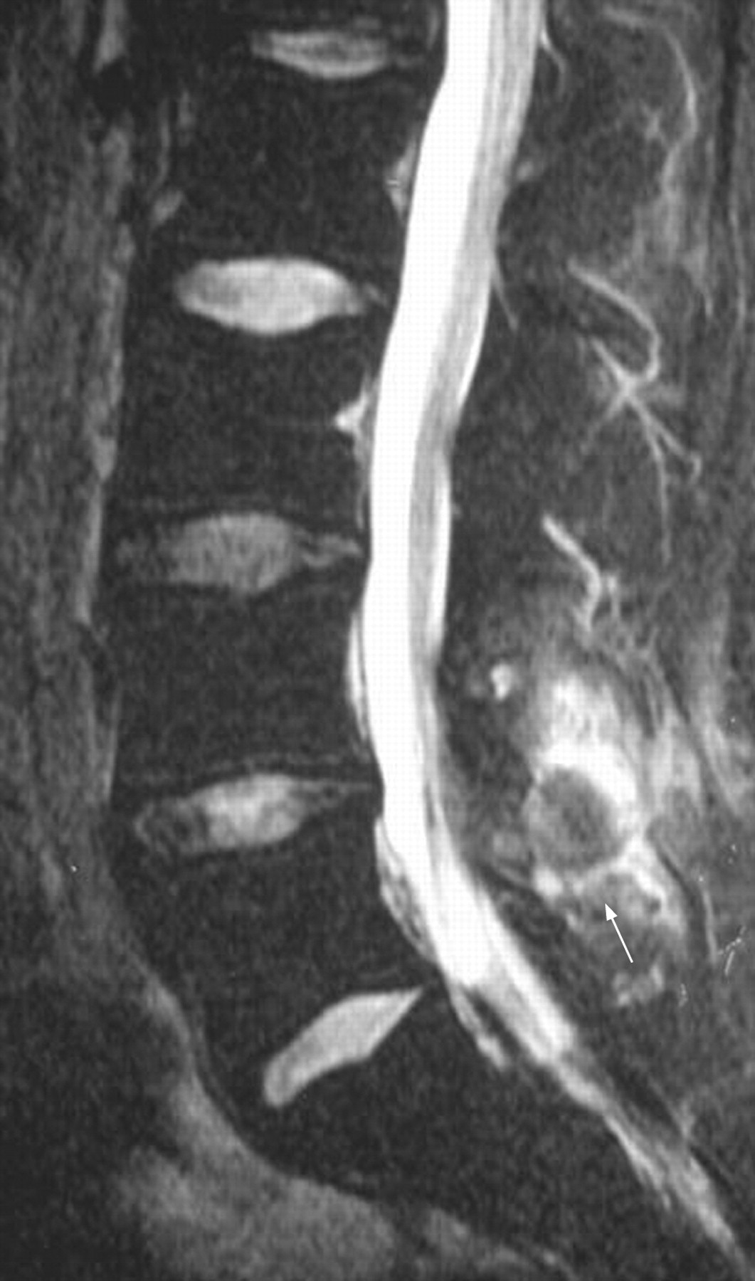
T2 fat-suppressed sagittal sequence of the lumbar spine of a 28-year-old HLA-B27-positive patient with a 24-week history of back pain and plantar fasciitis. The white arrow points towards oedema on the L4 facet area and associated soft tissue. The patient had a normal sacroiliac joint scan.
MRI scores at baseline did not differ between patients who were taking NSAIDs (n = 46, median (IQR) = 2.00 (1.00 to 7.00)); and those who were not (n = 8, median (IQR) = 2.00 (1.25 to 7.50); p = 0.952).
In the control groups BMO lesions were reported in 41% of subjects (9/22) with a total of 13 SIJ lesions reported in 27% (n = 6) and three spinal lesions (vertebral body = 2; interspinous ligament = 1) seen in 13.6% (n = 3) of subjects, respectively. Of these, two lesions were reported in the healthy volunteer group (one patient had one lesion at the SIJ and one patient had one vertebral body lesion) (fig 2). The total SIJ activity score differed significantly between the three groups (Kruskal–Wallis χ2 = 20.94, df = 2, p<0.001) with no scores above 1 (mild activity) being reported in the control group compared with scores of 2 (moderate activity) and 3 (severe activity) in 52.2% of the affected patient group as discussed above. Further analysis to assess the sensitivity and specificity of the grading of inflammation at the SIJs between the inflammatory and control groups showed that for a score >0 (grade 1) and for scores >1 (grade ⩾2) the sensitivities and specificities are 0.82 and 0.73 and 0.43 and 1.0, respectively.

(A) The coronal oblique sequence of the sacroiliac joint (SIJ) of a 35-year-old female patient with a short history of mechanical back pain showing grade 1 bone oedema in the inferior aspect of the sacral and ilial sides of the right SIJ (white arrow). (B) and (C) The SIJ and lumbar spine of two subjects from the healthy volunteer control group showing bone marrow oedema grade 1 on the right lower ilial quadrant and one lesion on the anterior inferior aspect of L1.
Correlation of MRI bone oedema and other parameters of disease activity
There were no significant correlations between the SIJ and LS BMO lesions and any measures of disease activity such as BASDAI, BASFI or CRP or with the disease duration in the IBP group. Likewise no association was seen between the VAS pain at night with the number and severity of lesions. Furthermore night pain did not differ between HLA-B27-positive and HLA-B27-negative patients. A normal MRI scan was seen in 14.8% of all patients at baseline (n = 8), of whom 50% were HLA-B27 positive.
Follow up MRI-SIJ
Follow-up MRI data were available for 34 patients for the SIJ and 33 for the LS. Considering total SIJ score and looking at change exceeding SDC, the majority of patients still had signs of inflammation as shown by the presence of BMO on MRI (73.5% of patients scored >0, of whom 19% scored grade ⩾2). However, there was a significant reduction in total SIJ activity score from baseline to follow-up (median baseline = 4.00 (IQR 1.00–9.75); median follow-up = 2.00 (0.00—4.00), Wilcoxon signed-rank test Z = −3.80, p<0.001), suggesting that although BMO lesions were present in the follow-up scans they were less active. The change in disease activity score was significantly correlated with the change of the VAS for night pain (Spearman’s rs = 0.497, p = 0.007). No patients showed deterioration in the activity or BMO score or developed new lesions at the SIJ in this time, although a change in location was noted in two patients who had BMO affecting mainly the right SIJ at baseline whereas the left side was more affected on follow-up (fig 3).

T2 FS coronal oblique sequences of the sacroiliac joint of an HLA-B27-positive 17-year-old man with a 15-week history of lower inflammatory back pain with alternate buttock pain and intermittent unilateral plantar fasciitis. (A) Baseline scan when symptoms were reported as more severe on the right side. (B) Follow-up scan at 12 months when his symptoms were reported to be predominantly on the left side. Treatment during this time consisted of non-steroidal anti-inflammatory drugs and physical therapy. The Bath Ankylosing Disease Activity Score was <2 at both visits. The white arrows point towards areas of severe bone marrow oedema, reflecting active inflammation.
Ten patients (five HLA-B27 positive) reported no symptoms of IBP on follow-up, of whom six were receiving regular NSAIDs and/or DMARDs (n = 2). Of this group (n = 10), eight patients had a positive MRI at follow-up with the majority showing grade 1 BMO (n = 6). Two patients who had grade 2 and 3 BMO (one patient (HLA-B27 positive) fulfilled criteria for AS and the other patient (HLA-B27 negative) for undifferentiated SpA).
Follow-up MRI- lumbar spine
In the LS, nine lesions were seen in 6/33 patients at baseline (three patients with one lesion each and three patients with two lesions each). At follow-up there was complete resolution of MRI lesions in five out of six patients with the other patient having one lesion unchanged. Another three patients (all HLA-B27 positive) with no lesions seen at baseline developed new lesions at follow-up (two patients with one lesion each and one with two lesions).
Relationship between HLA-B27 and MRI and clinical parameters of IBP
Both the number of affected SIJ regions and the total SIJ activity score were higher at baseline in HLA-B27-positive patients (Mann–Whitney test Z = −2.00, p = 0.045 and Z = −2.59, p = 0.009, respectively). Examination of the relationship between HLA-B27 and BMO, showed that persistence of BMO at follow-up was present in all the HLA-B27-positive patients who were affected at baseline (17/17) as opposed to 66.7% (8/12) of HLA-B27 negative with a positive baseline scan. This represents a clear association between the presence of HLA-B27 positivity and persistence of BMO lesions at either site at 1-year follow-up (Fisher exact test p = 0.02). No differences were seen in the LS between the number of active lesions and HLA-B27. Likewise, no significant association was seen between changes in LS scores and any measures of disease activity or CRP.
Discussion
This study used MRI in an inception IBP cohort to investigate the relationship between MRI-determined axial osteitis and HLA-B27. These findings show that although sacroiliitis is the most likely cause of early inflammatory low back pain, LS osteitis also occurs early and coexists with sacroiliitis. Furthermore, the severity of the osteitis as measured by its extent on MRI correlated with the presence of HLA-B27, but not with the severity of symptoms or clinical parameters including the CRP. Also the persistence of MRI-determined osteitis at 1 year was linked to HLA-B27, which further supports the assertion that the effect of this gene may be mediated in the bone19 20 rather than the synovium as previously thought.
This study reported IBP of very short duration and used MRI to report simultaneously on the SIJs and the LS, showing that inflammation occurs at both sites in a third of the patients. The traditionally held belief, based on radiographic findings, that disease starts at the SIJ21 and travels up the spine is therefore not supported by these results. In our study, MRI-determined acute LS skip lesions related to enthesitis were noted to coexist with sacroiliitis. In addition, it was found that spinal disease may exist in a subset of patients in the absence of SIJ involvement, suggesting that limited imaging of only one region may miss the diagnosis. However, our numbers are small and ideally the whole spine should have been studied. Recent reports have shown that the thoracic spine is commonly affected in early22 and established AS23 24 and it is known that neck pain can start early in the disease process. It is our subjective impression that MRI seems to correspond with the patients’ perception of location of pain, and that a clinically symptomatic area is likely to show abnormality when imaged, and indeed this has been recently shown by other authors.25
In this cohort MRI inflammation was present in 85% of symptomatic subjects at presentation, which is in accordance with some reports8 but not with others which showed a lower prevalence of 32%.26 There are a number of reasons for this. First our cohort is comparable to that of Puhakka et al8 as both are predominantly HLA-B27 positive (54% and 63%, respectively) male (62% and 58.5%, respectively) populations. In addition, IBP was defined according to Calin’s proposal, which requires four out of five criteria to be fulfilled. Therefore, it is likely that these patients were a highly selected group who belonged within the SpA spectrum of diseases. By contrast, the cohort reported by Heuft-Dorenbosch et al26 comprised a predominantly female-based population (62%) with a lower prevalence of HLA-B27 positivity (42%). Furthermore, the majority of patients (56%) fulfilled only three out of five IBP criteria. Nevertheless, it is interesting to note that the majority of active lesions at the SIJ were scored as mild and indeed no correlation was seen between presence of lesions at either the SIJ or LS with the severity of lesions at the SIJ and any parameters of disease activity such as CRP or BASDAI, which has also been noticed by other authors.11 27 This may reflect the small number of patients studied or the fact that disease was mild at this stage in the majority. Furthermore, most patients seen at follow-up had evidence of subclinical disease as shown by persistent MRI lesions despite clinical improvement This is relevant since methodologically these lesions represent true findings as they exceeded measurement error as represented by the SDC.28 These results support the long-held view that disease evolves slowly and can be symptomatically controlled in the short and medium term with conventional treatments such as NSAIDs. Longer, longitudinal follow-up of these patients will demonstrate whether MRI has prognostic implications in the diagnosis of early AS.
We have previously shown that HLA-B27 determines the severity of osteitis adjacent to the plantar fascia in early SpA.20 29 This study confirms these observations at the SIJ and is in accordance with other reports also in early disease.30 More importantly we have shown that HLA-B27 correlates with the persistence of disease as shown by MRI-determined BMO at 1-year follow-up. However, it must be emphasised that our findings of BMO do constitute a biomarker for active inflammation and cannot be considered the forerunner of the osseous changes identified by plain radiography as the relationship between inflammation and new bone formation in AS remains unclear31 and may indeed be uncoupled, as recently suggested.32 33 34
A shortcoming of our study was that our control group was heterogeneous. Although diffuse BMO at the SIJ has been reported in other conditions including trauma, infection and neoplasms, sacroiliitis is uncommon in the rest of the inflammatory arthropathies, such as rheumatoid arthritis and is even rarer in early disease so we did not have a non-SpA inflammatory group. However, our cohort did not have clinical features to suggest an alternative diagnosis to SpA and these findings confirm previous reports by other groups studying disease at different stages of evolution.4 Another important concern is the large number of patients defaulting at follow-up. Our impression is that despite methodological recall, patients felt that follow-up was no longer appropriate if symptoms had improved.
In summary this study evaluated the bone changes at the SIJ and LS in early IBP and showed that prominent BMO is common in early disease and occurs at both sites simultaneously. The severity and extent of this involvement correlates with HLA-B27 and MRI at 1 year shows persistent disease despite improvement in clinical outcomes. Further follow-up of this cohort will determine the predictive prognostic value of the BMO lesions.
Acknowledgments
We thank rheumatology colleagues in the Yorkshire region for patient referral and Professor Philip Conaghan for helpful comments on the manuscript.
REFERENCES
Footnotes
Competing interests None.
Ethics approval Ethics committee approval from Leeds Teaching Hospitals NHS Trust.
Linked Articles
- Miscellaneous