Article Text

Abstract
Objectives Spinal MRI is used to visualise lesions associated with axial spondyloarthritis (axSpA). The ASAS MRI working group (WG) updated and validated the definitions for inflammatory and structural spinal lesions in the context of axSpA.
Methods After review of the existing literature on all possible types of spinal MRI pathologies in axSpA, the group (12 rheumatologists and two radiologists) consented on the required revisions of lesion definitions compared with the existing nomenclature of 2012. In a second step, using 62 MRI scans from the ASAS classification cohort, the proposed definitions were validated in a multireader campaign by global (absent/present) and detailed (inflammation and structural) lesion assessment at the vertebral corner (VC), vertebral endplate, facet joints, transverse processes, lateral and posterior elements. Intraclass correlation coefficient (ICC) was used for analysis.
Results Revisions were made for both inflammatory (bone marrow oedema, BMO) and structural (fat, erosion, bone spur and ankylosis) lesions, including localisation (central vs lateral), extension (VC vs vertebral endplate) and extent (minimum number of slices needed), while new definitions were suggested for the type of lesion based on lesion maturity (VC monomorphic vs dimorphic). The most reliably assessed lesions were VC fat lesion and VC monomorphic BMO (ICC (mean of all 36 reader pairs/overall 9 readers): 0.91/0.92; 0.70/0.67, respectively.
Conclusions The lesion definitions for spinal MRI lesions compatible with SpA were updated by consensus and validated by a group of experienced readers. The lesions with the highest frequency and best reliability were fat and monomorphic inflammatory lesions at the VC.
- Spondylitis, Ankylosing
- Magnetic Resonance Imaging
- Inflammation
Data availability statement
Data are available upon reasonable request. Data are available upon reasonable request. All data relevant to the study are included in the article or uploaded as supplementary information.
Statistics from Altmetric.com
What is already known about this subject?
The usage of spinal MRI has increased and the understanding of how to interpret both inflammatory and structural spinal lesions in the context of clinical symptoms in axSpA and differential diagnoses has advanced significantly.
What does this study add?
The ASAS MRI working group revised the existing definitions of spinal MRI lesions where needed, to increase the understanding of their interpretation in the context of axSpA.
How might this impact on clinical practice or future developments?
These results may be used in ongoing efforts for re-evaluation of the definition of a ‘positive’ MRI of the spine in the context of axSpA versus non-SpA.
Introduction
Imaging of the sacroiliac joints (SIJ) and the spine are important tools for the correct recognition of axSpA.1 2 Radiographs have been used for decades for imaging of structural changes of both the SIJ and spine, which occur due to long-standing disease.3 With the introduction of MRI, the inflammatory nature of the disease was visualised and recognised as an objective manifestation of active disease.4 Imaging of the SIJ has been included in the classification criteria of axSpA as a key objective criterion, next to human leucocyte antigen B27.5 On the other hand, imaging of the spine, although more frequently performed in daily practice for identification of any cause of back pain, has not yet been included in these criteria. One main reason for this is that spinal changes on both MRI and radiography are considered to occur later in the course of the disease.6 Furthermore, although descriptions of spinal MRI lesions were published over 20 years ago and the first definition of a ‘positive MRI’ highly suggestive of axSpA was published by the ASAS/OMERACT MRI group almost a decade ago,2 the sensitivity and specificity of the definitions have been a matter of debate.7
Since that publication from 2012,2 the usage of MRI of the spine has increased and the understanding of how to define and interpret both inflammatory and structural spinal lesions in the context of clinical symptoms in axSpA and differential diagnoses has advanced significantly.7–11
Based on the progress with MRI interpretation in general and in the field of axSpA, in particular, the ASAS MRI working group (WG) decided to revise the existing axSpA-related definitions of these lesions where needed, in order to increase the understanding of spinal MRI interpretation in the disease. This publication presents the most recent update on this topic, not only dealing mainly with the updated definitions but also providing information on lesions where an update was not necessary, for completeness.
Methods
Preparatory steps
The ASAS MRI WG for the spine consisted of 12 rheumatologists and two radiologists. In a first step, a literature review on published descriptions on inflammatory and structural changes in the MRI of the spine was performed based on a PubMed search using the terms: spondyloarthritis, MRI, spine, inflammation, bone marrow oedema (BMO), erosion, sclerosis, fat metaplasia, ankylosis as a narrative review. The results were presented to the group in face-to-face meetings and discussed based on all findings reported in the assessed literature.
After presentation of the results to all ASAS members and incorporation of feedback by the ASAS assembly, the WG finalised the wording on the description of the lesions and agreed on a set of reference images for each lesion, in a virtual meeting. Furthermore, the group agreed on the study design and the study-specific interactive electronic case report form (eCRF).12 As a second step, the validation of the lesion descriptions was conducted in a multireader exercise with experts in the field of spinal MRI.
Image resources
Spinal MRIs were available from 62 patients, who were all participants in the ASAS classification cohort.
Usage of the eCRF for evaluation of spinal MRI lesions in the spine
Details on the eCRF platform have been published recently.13 More specifically, for the spinal application, information from the global impression was entered first as to whether the spine scan findings were consistent with axSpA, and whether there were degenerative lesions in the segments of the cervical and lumbar spine. After the global assessment, not only more detailed information on the presence of inflammatory but also postinflammatory structural changes for all possible lesion types were collected for each segment, taking into account the anatomical localisation of the lesions according to individual discovertebral units.
Statistical analysis
Descriptive statistics were performed for the frequencies of the different spinal lesions not only for each individual reader but also for the majority reader (≥5/9) and ≥2-reader data.
Reliability for the total number of the different types of lesions at the level of the patient was assessed by intraclass correlation coefficient (ICC 2.1 (two-way random effects, absolute agreement, single rater/measurement MedCalc V.12.6)).
Results
Overarching considerations and general consensus on MRI lesions in the spine
After review and discussion of the literature, the group decided on specific overarching principles for reviewing spinal MRIs in the context of axSpA (box 1). A major difference from previous reports is the subdivision of lesion definitions according to central and lateral slice anatomical locations in the thoracic and lumbar spinal segments on a sagittal MRI. The central sagittal slices include those that visualise the spinal canal. The pedicle may be partially seen but is not continuous between the vertebral body and posterior elements. The lateral sagittal slices are located lateral to the spinal canal. These slices do not include the visualisation of the spinal canal, and either the pedicle must be continuous between vertebral body and posterior elements, or the slice is lateral to the pedicle.
Assessments in spondyloarthritis International Society MRI Working Group consensus definitions for MRI lesions in the spine of patients with axial spondyloarthritis
A. Overarching principles
All definitions of inflammatory lesions relate to their appearance on the water-sensitive sagittal T2-weighted fat-suppressed (T2FS) or sagittal short tau inversion recovery (STIR) images in the sagittal orientation. In both, an increased water content is seen as an increased signal intensity.
All definitions of structural lesions relate to their appearance on the fat-sensitive sagittal T1-weighted (T1W) MR images in the sagittal orientation.
The appearance of all lesions must be highly suggestive of spondyloarthritis.
The term ‘increased signal in bone marrow’ refers to a signal intensity higher than the ‘normal bone marrow signal’. The bone marrow signal in the centre of the vertebra, if normal, constitutes the reference for designation of normal signal or, alternatively, in the centre of the closest available normal vertebra.
Based on anatomical location, the images of the thoracic and lumbar spine on a sagittal MRI scan may be divided into ‘central’ and ‘lateral’ slices, which are defined as follows:
Central sagittal slices: the sagittal slices that include the spinal canal. The pedicle may be partially seen but is not continuous between the vertebral body and posterior elements.
Lateral sagittal slices: the sagittal slices that are located lateral to the spinal canal. These slices do not include the spinal canal, and either the pedicle must be continuous between vertebral body and posterior elements or the slice is lateral to the pedicle.
The maximum sagittal slice thickness is 4 mm.
B. MRI spine lesions indicating activity
These observations are made on MRI sequences that are sensitive for the detection of disease activity such as T2-weighted sequences with fat suppression that are sensitive for free water such as STIR or T2FS or T1W sequences with fat suppression that are sensitive for contrast enhancement such as T1FS post-Gd.
Inflammatory lesions
These can be divided into:
Vertebral body inflammatory lesion:
Vertebral corner inflammatory lesion (also known as anterior/posterior spondylitis): increased signal in bone marrow in a water-sensitive sequence at the vertebral corner, in at least two continuous sagittal slices. These can be subdivided into anterior and posterior vertebral corner lesions. There are two types:
Regular corner lesion or type A: increased signal extends to the corners.
‘Irregular’ corner lesion or type B: increased signal does not cover the whole corner but extends to both the endplate and the anterior/posterior border of the vertebra. Notes: in the corner itself often an erosion, sclerosis or a fat lesion is present. If inflammation (bone marrow oedema) is only visible on one slice, a type B lesion may be scored on that single slice, provided the structural component of the lesion is visible in at least two slices. In all other circumstances, the appearance of the type B lesion must be present on two or more slices.
Vertebral endplate inflammatory lesion including the intervertebral disc (also known as aseptic spondylodiscitis): increased signal in bone marrow in a water-sensitive sequence adjacent to the vertebral endplate that involves the vertebral endplate but not the vertebral corner.
Thoracic lateral inflammatory lesion (a lateral inflammatory lesion located in the posterior part of the slice is also known as arthritis of the costovertebral joints) (applies to thoracic spine only). Increased signal in bone marrow on STIR/T2FS sequence adjacent to the endplate in at least one lateral sagittal slice.
Vertebral inflammatory lesions not involving the vertebral body
Facet joint inflammatory lesion (also known as facet joint arthritis): increased signal in bone marrow in at least one sagittal slice in a water-sensitive sequence in at least one facet of a facet joint.
Posterior element inflammatory lesion (including enthesitis of spinal ligaments and costotransverse joint inflammation): increased signal in bone marrow in at least one sagittal slice in a water-sensitive sequence in one of the other posterior elements at which there are ligamentous or muscular attachments, or at the costotransverse joint (the pedicle, facet processes and pars interarticularis are excluded).
C. MRI spine lesions indicating structural change
These observations are made on MRI sequences that are sensitive for the detection of structural change. Most of the observations can only be seen clearly on sequences sensitive for fat signal, specifically T1W spin echo without fat suppression.
Bone erosion: full-thickness loss of the dark appearance of cortical bone and loss of normal bright appearance of adjacent bone marrow on T1w images in at least one sagittal slice. Only erosions involving the vertebral corners are assessed. Erosions can be subdivided into anterior and posterior corner erosions.
Focal fat lesion: focal increased signal in bone marrow on T1w images in at least two sagittal slices. Only fat lesions involving the vertebral corners are assessed. Fat lesions can be subdivided into anterior and posterior corner fat lesions.
Bone spur in the direction of the anterior or posterior longitudinal ligament (also known as syndesmophytes): Bright signal on T1w images extending vertically from the vertebral corner towards the adjacent vertebral corner, seen in at least one sagittal slice. Bone spurs do not reach the adjacent vertebra and can be subdivided into anterior and posterior corner bone spurs (located in anterior and posterior corners, respectively). Notes: bone spurs should not be scored as related to SpA (ie, syndesmophytes) in the presence of disc degeneration.
Ankylosis: bright signal on T1w images extending from a vertebra and being continuous with the adjacent vertebra. This can be divided into:
Vertebral corner ankylosis: ankylosis involving the vertebral corner, in at least one sagittal slice. This can be subdivided into anterior and posterior corner ankylosis (located in anterior and posterior corners, respectively).
Vertebral endplate ankylosis: ankylosis involving the endplate, but neither the anterior nor the posterior vertebral corner.
Facet joint ankylosis: ankylosis of a facet joint.
Lesion definitions
Active lesions
These are divided into lesions involving or not involving the vertebral bodies (figures 1 and 2). Definitions of lesions are provided in box 1. For vertebral body lesions, active inflammatory lesions are considered as present if BMO is located at the VCs or endplates. The terms BMO and osteitis are considered to be equivalent. Similar to the previous publication,2 it was agreed (based on expert opinion on the morphology of the lesion and taking into account possible image artefacts) that each of the inflammatory lesions described has to be visible in at least two or more consecutive sagittal slices. This rule does not apply to: (1) lateral slices and facet or posterior element lesions, which can be considered present on a single slice and (2) dimorphic lesions (explanation see below), which may be considered present on a single slice, provided their structural component is visible in at least two slices, while in all other circumstances, their appearance must be present on two or more slices (box 1).

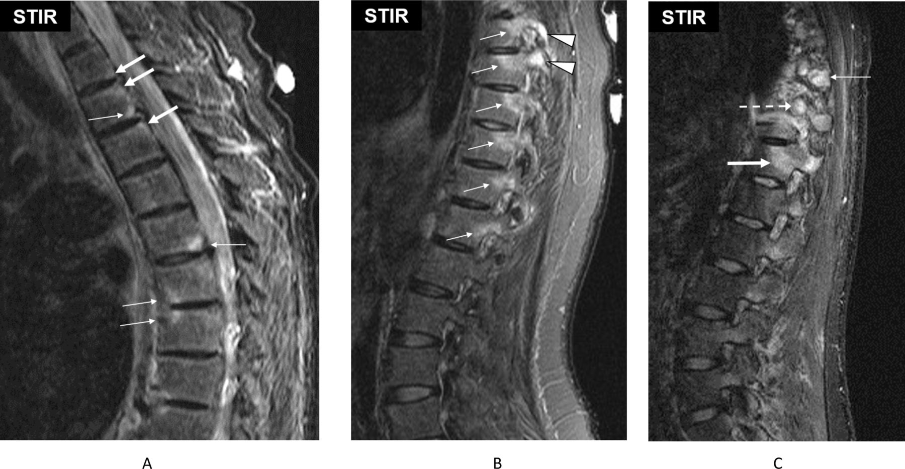
Signs of active changes in the lumbar spine of two patients with axial spondyloarthritis: (A) Anterior and posterior spondylitis with monomorphic (thick arrows) and dimorphic (thin arrows) lesions. (B) Thoracic lateral inflammatory lesions (arrows). Facet joint lesions (arrowheads). (C) Lateral inflammatory lesion (thick arrow), inflammation in the rib (dashed arrow), inflammation in the transverse process (thin arrow). Provided courtesy of the Canada-Denmark MRI Working Group (Lambert et al 9).

Signs of active changes in the lumbar spine of a patient with axial spondyloarthritis: Examples of anterior vertebral corner inflammatory lesions are prominent in the thoracic spine and more subtle in the lumbar spine (arrows), with an area of spondylodiscitis at L1–L2 (asterisk). Provided courtesy of the Canada-Denmark MRI Working Group (Lambert et al 9).
Inflammatory lesions are specified for the different anatomical localisations, such as the VCs (anterior/posterior corner inflammatory lesions, also known as anterior or posterior spondylitis, figures 1 and 2). Inflammatory activity at the VCs is subdivided into two types (box 1, figure 1): in a monomorphic corner lesion, the increased inflammatory signal extends to the cortex of the corner. In a dimorphic corner lesion, the increased inflammatory signal does not extend to the cortex of the corner but does extend to both the endplate and the anterior/posterior border of the vertebra. At the corner itself, there may be an erosion, sclerosis or a fat lesion.
Additional lesions affecting the vertebral bodies are the vertebral endplate inflammatory lesion (figure 2), and the thoracic lateral inflammatory lesion (a lesion located posteriorly in a lateral slice is also known as arthritis of the costovertebral joints) (figure 1), which is only recorded for the thoracic spine (box 1).
Inflammatory lesions that are not involving the vertebral body include the facet joint inflammatory lesion (also known as facet joint arthritis) and the posterior element inflammatory lesion (including enthesitis of spinal ligaments) (figure 1), but excluding the pedicle, facet processes and pars interarticularis.
Structural lesions
Structural lesions refer to the clear presence of typical findings such as fat lesions, erosions, sclerosis, syndesmophytes or ankylosis located at the vertebrae (box 1). All types of structural lesions may present solely or accompanied/surrounded by BMO (box 1, figure 2). Most of the observations can only be seen clearly on sequences sensitive for fat signal, specifically T1-weighted (T1W) spin echo without fat suppression. An update on structural lesions was felt necessary for erosions, syndesmophytes and ankylosis, while the definition of fat lesions remained unchanged.2
Erosions
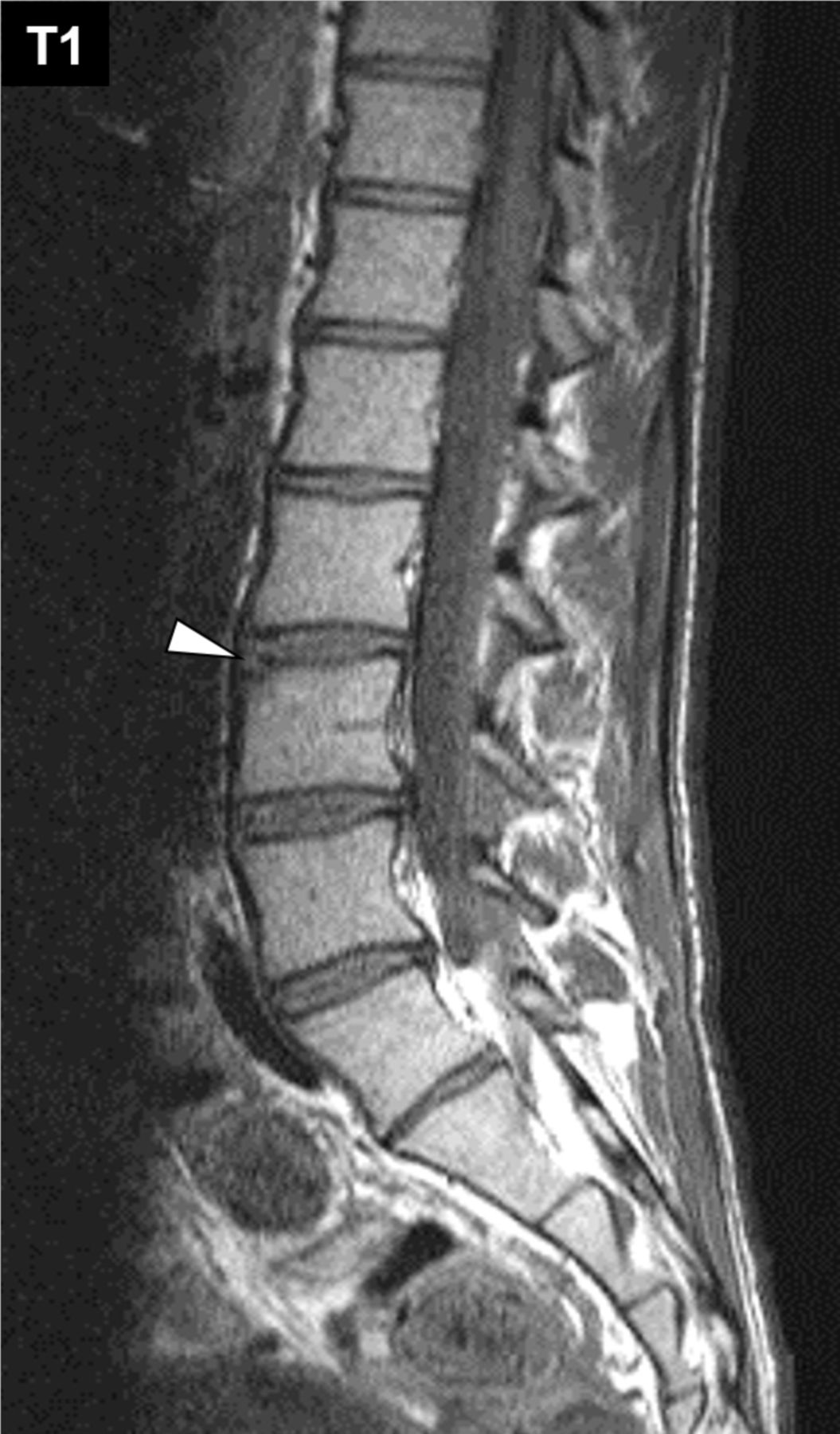
Erosion is defined as full-thickness loss of the dark appearance of cortical bone and loss of the normal bright appearance of adjacent bone marrow on T1w images in at least one sagittal slice. Only erosions involving the VCs are assessed and these can be subdivided into anterior and posterior corner erosions. Although erosions may affect vertebral endplates, these are not considered sufficiently specific for axSpA to warrant inclusion in the list of axSpA-associated lesions (box 1, figure 3).

Signs of structural change in the lumbar spine of patients with axial spondyloarthritis: vertebral corner erosion (arrowhead) in the lumbar spine. Provided courtesy of the Canada-Denmark MRI Working Group (Østergaard et al 8).
Bone spurs/syndesmophytes
This description includes the bony outgrowths at the anterior, posterior or lateral corners of vertebral bodies that do not reach the adjacent vertebra. Their origin of growth is at the attachment site of the annulus fibrosus (box 1, figure 4). These are defined as bright signal on T1w images extending vertically from the VC towards the adjacent VC, seen in at least one sagittal slice. Bone spurs should not be scored as related to SpA (ie, syndesmophytes) in the presence of disc degeneration.

Signs of structural changes in the lumbar spine of patients with axial spondyloarthritis: bone growth with origin at the attachment site of the annulus fibrosus of different size (arrows) including a segment with bridging syndesmophytes/ankylosis (arrowhead) or within the intervertebral disc (asterisk). Provided courtesy of the Canada-Denmark MRI Working Group. (Østergaard et al 8). STIR, sagittal short tau inversion recovery.
Ankylosis
This finding represents the bony fusion at the attachment sites of the annulus fibrosus (bridging syndesmophytes) and/or bony fusion across the intervertebral disc (box 1, figure 4) or across apophyseal joints (figure 5) or costovertebral joints (figure 5). Vertebral is defined as bright signal on T1w images extending from a vertebra and being continuous with the adjacent vertebra on at least a single sagittal slice.

Signs of structural changes in the thoracic spine of patients with axial spondyloarthritis: Facet joint (asterisks) and posterior intervertebral ankylosis (arrows). Image provided courtesy of Canada-Denmark MRI Working Group (Østergaard et al 8). STIR, sagittal short tau inversion recovery.
Fat lesions
These lesions are defined as focal increased signal in bone marrow on T1w images in at least two consecutive sagittal slices. Similar to bone marrow oedema, it was agreed (based on expert opinion on the morphology of the lesion and taking into account possible image artefacts) that each of the lesions described has to be visible in at least two consecutive sagittal slices. Only fat lesions involving the VCs are assessed and these can be subdivided into anterior and posterior corner fat lesions (box 1, figure 6). Although fat lesions may occur adjacent to vertebral endplates, these are not considered sufficiently specific for axSpA to warrant inclusion in the list of axSpA-associated lesions.

Signs of structural changes in the lumbar spine of a patient with axial spondyloarthritis: fat lesions in the anterior and posterior vertebral corners. Provided courtesy of the Canada-Denmark MRI Working Group (Østergaard et al 8). STIR, sagittal short tau inversion recovery.
Sclerosis
Similar to fat lesions, sclerosis also represents a stage of local tissue transformation due to chronic occurrence of inflammation and is seen at the area of the VCs. It is defined as low signal at a VC on all MRI sequences, but the lesion is infrequent, the MRI appearance is hard to interpret, and it is not considered sufficiently specific for axSpA to warrant inclusion in the list of axSpA-associated lesions.
Degenerative lesion of the intervertebral disc
In parallel to the typical lesions suggestive of axSpA, degenerative disc lesions may occur. The agreed definition for such lesions is the presence of irregularity of both vertebral endplates and reduction of intervertebral disc height by ≥50%. The presence of any of the inflammatory and structural lesions described above at the site of a degenerating disc lesion should not be regarded as indicative of axSpA.
Frequency of pathologic lesions and reliability of spinal lesion identification
Based on the agreement of either ≥2/9 or ≥5/9 readers, the most frequently observed lesions were the monomorphic BMO corner lesion and the corner fat lesion (table 1).
Frequency of the different lesion types and number of lesions based on the agreement of ≥2/9 and the majority (≥5/9) readers
For the reliability of lesion assessment, agreement of all 36 reader pairs and for all 9 readers for fat lesions was (0.88) (0.08) and 0.87 (0.82–0.91), respectively, and for BMO 0.69 (0.12) and 0.68 (0.58–0.77), respectively (table 2).
Single measures intraclass correlation coefficient for absolute agreement of all reader pairs and all nine readers for the different spinal lesions
On the level of the patient, agreement of reader pairs (mean ICC (SD)) and for all nine readers (ICC (95% CI) for fat lesions at VCs was 0.91 (0.06) and 0.92 (0.89–0.94), respectively. In comparison, agreement for monomorphic BMO lesions was 0.70 (0.10) and 0.67 (0.60–0.76), respectively, while agreement for dimorphic BMO lesions was much lower (table 2).
Discussion
The main aim of this publication was the update of the definitions of spinal lesions related to axSpA, by the ASAS MRI WG.
The characteristic MRI findings in the spine of patients with axSpA were described over 20 years ago and definitions of spinal MRI lesions were first published in 2009.8 9 The first definition of a positive spinal MRI for inflammation and structural changes was made by ASAS in 2012,2 arising from the evidence that inflammation in the spine may also occur in parallel and also before or even without inflammation in the SIJ. In addition to that consensus statement, several other studies have been published in the meantime7 10 11 partly confirming and partly questioning the former definitions, and identifying the potential for misleading interpretation if imaging is assessed without the clinical context.14 15 One reason for this was the knowledge gained on the relationship between lesions found on MRI and subsequent structural progression on conventional radiographs.16–18 Another reason was the practical aspect arising from the technical improvement of MR image quality over time,19 which provides improved insight into the lesions found in patients with axSpA in comparison to patients with chronic back pain without a diagnosis of axSpA. One step towards a more precise terminology and validation of the SpA-related lesions was the recently published update of definitions and validation of the MRI lesions of the SIJ for patients with SpA by the ASAS MRI WG.13 With the present paper, our group has completed a set of updated definitions for all the relevant axial MRI lesions in SpA.
The present analysis is based on the evaluation of MRIs from the ASAS classification cohort by international experts, who are all full ASAS members with extensive experience in the reading of such images. The group of experts agreed, based on20 the best possible wording for the definition of all known types of spinal lesions for activity (bone marrow oedema with and without concomitant structural lesions) and structural (fat, erosions, sclerosis, ankylosis) findings. The wording of definition of localisation (central, or lateral, in the vertebral body or in the posterior elements, that is, outside the vertebral bodies), extension (VC vs vertebral endplate) and extent (minimum number of slices needed for identifications of lesions) of these lesions was also agreed on. Overall, in comparison to the previous publication,2 updated definitions are now provided for both the active and the structural spinal lesion types. For the active lesions, the anatomic localisation was considered, now including not only the VC with the different types of appearance (monomorphic and dimorphic) but also the lesions located at the endplate, the lateral vertebral region, the facet joints and the posterior elements. For the structural lesions, an update was felt necessary for erosions, syndesmophytes and ankylosis, leaving the definition of fat lesions unchanged.2 Finally, for bone spurs, which occur in the longitudinal ligaments and the tissue intimately attached to them, it is well known that they are less well detected on conventional MRI than on conventional radiographs or CT. In addition, especially for the identification on MRI in contrast to the identification on conventional radiographs or CT, the presence of syndesmophytes may not necessarily be interpreted as a reliable sign of spondyloarthritis.20
The statistical analysis of the MRI evaluation confirmed that not only inflammatory but also structural lesions are frequently observed in the spine of patients with axSpA. These lesions seem to be most frequently located at the VC area, consisting either of fat or bone marrow oedema. In addition, the VC inflammatory lesion type that was clearly more frequently and more reliably observed was the monomorphic BMO lesion, where the inflammatory signal extends to the VC, in contrast to the dimorphic BMO lesions, where the signal does not cover the whole corner but extends to both the endplate and anterior or posterior border of the vertebra. This is an important finding, since these lesions especially have been reported to be associated with the highest risk for radiographic progression in follow-up examinations of patients with radiographic axSpA.16 17 The lower frequency of the dimorphic BMO lesion in our analysis compared with previous reports is likely because the ASAS classification cohort is a cohort of patients at an early stage of their disease.21 In addition, it also needs to be taken into account that dimorphic lesions may also be less reliably detected because of their complex morphology and the fact that the short tau inversion recovery signal is often of low intensity, making it difficult to detect these lesions, which often reflect resolving inflammation. Fat lesions and monomorphic BMO lesions were the lesion types with the highest reliability of detection between experts. This result was independent of the number of experts who had to agree on the presence of these lesions, suggesting a possibly similar accuracy of their detection in daily practice settings.
Interestingly, beyond fat and monomorphic BMO lesions, all other lesions assessed were observed far less frequently, as expected for an inception cohort of patients referred with undiagnosed back pain and suspicion of axSpA. The lower degree of reliability at least partly reflects this lower frequency of detection. This was especially true for lesions in the more posterior parts of the spine (posterior VCs and facet joints). The posterior parts of the spine have been reported to be more frequently affected by inflammatory lesions in patients diagnosed at a young age.22 An explanation for the differences between these findings and the results reported here may be because, despite the early stage of their disease in the ASAS classification cohort, the mean age of these patients was still not different from other studies with axSpA patients. Such data are also consistent with studies that showed no differences in the mean age at diagnosis independent of the stage of radiographic axSpA.22
In summary, this publication provides a consensus-based update of the definitions for spinal MRI lesions of patients referred with undiagnosed back pain and with clinical suspicion of axSpA. The most frequent and reliably detected lesions were fat lesions and monomorphic BMO VC lesions, while posterior elements were much less evident in this cohort of patients with undiagnosed back pain referred to a rheumatologist. These results not only confirm the observation that BMO and fat lesions are important in the identification of pathologic findings when evaluating spinal MRIs but may also be used in ongoing efforts for re-evaluation of the definition of a ‘positive’ MRI of the spine in the context of axSpA versus non-SpA.
Data availability statement
Data are available upon reasonable request. Data are available upon reasonable request. All data relevant to the study are included in the article or uploaded as supplementary information.
Ethics statements
Patient consent for publication
Ethics approval
Not applicable.
Acknowledgments
We thank Joel Paschke of CARE Arthritis for development of the web-based ASAS MRImagine eCRF and scoring interface, for processing of MR images for reading online and for image data cleaning and processing. We thank Matthew Maksymowych and Mikhail Protopopov for processing of MR images for reading online.
References
Footnotes
Handling editor Josef S Smolen
Twitter @pedrommcmachado
Correction notice This article has been corrected since it published Online First. The last author's name has been corrected.
Contributors XB, MO, RGWL, IE, PMM, SJP, UW MdH read the MRIs. XB and WM drafted the manuscript. All authors contributed to the design of the study, review of study data, drafting of the final manuscript and agreed to the final version of the manuscript. WM is the guarantor.
Funding The authors have not declared a specific grant for this research from any funding agency in the public, commercial or not-for-profit sectors.
Competing interests XB: Consulting fees: Abbvie, BMS, Eli-Lilly, Galapagos, Janssen, MSD, Novartis, Pfizer, Roche, Sandoz, Sanofi, UCB, Payment or honoraria for lectures: Abbvie, BMS, Eli-Lilly, Galapagos, Janssen, MSD, Novartis, Pfizer, Roche, Sandoz, Sanofi, UCB, Advisory Board: Abbvie, Eli-Lilly, Galapagos, Janssen, MSD, Novartis, Pfizer, UCB. Leadership role: Editorial Board Member of Annals of Rheumatic Diseases, ASAS President. MØ: Consulting fees: Abbvie, BMS, Celgene, Eli-Lilly, Galapagos, Gilead, Hospira, Janssen, Merck, Novartis, Pfizer, UCB. Payment or honoraria for lectures: Abbvie, BMS, Eli-Lilly, Galapagos, Gilead, Janssen, Merck, Novartis, Pfizer, UCB. RGL: Consulting fees: Calyx, CARE Arthritis Ltd., Image Analysis Ltd. IE: Payment or honoraria for lectures: Abbvie, Novartis. PMM: consulting fees: Abbvie, BMS, Celgene, Eli Lilly, Galapagos, Janssen, MSD, Novartis, Orphazyme, Pfizer, Roche and UCB. Other financial or non-financial interests: Supported by the National Institute for Health Research (NIHR) University College London Hospitals (UCLH) Biomedical Research Centre (BRC). SJP: Consulting fees: Abbvie, UCB, Novartis (paid to the institution). Payment or honoraria for lectures: MSD, Pfizer, Abbvie, UCB, Novartis (paid to private account and to institution). Support for attending meetings: MSD, Pfizer, Abbvie, Novartis, Boehringer Ingelheim. Participation to Advisory Committee: Novartis, UCB, Abbvie. MdH: Grants: FWRO/FRSR, Leadership role: EDULAR Advocacy Committee. JS: Consulting fees: AbbVie, Novartis, UCB. Payment or honoraria for lectures: Abbvie, Merck, Novartis. Participate on Advisory Board: Abbvie, DP: Grants: AbbVie, Eli Lilly, MSD, Novartis, Pfizer. Consulting fees: AbbVie, Biocad, Eli Lilly, Gilead, GlaxoSmithKline, Janssen, MSD, Moonlake, Novartis, Pfizer, Samsung Bioepis, UCB. Payment or honoraria for lectures: AbbVie, Bristol-Myers Squibb, Eli Lilly, Janssen, MSD, Medscape, Novartis, Peervoice, Pfizer, and UCB. Participation on Advisory Board: AbbVie, Biocad, Eli Lilly, Gilead, GlaxoSmithKline, Janssen, MSD, Moonlake, Novartis, Pfizer, Samsung Bioepis, and UCB. MR: Consulting fees: Abbvie, Eli Lilly, Novartis, Pfizer, UCB. Payment or honoraria for lectures: Abbvie, BMS, Eli Lilly, Janssen, MSD, Novartis, Pfizer, Roche, UCB. Support for attending meetings: Galapagos, Janssen, Novartis, Abbvie. DvdH: Consulting fees: AbbVie, Bayer, BMS, Cyxone, Eisai, Galapagos, Gilead, Glaxo-Smith-Kline, Janssen, Lilly, Novartis, Pfizer, UCB. Director of Imaging Rheumatology bv. Leadership role: Associate Editor of Annals of Rheumatic Diseases, Editorial Board Member Journal of Rheumatology, Advisory Committee RMD Open. RL: Grants: Research grants from Galapagos, AbbVie, Novartis, UCB (paid to institution). Consulting fees: AbbVie, Bristol-Myers Squibb, Celgene, Eli Lilly, Galapagos, Gilead, GlaxoSmithKline, Janssen, Merck, Novartis, Pfizer, Roche, UCB (paid to own company). Payment or honoraria for lectures: AbbVie, Bristol-Myers Squibb, Celgene, Eli Lilly, Galapagos, Gilead, GlaxoSmithKline, Janssen, Merck, Novartis, Pfizer, Roche, UCB (paid to own company). Participation on Advisory Board: UCB, AbbVie, Galapagos, Gilead, Eli Lilly, Jansen, Novartis, Pfizer (paid to own company). Data safety Monitoring Board: UCB (no compensation). Director of Rheumatology Consultancy. Partner of ‘Reumatologie Maatschap Sittard/Heerlen’ (paid to own company). Leadership role: Council Member and EULAR's chair of quality of care. Member of Editorial Board of Annals of Rheumatic Diseases. WPM: Grants: Abbvie, Galapagos, Novartis, Pfizer, UCB. Consulting fees: Abbvie, Boehringer Ingelheim, Celgene, Lilly, Novartis, Pfizer, UCB. Payment or honoraria for lectures: Abbvie, Janssen, Lilly, Novartis, Pfizer, UCB. Leadership role: SPARTAN Board of directors. Chief Medical Officer of CARE Arthritis Ltd.
Patient and public involvement Patients and/or the public were not involved in the design, or conduct, or reporting, or dissemination plans of this research.
Provenance and peer review Not commissioned; externally peer reviewed.