Article Text

Abstract
Background Previously our group described in X-ray studies bone alterations in NOD mice affected with Sjögren's syndrome (SS), characterized by the radiopacity decrease of the tibia cortical layers, compared to controls. These results would suggested that NOD mice with SS could developed disorders in tibial histological architecture. In the current literature, have not been described accurately bone disorders related to the SS.
Objectives To assess histologically the tibial metaphysis area of non-obese diabetic mice with SS, compared to controls NOD without SS.
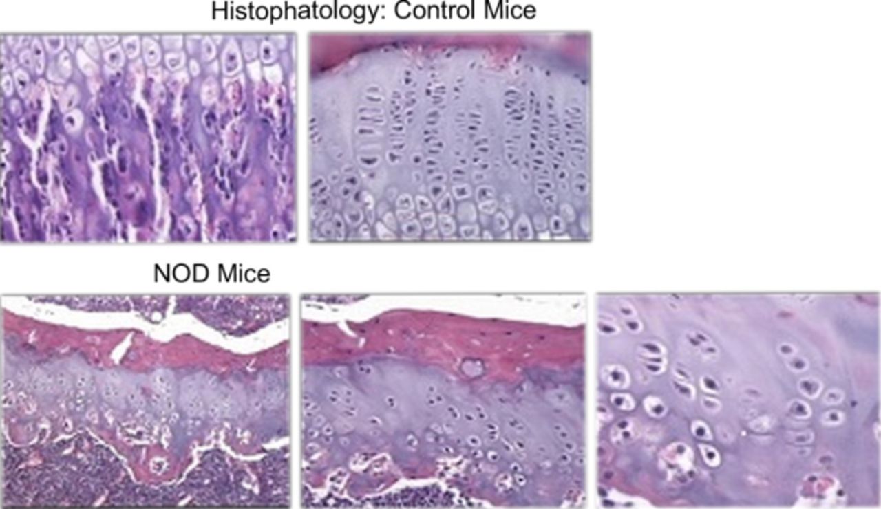
Methods We used 10 NOD female mice with SS (group SS) and 10 controls NOD without SS (group C, control). They were cared in the animal facilities of the Faculty of chemistry of the UNC. At 4 months of age (weight 80 g ± 10) mice were anesthetized with ketamine-xylazine, both tibias were extracted, and then they were taken to euthanasia. The bones were fixed in formalin buffer, decalcified with EDTA and processed for its inclusion in paraffin; cuts were oriented to the longitudinal axis of the tibia. The samples obtained were stained with hematoxilin and eosin and was assessed qualitatively the tibial metaphysis area histology of both groups.
Results In the SS group an histological architecture alteration was demonstrated in the cartilage and bone trabeculae, which less trabeculae, with less length, as well as sealing trabeculae, when they were compared to the C group.

Conclusions The radiographic findings of the previous study in which it was observed a decrease in the tibial cortical bone density and the alterations in the histological architecture detected in the tibial metaphysis area in this qualitative preliminary study, we would suggest that NOD with SS mice generate changes in bone tissue. It's necessary to carry out histomorphometric studies in 2D and immunostaining techniques of quality and activity of bone tissue, with the purpose of objectify the response in NOD mice with SS.
Gravani F, Papadaki I, Antypa E, Nezos A, Masselou K, Ioakeimidis D, Koutsilieris M, Moutsopoulos HM, Mavragani CP.
Yin H, Cabrera-Perez J, Lai Z, Michael D, Weller M, Swaim WD, Liu X, Catalán MA, Rocha EM, Ismail N, Afione S, Rana NA, Di Pasquale G, Alevizos I, Ambudkar I, Illei GG, Chiorini JA
El Ati Z, Fatma LB, Boulahya G, Rais L, Krid M, Smaoui W, Maiz HB, Beji S, Zouaghi K, Moussa FB
Acknowledgement We are grateful whit Secyt subsidy UNC
Disclosure of Interest None declared