Article Text

Abstract
Background Therapeutic monoclonal antibodies (mAbs) are used to treat patients with a wide range of disorders including autoimmune diseases. To perform pharmacokinetics analyses, they are commonly quantified using immunoassays (such as ELISAs) based on the specific affinity of the mAb to its target antigen. However the development of these tests is long and fastidious. A new method which could quantify any mAb without relying on their antigen specificity would be useful. Heavy and light chains from each therapeutic mAb have a specific molecular weight based on their unique amino acid sequence. New generation mass spectrometers have the mass accuracy and resolution to identify distinct mAbs using mass measurements and can quantify large peptides such as light chains in the serum of patients with high precision.
Objectives To develop a new method based on mass spectrometry (MS) to quantify rituximab in the serum of patients treated for an autoimmune disease, and to compare its performance to a traditional ELISA test.
Methods We analyzed 775 serum samples from the 99 patients included in the Rituximab for ANCA-associated vasculitis (RAVE) trial who received rituximab. Serum was first enriched for IgG then reduced to dissociate heavy and light chains. The molecular weight of each serum light chain was analyzed by microflow-liquid chromatography (LC) followed by ESI-Q-TOF MS. Rituximab was quantified using a unique software package developed by our group. Rituximab quantification by ELISA was performed as previously described.
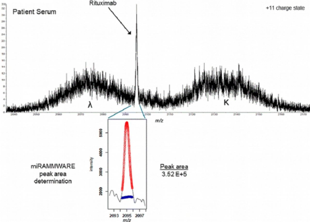
Results The peak corresponding to rituximab light chain molecular mass was easily recognized and quantified using our method (figure). The performance parameters of our assay were within acceptable limits (coefficients of variation: intra-day 5.0%, inter-day 14.1% at 50 μg/mL and 14.8% at 200 μg/mL; acceptable linearity from 0 to 250 μg/mL of rituximab; and lower limit of quantification of 14.3 μg/mL). The rituximab concentrations determined by MS were in good agreement with those found by ELISA (R2=0.853/m=0.70). In addition, endogenous monoclonal and oligoclonal light chain immunoglobulins secreted into circulation during the course of treatment were documented using the LC-MS assay.

Conclusions Our LC-MS assay was able to quantify a therapeutic mAb in a large cohort of patients in a clinical trial without the need for specific reagents. Our method could be applied to the quantification of any therapeutic mAb. This fact, coupled with the ability to phenotype a patient's polyclonal repertoire in the same analysis further shows the potential of this innovative approach to mAb analysis.
Disclosure of Interest None declared