Tibialis posterior dysfunction: a common and treatable cause of adult acquired flatfoot
BMJ 2004; 329 doi: https://doi.org/10.1136/bmj.329.7478.1328 (Published 02 December 2004) Cite this as: BMJ 2004;329:1328
- Julie Kohls-Gatzoulis (gatzoulis{at}ntlworld.com), specialist registrar orthopaedic and trauma surgery1,
- John C Angel, consultant orthopaedic surgeon1,
- Dishan Singh, consultant orthopaedic surgeon1,
- Fares Haddad, consultant orthopaedic surgeon2,
- Julian Livingstone, podiatrist3,
- Greg Berry, consultant orthopaedic surgeon4
- 1 Royal National Orthopaedic Hospital, Stanmore HA7 4LP
- 2 Middlesex Hospital, London W1N 8AA
- 3 Barnet General Hospital, Barnet EN5 3DJ
- 4 McGill University Health Centre, Hôpital Général de Montréal 1650, Avenue Cedar, Montreal, Canada H3G 1A4
- Correspondence to: J Kohls-Gatzoulis
- Accepted 14 September 2004
Introduction
Adults with an acquired flatfoot deformity may present not with foot deformity but almost uniformly with medial foot pain and decreased function of the affected foot (for a list of causes of an acquired flatfoot deformity in adults, see box 1).1 Patients whose acquired flatfoot is associated with a more generalised medical problem tend to receive their diagnosis and are referred appropriately. However, in patients whose “adult acquired flatfoot deformity” is a result of damage to the structures supporting the medial longitudinal arch, the diagnosis is often not made early.2 These patients are often otherwise healthier and tend to be relatively more affected by the loss of function resulting from an acquired flatfoot deformity. The most common cause of an acquired flatfoot deformity in an otherwise healthy adult is dysfunction of the tibialis posterior tendon, and this review provides an outline to its diagnosis and treatment.
Sources and selection criteria
We seached PubMed for publications by using the keywords “flatfoot” and “tibialis posterior dysfunction”.
Tibialis posterior dysfunction: a common condition
Tibialis posterior dysfunction is well recognised by orthopaedic surgeons specialising in foot and ankle surgery and by podiatrists. However, greater general awareness of this condition is required,2 as most patients presenting to a general practitioner receive a diagnosis of ankle sprain or arthritis. By the time most patients present to a specialist foot and ankle clinic they have had the condition for several years and have consulted numerous doctors.3 Even general orthopaedic surgeons and physiotherapists often miss the diagnosis.3 However, tibialis posterior dysfunction need not remain a “specialist diagnosis” as it is usually diagnosed without any investigations, from a history and physical examination.2 Many patients benefit from relatively simple treatment, such as orthotic devices.4 Population based studies to identify the prevalence of tibialis posterior dysfunction are under way. In elderly people the condition may reach a prevalence of 10% in women5 and it is often seen in general practice (unpublished data).
Tibialis posterior tendon: the key dynamic support of the medial longitudinal arch of the foot
Understanding the importance of the tibialis posterior tendon for the normal foot clarifies the role that the dysfunctional tibialis posterior has in the development of an acquired flatfoot. This tendon courses just posterior to the medial malleolus inserting into the navicular tuberosity (on the medial aspect of the foot) and the mid-part of the plantar aspect of the tarsus. The tibialis posterior tendon is the primary dynamic stabiliser of the medial longitudinal arch,6 and its contraction results in inversion and plantar flexion of the foot and serves to elevate the medial longitudinal arch, which locks the mid-tarsal bones, making the hindfoot and midfoot rigid.7 This later action allows the gastrocnemius muscle to act with much greater efficiency during gait. Without the tibialis posterior, the other ligaments and joint capsules gradually become weak, and thus flatfoot develops. Furthermore, without the tibialis posterior the gastrocnemius is unable to act efficiently, and therefore gait and balance are seriously affected.
Summary points
Dysfunction of the tibialis posterior tendon is a common condition and a common cause of acquired flatfoot deformity in adults
Women older than 40 are most at risk
Patients present with pain and swelling of the medial hindfoot. Patients may also report a change in the shape of the foot or flattening of the foot
The foot develops a valgus heel (the heel rotates laterally when observed from behind), a flattened longitudinal arch, and an abducted forefoot
Conservative treatment includes non-steroidal anti-inflammatory drugs, rest, and immobilisation for acute inflammation; and orthoses to control the more chronic symptoms
Surgical treatment in the early stages is hindfoot osteotomy combined with tendon transfer
Arthrodesis of the hindfoot, and occasionally the ankle, is required in the surgical treatment of the later stages of tibialis posterior dysfunction
Box 1: Causes of an adult acquired flatfoot
Neuropathic foot (Charcot foot) secondary to:
Diabetes mellitus
Leprosy
Profound peripheral neuritis of any cause
Degenerative changes in the ankle, talonavicular or tarsometatarsal joints, or both, secondary to:
Inflammatory arthropathy
Osteoarthropathy
Fractures
Acquired flatfoot resulting from loss of the supporting structures of the medial longitudinal arch:
Dysfunction of the tibialis posterior tendon Tear of the spring (calcaneoanvicular) ligament (rare)
Tibialis anterior rupture (rare)
Painful flatfoot can have other causes, such as tarsal coalition, but as such a patient will not present with a change in the shape of the foot these are not included here.
Box 2: Symptoms suggesting tibialis posterior dysfunction
Pain and/or swelling behind the medial malleolus and along the instep
Change in foot shape
Decrease in walking ability and balance
Ache on walking long distances
Pathogenesis
Histological examination has shown that, rather than tendinitis (tendon inflammation), the process is one of tendinosis (tendon degeneration), and the tibialis posterior tendon becomes fibrotic through a process of repeated microtrauma.8 Some controversy remains about the underlying aetiology of this tendinosis.9–15 A poor blood supply to the tendon has been identified as it courses posterior to the medial malleolus.9 Mechanical factors must be at play that predispose the tendon to progressive fibrosis.10 A growing body of research proposes that abnormal forces arise from even mild flatfootedness, resulting in lifelong greater mechanical demands on the tibialis posterior than in a normal foot.11–14 Another possible mechanical aetiology is overpull of the opposing peroneus brevis muscle.15 Rarely, rupture of the tibialis posterior tendon has also been associated with a traumatic event such as ankle fracture,16 ankle sprain,17 or a direct blow to the tendon.18 It is important to point out that, although the tibialis posterior tendon may rupture, this is not necessary for tibialis posterior dysfunction to develop.10 Likewise, iatrogenic loss of the tibialis posterior tendon does not always lead to an acquired flatfoot deformity.14 15
Risk factors
Middle aged women are most commonly affected, and the prevalence is known to increase with age.19 20 Pes planus (flatfoot),11–14 hypertension,19 diabetes mellitus,19 steroid injection around the tendon,7 and seronegative arthropathies7 21 have all been identified as risk factors in patients with insufficiency of the tibialis posterior tendon.
Classification
Johnson and Strom's classification describes the progression of tibialis posterior dysfunction, and also serves as a guide to management.22 In stage I dysfunction, the tendon is still intact and to some degree functioning, but it is inflamed. In stage II dysfunction, the tendon has become dysfunctional and the foot has developed acquired flatfoot, but the deformity is passively correctable. In stage III dysfunction, the foot deformity has become fixed, and degenerative changes are seen in the subtalar joint. Stage IV was not described by Johnson and Strom but has been added subsequently by Myerson.7 Stage IV dysfunction occurs when degenerative changes are also present in the ankle joint.
Diagnosing tibialis posterior dysfunction
History
In stage I, patients typically present with an insidious onset of vague pain in the medial foot and swelling behind the medial malleolus along the course of the tendon (fig 1).2 5 Patients usually have no history of acute trauma. As the condition progresses to stage II, patients have more complaints related to loss of function and change in the shape of the foot. They report either a unilateral worsening of a pre-existing bilateral flatfoot or a newly acquired flatfoot deformity.22 We tested survey questions and found that medial pain or swelling behind the medial malleolus together with a change in foot shape picked up 100% of patients with tibialis posterior dysfunction and had a specificity of 98% (box 2).5 Also the patient may have a feeling of instability, a limp, a restricted walking distance, and an inability to walk on uneven surfaces. This also leads to an increased awareness of other foot pathologies, such as bunions, hallux rigidus, and metatarsalgia, which may be the reason why the patient seeks medical attention. In the later stages, as the flatfoot deformity worsens, patients have less medial pain and swelling and may even have lateral hindfoot pain secondary to impingement of the fibula on the sinus tarsi.20

Arrows indicate swelling along the tibialis posterior tendon
Box 3: Definitions
Valgus: Angulation of an extremity at a joint or fracture site such that the distal part deviates away from the midline
Varus: Angulation of an extremity at a joint such that the distal part deviates toward the midline
Examination
In stage I tibialis posterior dysfunction the signs are of swelling and tenderness behind and below the medial malleolus (along the course of the tendon), and some weakness or pain with inversion of the foot (fig 2).2 The patient may have some difficulty rising on one heel only, or weakness after multiple heel rises.7 20

Left tibialis posterior dysfunction deformity is easily visible. The medial longitudinal arch is flattened. The left heel is in valgus. Also visible is the “too many toes sign,” which results from abduction of the left forefoot
In the later stages the patient may have less swelling and pain but will have developed an acquired flatfoot deformity.2 Looking at the standing patient from the back best shows the asymmetry of a unilateral acquired flatfoot deformity (fig 2). The affected heel is in a valgus position (box 3), and flattening of the medial longitudinal arch and forefoot abduction are visible. This leads to the commonly quoted “too many toes sign” where more than the normal (one and a half to two) toes are seen along the lateral border of the foot.2 In later stages the tendon may not be palpable at its insertion site (the navicular tuberosity)1; this is best assessed with the patient attempting to invert and point the foot downwards. The most commonly applied functional test is the single heel rise (fig 3)2: patients with tibialis posterior dysfunction are unable to perform an ipsilateral unsupported single heel rise (to go from a flatfooted stance to standing on only the toes of their affected foot).

(top) Unsupported single heel rise on patient's good (right) side. (bottom) Attempt to perform a single heel rise on the affected left side. The patient was unable to do so unsupported but for the purpose of this photo was allowed to lean forward and support herself on the counter in front of her. The heel of the left foot has not inverted into varus

(top) Unsupported single heel rise on patient's good (right) side. (bottom) Attempt to perform a single heel rise on the affected left side. The patient was unable to do so unsupported but for the purpose of this photo was allowed to lean forward and support herself on the counter in front of her. The heel of the left foot has not inverted into varus
Someone with a normal foot can perform a single heel rise eight to 10 times, but by stage II patients are unable (or barely able) to perform a single unsupported heel rise. Even if the patient is supported to facilitate the attempted heel rise, the heel will not invert as it normally does (fig 3).16 In stages III and IV, in addition to the findings in stage II, the flatfoot deformity has become fixed. This is examined by grasping the heel and attempting to correct the valgus of the hindfoot (box 4).
Imaging
The diagnosis of tibialis posterior dysfunction is essentially clinical. However, plain radiographs of the foot and ankle are useful for assessing the degree of deformity and to confirm the presence or absence of degenerative changes in the subtalar and ankle articulations. The radiographs are also useful to exclude other causes of an acquired flatfoot deformity.1 The most useful radiographs are bilateral anteroposterior and lateral radiographs of the foot and a mortise (true anteroposterior) view of the ankle. All radiographs should be done with the patient standing (figs 4, 5).20 In most cases we see no role for magnetic resonance imaging or ultrasonography, as the diagnosis can be made clinically.

Before surgery (top) and five years after surgery (bottom) for a patient with stage II tibialis posterior dysfunction. The surgical reconstruction included a Cobb split tibialis anterior tendon transfer and a Rose varus calcaneal osteotomy

Before surgery (top) and five years after surgery (bottom) for a patient with stage II tibialis posterior dysfunction. The surgical reconstruction included a Cobb split tibialis anterior tendon transfer and a Rose varus calcaneal osteotomy
Stages of tibialis posterior dysfunction and treatment options

Stage III tibialis posterior dysfunction before and after surgery (a triple arthrodesis). (top) Preoperative film shows a plantar flexed talus (arrows point to head of talus) and loss of arch contour and height. (bottom) Postoperative film shows union and reconstitution of the arch
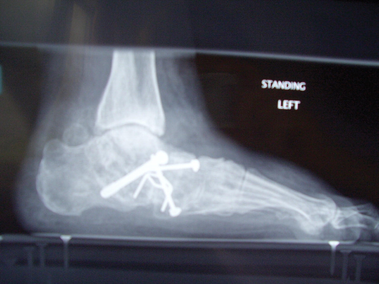
Stage III tibialis posterior dysfunction before and after surgery (a triple arthrodesis). (top) Preoperative film shows a plantar flexed talus (arrows point to head of talus) and loss of arch contour and height. (bottom) Postoperative film shows union and reconstitution of the arch
Box 4: How to examine for tibialis posterior dysfunction
Both of patient's legs visible from knee down
Observe heel alignment with patient standing with feet shoulder width apart, feet parallel. (Heel becomes valgus, arch collapses, and forefoot adducts in cases of tibialis posterior dysfunction)
Inspect for swelling behind medial malleolus
Ask patient to stand on tiptoes. Normally the heel should bend inwards. A patient with tibialis posterior dysfunction will have great difficulty standing on tiptoes, and the heel will not bend inwards
Ask the patient to perform 10 unsupported heel rises on each leg. A patient with tibialis posterior dysfunction will not be able to do this
Palpate along the tibialis posterior tendon for tenderness
Test tibialis posterior tendon for power. Ask the patient to bring foot into an inverted and plantar flexed position from an everted and dorsiflexed position against your resistance
Examine for hindfoot movement. In stages I and II, the foot is supple and the flatfoot deformity can be corrected by rotating the heel inwards (the arch of the foot will be reconstituted). In stage III and IV subtalar arthritis is present, and movement of the subtalar joint will be lessened and painful. Additionally in stage IV, ankle arthritis has set in and the ankle becomes stiff and painful
Treatment
Conservative treatment
Conservative treatment is indicated for nearly all patients initially before surgical management is considered.4 The key factors in determining appropriate treatment are whether acute inflammation and whether the foot deformity is flexible or fixed. However, the ultimate treatment is often determined by the patients, most of whom are women aged 40 or older. Compliance can be a problem, especially in stages I and II. It helps to emphasise to the patients that tibialis posterior dysfunction is a progressive and chronic condition and that several fittings and a trial of several different orthoses or treatments are often needed before a tolerable treatment is found.
Stages I and II: the flexible foot
Any acute inflammation surrounding the sheath of the tibialis posterior tendon should be dealt with before the chronic aspect of the condition is treated. A period of four to eight weeks of immobilisation in a plaster cast below the knee or a removable boot (such as those made by Aircast) may be required to control accompanying inflammation.7 20 23 In conjunction, RICE (rest, ice, compression, and elevation) and anti-inflammatories can be used, but steroid injection is contraindicated.7
Footwear has an important role, and patients should be encouraged to wear flat lace-up shoes, or even lace-up boots, which accommodate orthoses. Stage I patients may be able to manage with an off the shelf orthosis (such as an Orthaheel or Formthotics). They can try a laced canvas ankle brace before moving to a casted orthosis. The various casted, semirigid orthoses support the medial longitudinal arch of the foot and either hold the heel in a neutral alignment (stage I) or correct the outward bent heel to a neutral alignment (stage II). This approach is meant to serve several functions: to alleviate stress on the tibialis posterior; to make gait more efficient by holding the hindfoot fixed; and thirdly, to prevent progression of deformity.23 Devices available to do this are the orthosis of the University of California Biomechanics Laboratory, an ankle foot orthosis, or a removable boot. When this approach has been used, two thirds of patients have good to excellent results.4
Stage III and IV: the rigid flatfoot
Overall, inflammation tends to be a less common feature of presentation with the rigid acquired flatfoot. Treatment revolves around accommodating the deformity (rather than attempting to correct it) with customised moulded rigid orthoses, used in conjunction with appropriate footwear (often custom made), as the foot deformity worsens.
Resources for patients and doctors
http://www.footcaremd.com/—Designed for patients and run and maintained by the American Orthopaedic Foot and Ankle Society. The information about tibialis posterior dysfunction can be found under the heading “progressive flat foot”
http://www.bofss.org.uk/—Designed for both patients and doctors and is run by the British Orthopaedic Foot Surgery Society and gives good general advice about foot care
http://www.orthoteers.co.uk/—A good site for doctors. Click on flatfeet (in the paediatrics section) and then on tibialis posterior insufficiency
www.rcsed.ac.uk/fellows/jbarrie/hyperbook—The website for the Blackburn Foot and Ankle Hyperbook. Clicking on “elective foot and ankle problems” and then on “tibialis posterior insufficiency” links you to a comprehensive list of causes, complaints, examinations, and stages
Surgical treatment
Good to excellent results for more than 80% of patients have been reported at five years' follow up for the surgical interventions recommended below.7 20 However, the postoperative recovery is a lengthy process, and most surgical procedures require patients to wear a plaster cast for two to three months. Although many patients report that their function is well improved by six months, in our experience a year is required to recover truly and gain full functional improvement after the surgery. Clearly, some patients are not candidates for such major reconstructive surgery.
Stage I—In individuals with notable flatfoot whose symptoms do not resolve with conservative measures, a calcaneal osteotomy may be considered. This aims to correct the underlying foot deformity, to attempt to preserve the foot's function,7 in conjunction with either surgical debridement of the tendon24 or tendon transfer.
Stage II—Consensus is growing that surgical treatment should entail tendon transfer in combination with corrective osteotomy.7 22 The rationale behind this approach is that the osteotomy is required to correct the bony architecture of the foot in order to optimise the biomechanics of the reconstructed tibialis posterior tendon. Various osteotomies of the calcaneus can correct the bony alignment7 22 25 and may be augmented with a lengthening of the lateral column. The tendons used to reconstruct tibialis posterior are either a split tibialis anterior tendon (fig 4) or flexor digitorum longus.
Stage III—The goal of surgical treatment is to correct the deformity and alleviate pain through a triple arthrodesis of the subtalar, calcaneocuboid, and talonavicular articulations (fig 5).
Stage IV—This has been reached when additional degenerative changes are present in the ankle joint. The salvage treatment at this stage is usually a pantalar arthrodesis (ankle, subtalar, calcaneocuboid, and talonavicular articulations).
Conclusions
The tibialis posterior muscle is the key dynamic support of the medial longitudinal arch of the foot. When it fails progressively, the arch slowly collapses, the heel drifts into valgus, and the forefoot gradually abducts, resulting in painful acquired flatfoot. Tibialis posterior dysfunction is often misdiagnosed as a chronic ankle sprain, osteoarthritis, or collapsed arch as a result of ageing or obesity, and it leaves the patient debilitated. Prompt diagnosis prevents frustration for the patient and allows treatment to be started at an earlier, more easily managed stage (fig 6). The diagnosis of tibialis posterior tendon dysfunction is largely a clinical one. An increased awareness of the existence of tibialis posterior should serve to help patients with earlier referral and treatment and by limiting the amount of disability they experience.

Footnotes
-
 A film showing left sided tibialis posterior dysfunction is on bmj.com. Please note that the file is very large, so you will probably need broadband to watch this film
 -
Contributors JK-G wrote the paper. JL gave podiatric advice. FH and GB are involved in collaborative research on tibialis posterior dysfunction and have worked with senior authors JCA and DS. DS is guarantor.
-
Funding None.
-
Competing interests None declared.