Article Text

Statistics from Altmetric.com
Possible roles of cortisol and melatonin
It is well known that some clinical signs and symptoms of rheumatoid arthritis (RA) vary within a day and between days, and the morning stiffness seen in patients with RA has become one of the diagnostic criteria of the disease (fig 1).1
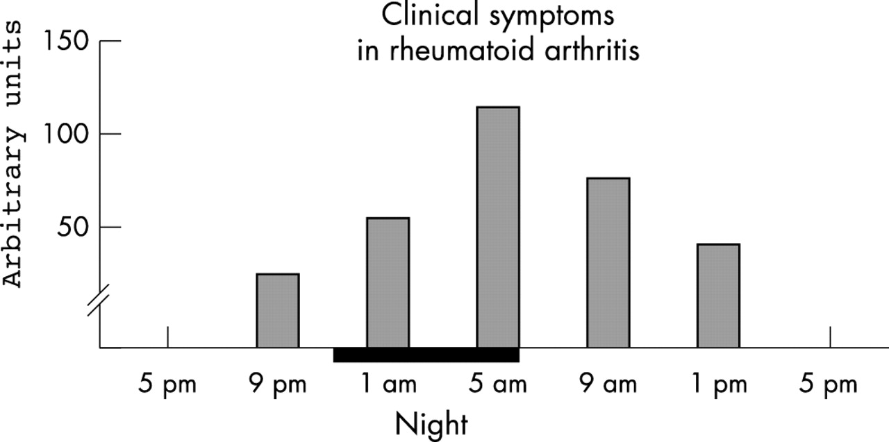
Clinical signs and symptoms of articular inflammation in patients with RA change consistently as a function of the hours of the day: pain and joint stiffness are greater after waking up in the morning than in the afternoon or evening.
Among the clinical signs of joint inflammation in patients with RA, the intensity of pain changes consistently as a function of the hours of the day: pain is greater after waking up in the morning than in the afternoon or evening.2 In patients with RA circadian variations are also found in joint swelling and finger size and these symptoms are in phase with the circadian rhythm of pain. The RA rhythms differ in phase by about 12 hours from the circadian changes of left and right hand grip strength: a greater grip strength is seen when joint circumferences and the subjective ratings of stiffness and pain are least and vice versa.3
“Clinical signs and symptoms in RA depend on the time of day”
Therefore, clinical signs and symptoms in RA show a rhythm that seems driven by a biological clock.
Biological rhythms in experimental inflammation
Biological rhythms have been seen in different models of inflammation, and maximal inflammation occurred during the activity period of the animals—that is, between midnight and 8 00 am.4
Biological rhythms with a periodicity longer than 24 hours have also been detected, and a circaseptan rhythm (almost seven days) of paw oedema, over a period of 30 days, was observed, with peak of inflammation every 6–7 days.5 Furthermore, circannual variations have been identified in different models of inflammation showing that maximal articular oedema was significantly larger in spring and lowest in winter.6 A time dependent change of blood flow at the inflammatory site may also explain the circadian variations in experimental oedema; some studies in rat models showed that the blood flow was greater in the night and lower in the morning.2
The mechanisms of the time dependent variations of the inflammatory reaction are complex and include several systems of mediators (that is, histamine, bradikinin, prostaglandin, and mainly, pro- and anti-inflammatory cytokine production). However, the circadian changes in the metabolism or secretion of endogenous corticosteroids are certainly implicated in the time dependent changes seen during the inflammatory response. This assumption is supported by data showing that adrenalectomy abolished the circadian variation in the rate of formation of experimental oedema and that this was restored by hydrocortisone administration.7 Other data indicate that the inflammatory response, produced by different agents, was in phase opposition to the cortisol rhythm: less oedema was obtained when the plasma cortisol levels were higher and vice versa.4,6
More recently, melatonin (MLT), another circadian hormone that is the secretory product of the pineal gland, has been found to be implicated in the time dependent inflammatory reaction, with effects opposite to those of cortisol.8 In several species, pinealectomy or any other experimental procedure that inhibits MLT synthesis and secretion induces a state of immunodepression, which is counteracted by MLT replacement.9–11 In general, MLT displays an immunoenhancing effect.12 MLT can activate T lymphocytes, monocytes, NK cells, and even neutrophils, activates antibody dependent cellular cytotoxicity, and enhances antibody responses in vivo.10 In animal models, as well as in human and in vitro experiments, MLT enhances inflammatory cytokine and nitric oxide production.13,14 In addition, the in vitro effects exerted by glucocorticoids on the immune function seem modulated by MLT in physiological to pharmacological concentrations.15
Cortisol and melatonin regulate circadian cytokine production
In adult primates, only visible light (400–700 nm) is received by the retina. This photic energy is then transduced and delivered to the visual cortex and, by an alternative pathway, to the suprachiasmatic nucleus, the hypothalamic region that directs circadian rhythms. Visible light exposure also modulates the pituitary and pineal glands, leading to neuroendocrine changes. MLT, norepinephrine, and acetylcholine decrease with light activation, whereas cortisol, serotonin, γ-aminobutyric acid, and dopamine levels increase.16
Therefore, ocular light seems to be the predominant modulator and major determinant of circadian rhythm for many neurohormones, with cortisol and MLT showing an opposite response to the light. The light conditions in the early morning have a strong impact on the morning cortisol peak, whereas direct inhibitory effects of light on pineal activity may contribute to phasing of the onset and termination of MLT production in a strictly nocturnal pattern.9,17
“Melatonin counteracts the effects of cortisol”
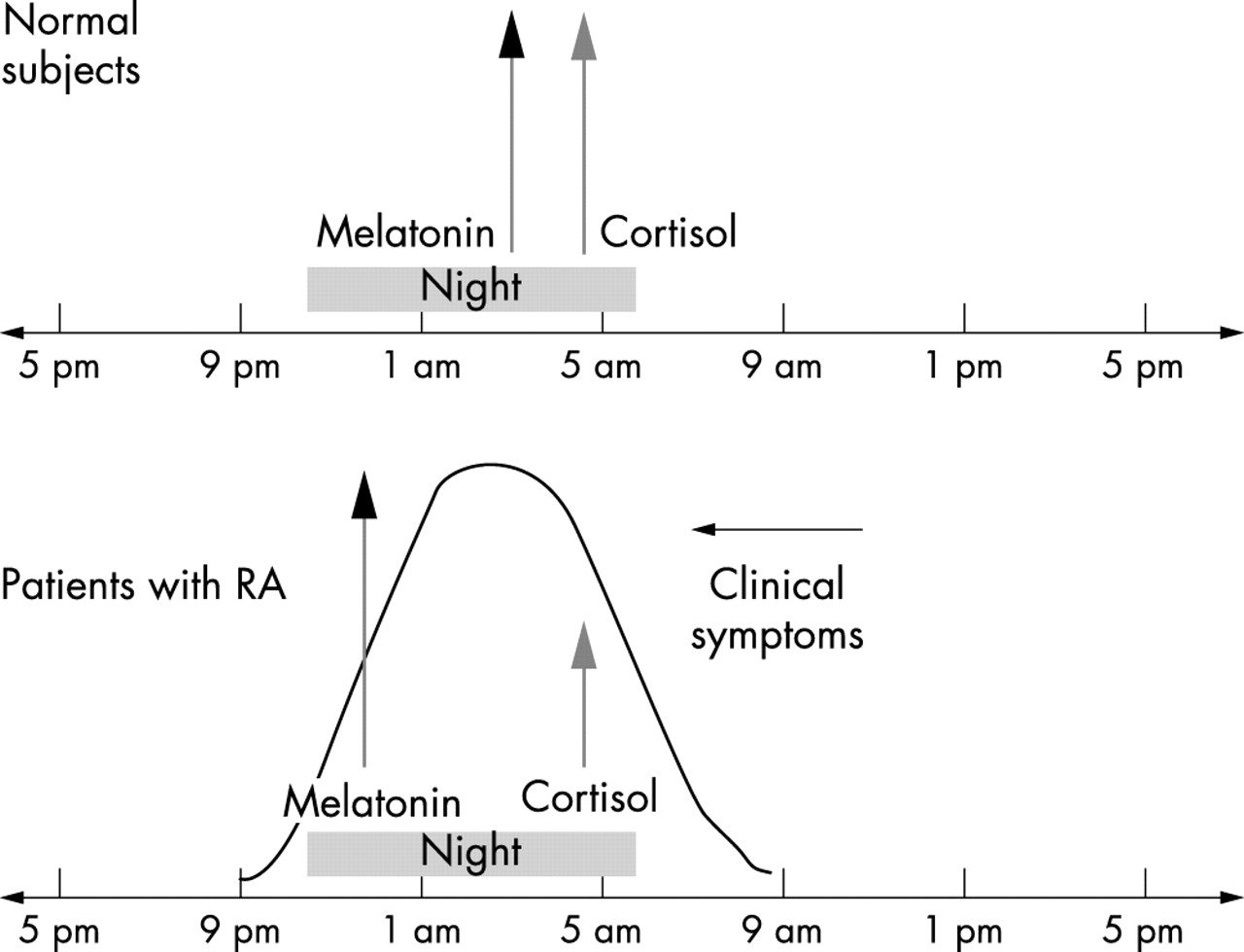
Recently, a diurnal rhythmicity in healthy humans between cellular (Th1 type) or humoral (Th2 type) immune responses has been found and related to immunomodulatory actions of cortisol and MLT (fig 2).18 In particular, the production of the interferon γ (IFNγ; type 1) and interleukin 10 (IL10; type 2) in human whole blood stimulated with lipopolysaccharide or tetanus, as well as the IFNγ/IL10 ratio, exhibited a significant diurnal rhythmicity. The IFNγ/IL10 ratio peaked during the early morning and correlated negatively with plasma cortisol and positively with plasma MLT; the IFNγ/IL10 ratio decreased by >70% after the administration of oral cortisone acetate (25 mg). Therefore, these findings support the concept that plasma cortisol and possibly MLT seem to regulate diurnal variations of cytokine production.

A diurnal rhythmicity in healthy humans between cellular (Th1 type) or humoral (Th2 type) immune responses has been found and related to the immunomodulatory effects exerted by cortisol and melatonin, respectively.
In normal subjects, MLT peaks at about 3 am, whereas cortisol peaks at about 4 am.19 Interestingly, IL1, IL6, and soluble IL2 receptors peak at 1–4 am and are low throughout the day.20 MLT stimulates IL1 and IFNγ production by human monocytes, and serum IL2 increases during the night concomitantly with the rise in MLT; MLT also seems to enhance IL2 immunomodulating effects.21–23 In addition, MLT increases the production of IL12 and nitric oxide by cultured human synovial macrophages, enhances IL2, IL6, and IFNγ production by human circulating CD4+ lymphocytes, and up regulates the level of gene expression of tumour necrosis factor α (TNFα) and macrophage-colony stimulating factor.14,24,25
On the contrary, cortisol was found to be negatively correlated with the IFNγ/IL10 ratio, and cortisone administration markedly reduced this ratio with a clear causal relationship.18 Furthermore, similarly to IFNγ and IL1, TNFα and IL12 also exhibit distinct diurnal rhythms that peak in the early morning, and these changes are inversely related to the rhythm of plasma cortisol.26
In conclusion, because IFNγ and IL10 might be considered markers of cellular (type 1) and humoral (type 2) immunity, respectively, these circadian studies suggest that there is a bias towards cellular immunity during the night and early morning (peak of MLT) when the IFNγ/IL10 ratio is high and, conversely, a relative bias towards humoral (type 2) immunity during the rest of the day (cortisol effects) (fig 2).27
Cortisol and melatonin effects on circadian rhythms in rheumatoid arthritis
The inflammatory cytokines (that is, IL6, IL1, TNFα), as soluble products of the activated immune system, stimulate in the central nervous system, the production of corticotrophin releasing hormone (CRH) in the hypothalamus: CRH release leads to pituitary production of adrenocorticotrophic hormone (ACTH), followed by glucocorticoid secretion by the adrenal cortex.28,29
These components constitute the hypothalamic-pituitary-adrenocortical axis (HPA).
Recently, intact ACTH secretion, but impaired cortisol response in patients with active RA has been described and this observation was consistent with a relative adrenal glucocorticoid insufficiency, the latter already suggested 40 years earlier.30,31 Increased HPA axis function is a normal response to the stress of inflammation and may be mediated by central and peripheral actions of circulating cytokines.
Besides IL1 and TNFα, IL6 seems to be a major factor mediating interactions between the activated immune system and both the anterior pituitary cells (central) and the adrenal (peripheral) steroidogenesis. However, recent studies in patients with RA have shown that the overall activity of the HPA axis remains inappropriately normal (or low) and is apparently insufficient to inhibit ongoing inflammation, at least in patients with early untreated arthritis.32,33
In particular, in the early morning hours, an earlier surge of plasma ACTH and cortisol was seen in patients with RA, who at the same time had significantly increased IL6 levels and a pronounced circadian variation of plasma levels in comparison with healthy subjects.34
In addition, in the patients with RA, a positive temporal correlation was found between plasma IL6 levels and ACTH/cortisol, with raised levels of IL6 before the increases of ACTH and cortisol by one and two hours, respectively.34 In the same patients, a negative effect of cortisol upon IL6 was found, exerted with a delay of five hours, confirming that the HPA axis in RA is apparently insufficient to inhibit ongoing inflammation.
More recently, another study showed a significantly altered secretion of adrenal androgens in premenopausal patients with RA not treated with glucocorticoids.35 In the same study, low plasma levels of the adrenal androgen dehydroepiandrosterone (DHEA) and its sulphate metabolite were found to be significantly correlated with early morning low cortisol concentrations and high basal levels of IL6 in patients with RA.35 Early morning IL6 peak values were recently found to be higher in patients with RA than in controls, and significantly correlated with morning C reactive protein levels and Ritchie’s index.36 The observation of reduced DHEA production, combined with normal cortisol production during oCRH and ACTH testing, further support the concept of the presence of an adrenal hypofunction in patients with active RA.37
IL6 had a strong effect on steroid release and may be one of the factors controlling the long term adrenal response to stress, because this cytokine can act synergistically with ACTH on the adrenal cells to stimulate the release of corticosterone.38,39 Therefore, the reduced cortisol and adrenal androgen secretion, seen during testing in patients with RA not treated with glucocorticoids, should be clearly regarded as a “relative adrenal insufficiency” in a sustained inflammatory process, as shown by the high IL6 levels.40
“Low dose prednisolone has more effect on RA at 2 am than at 7.30 am”
In a very recent investigation on salivary cortisol levels in patients with recent onset RA, it was found that afternoon cortisol concentrations in patients with high disease activity did not drop, as did the cortisol levels in healthy controls and patients with RA with low disease activity.41 These findings further indicate that activation of the HPA axis in RA is evident, but insufficient. The lower than required production of cortisol supports the efficacy of low dose glucocorticoid “replacement therapy” in patients with RA.42 From recent studies, it seem that administration of low doses of glucocorticoid with a rather short biological half life may improve acute RA symptoms if the administration precedes the period of circadian flare in inflammatory activity, as defined by enhanced IL6 synthesis.43 In fact, administration of low doses of prednisolone at 2 00 am was found to induce significantly more favourable effects on RA clinical symptoms than administration at 7 30 am.43 Therefore, timing of low dose glucocorticoid administration should be adapted to the biological rhythms of the inflammatory process in RA (that is, rhythms of inflammatory cytokines such as IL6).44
Very recent studies have evaluated MLT levels in patients with RA, together with an analysis of circadian variations.45 Interestingly, MLT serum levels at 8 pm and 8 am were found to be significantly higher in patients with RA than in controls (p<0.05). The differences were greater in the older patients with RA (age >60 years) than in the younger ones. Both in patients with RA and healthy subjects, MLT levels progressively increased from 8 pm to the early hours of the morning, but reached a peak in patients with RA at 12 pm, at least two hours before that in controls (fig 3). Subsequently, in patients with RA, MLT concentrations reached a plateau, lasting 2–3 hours; this effect was not evident in the controls. After 2 am, MLT levels decreased similarly both in patients with RA and healthy subjects. The results of the study confirm the existence of a nocturnal rhythm of MLT also in patients with RA. However, the peak appears earlier in the night and lasts longer in the early morning than in healthy controls.45
A lower than expected cortisol secretion as seen during testing in patients with RA, should be clearly regarded as a “relative adrenal insufficiency” in the setting of a sustained inflammatory process (that is, high IL6 serum levels).
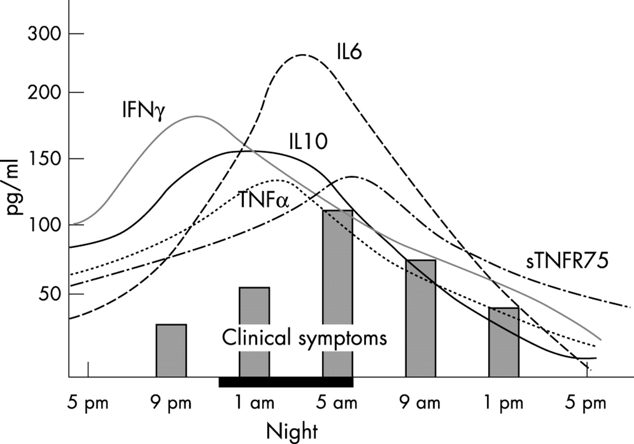
As previously discussed, IFNγ and IL2, as well as IL1, IL6, IL12, and TNFα production (Th1 cytokines) reach peak levels during the night and in the early morning, when MLT serum levels are highest and plasma cortisol levels are lowest.46 Therefore, MLT may play a part in the induction of a more active inflammatory response during the night, at least in patients with RA, because the disease is considered to be a Th1-cytokine driven immune disease (fig 4).45 At that time the lower than expected levels of cortisol seen in patients with RA are less efficient in counteracting the effects of MLT.

IFNγ, IL2, as well as IL1, IL6, IL12, and TNFα production (Th1 cytokines) reach a peak during the night and early morning, when MLT serum levels are highest and plasma cortisol levels the lowest. Therefore, MLT may be implicated in a more active inflammatory response during the night, and the clinical symptoms follow this rhythm in patients with RA.
Recently, MLT has been found at a rather high concentration in the synovial fluids of patients with RA, and binding sites for MLT have been detected in synovial macrophages.47,48
Conclusions
An altered functioning of the HPA axis and of the pineal gland seems to be an important factor in the perpetuation of clinical circadian symptoms in patients with RA. The clinical symptoms show a circadian variation, with joint stiffness and pain being more prominent in the early morning.
Consistently, human proinflammatory cytokine production exhibits a diurnal rhythmicity, with peak levels during the night and early morning when plasma cortisol level is lowest and MLT level is highest.
In particular, Th1 type cytokines that are mainly involved in RA, significantly increase, with an earlier peak in relation to altered peaking of both cortisol and MLT. An inappropriate low secretion of cortisol is a typical feature of the inflammatory disease in patients with RA. On the contrary, the nocturnal rhythm of MLT shows an earlier peak and a longer peak duration in the early morning in patients with RA than in normal subjects.
Therefore, an imbalance between anti-inflammatory effects exerted by cortisol and proinflammatory effects exerted by MLT during the night seems evident in patients with RA and suggests that this imbalance may have a crucial pathogenic role in RA, and may also drive the circadian rhythm of the clinical symptoms (that is, morning stiffness and pain).49
The relief of the clinical symptoms in RA, including the anti-inflammatory efficacy exerted by morning low dose corticosteroid “replacement” therapy, seems to confirm the importance of these circadian mechanisms. Inhibitors of MLT synthesis or MLT antagonists may in the future be considered as further possible therapeutic tools, at least in severe cases of RA.
Possible roles of cortisol and melatonin