Article Text

Abstract
Objective: To describe the osteoarthritis study population of CHECK (Cohort Hip and Cohort Knee) in comparison with relevant selections of the study population of the Osteoarthritis Initiative (OAI) based on clinical status and radiographic parameters.
Methods: In The Netherlands a prospective 10-year follow-up study was initiated by the Dutch Arthritis Association on participants with early osteoarthritis-related complaints of hip and/or knee: CHECK. In parallel in the USA an observational 4-year follow-up study, the OAI, was started by the National Institutes of Health, on patients with or at risk of symptomatic knee osteoarthritis. For comparison with CHECK, the entire cohort and a subgroup of individuals excluding those with exclusively hip pain were compared with relevant subpopulations of the OAI.
Results: At baseline, CHECK included 1002 participants with in general similar characteristics as described for the OAI. However, significantly fewer individuals in CHECK had radiographic knee osteoarthritis at baseline when compared with the OAI (p<0.001). In contrast, at baseline, the CHECK cohort reported higher scores on pain, stiffness and functional disability (Western Ontario and McMaster osteoarthritis index) when compared with the OAI (all p<0.001). These differences were supported by physical health status in contrast to mental health (Short Form 36/12) was at baseline significantly worse for the CHECK participants (p<0.001).
Conclusion: Although both cohorts focus on the early phase of osteoarthritis, they differ significantly with respect to structural (radiographic) and clinical (health status) characteristics, CHECK expectedly representing participants in an even earlier phase of disease.
Statistics from Altmetric.com
Osteoarthritis is the most common diagnosis made in older patients with knee or hip pain. The diagnosis can be based on symptoms, signs and radiographic findings, and as such can be defined by various sets and combinations of criteria.1,2 The prognosis of osteoarthritis for the individual patient is uncertain; the course of symptoms, clinical signs, disability and radiographic changes is difficult to predict.3 Besides, it has been demonstrated that there is inconsistency between the radiographic change and severity of joint pain with accompanying disability.4 Clearly, to understand more about the disease and its course, large independent detailed observational studies starting (very) early in the stage of the disease are necessary.
Therefore, in The Netherlands, a prospective 10-year follow-up study was recently initiated by the Dutch Arthritis Association in order to establish the onset and progression of osteoarthritis in participants with early complaints of hip and/or knee pain: CHECK (Cohort Hip and Cohort Knee), using the International Classification of Functioning, Disability and Health (ICF) as a conceptual framework. The ICF model provides an integrative framework, combining biological, psychological and social aspects of health and disease.5 The objective of CHECK is to study the course of complaints, the mechanisms that cause joint damage and to identify markers for the diagnosis and course of joint damage, as well as to identify prognostic factors that predict and explain the course of osteoarthritis. In parallel, an observational study on osteoarthritis was initiated by the National Institutes of Health: the Osteoarthritis Initiative (OAI). This 4-year follow-up study will create a public archive of data, biological samples and joint images to study the natural history of, and risk factors for, the onset and progression of knee osteoarthritis. In fact, both initiatives, CHECK and the OAI, seek answers to the same questions in comparable populations. In the present report the CHECK population is described at baseline and compared with relevant subpopulations of the OAI to provide a basis for further research and a comparison of both cohorts in the future.
Methods
CHECK cohort
Design
From October 2002 to September 2005 a cohort was formed of 1002 participants with pain and/or stiffness of the knee and/or hip, which is to be followed prospectively for a period of at least 10 years. Nationwide, 10 general and academic hospitals in The Netherlands are participating, located in urbanised and semi-urbanised regions. The study was approved by the medical ethics committees of all participating centres, and all participants gave their written informed consent before entering the study.
Study population
General practitioners (GP) in the surroundings of the participating centres were invited to refer eligible persons to these centres. All patients that visited the GP on their own initiative, potentially fulfilling the inclusion criteria, were referred to one of the 10 participating centres. In addition, participants were recruited through advertisements and articles in local newspapers and on the Dutch Arthritis Association website. The physicians in the participating centres checked whether referred patients as well as patients from their outpatient clinics fulfilled the inclusion criteria.
Inclusion criteria
Individuals were eligible if they had pain and/or stiffness of the knee and/or hip, were aged 45–65 years, and had never or not longer than 6 months ago visited the GP for these symptoms for the first time.
Exclusion criteria were: any other pathological condition that could explain the existing complaints (eg, other rheumatic disease, previous hip or knee joint replacement, congenital dysplasia, osteochondritis dissecans, intra-articular fractures, septic arthritis, Perthes’ disease, ligament or meniscus damage, plicasyndrome, Baker’s cyst) or co-morbidity that did not allow physical evaluation and/or follow-up of at least 10 years, malignancy in the past 5 years and inability to understand the Dutch language.
Baseline measurements
Variables categorised according to the ICF model (table 1).
Assessment of the variables, categorised according to the dimensions of the ICF, comparison of CHECK and OAI
Body function and structures: articular and kinesiological factors
To assess cartilage and bone at baseline, imaging techniques were employed and samples of blood and urine collected. During follow-up this is also done at 2, 5, and 10 years. At baseline both knees and hips were analysed in all participants, independent of symptoms and signs.
Radiographs of the tibiofemoral joints (TFJ) were made by a weight-bearing posteroanterior (PA) view, semi-flexed (7–10°) according to Buckland-Wright and colleagues.6,7,8 Radiographs of the patellofemoral joints were made by a single standing mediolateral view in 30° flexion and a skyline (inferior superior) view in 30° flexion.9,10 For the hip, weight-bearing anteroposterior (AP) radiographs of the pelvis were made.11,12 In addition, a weight-bearing single faux profile radiograph of both hips was obtained.13
All radiographs were made without fluoroscopy, and were digitalised and centrally stored. Radiographs of PA TFJ and AP pelvic views at baseline were scored according to Kellgren and Lawrence (K&L).14 Blood and urine samples were collected, using a standardised protocol at all sites. Multiple aliquots of serum, plasma and urine were centrally stored at −80°C. DNA was collected at baseline and was stored at −20°C.
Kinesiological factors (table 1) were assessed each year by a protocol that was established to measure clinical features of knee, hip, and hands.
Body function and structures: pain, stiffness, and fatigue
Questionnaires were selected based on the following criteria: validated in participants with osteoarthritis; demonstrated reliability, validity and, if applicable, responsiveness; the questionnaire is internationally accepted, is available in the Dutch language and has a high feasibility. Questionnaires are administered annually.
The Western Ontario and McMaster osteoarthritis index (WOMAC),15,16,17 a questionnaire with well-known and good clinimetric properties and recommended by OMERACT is utilised to measure pain (five items), stiffness (two items) and physical functioning (see below).18,19 The five point Likert version of the WOMAC was used; item responses range from “none” to “extreme” and are summed to produce subscales (pain 0–20, stiffness 0–8, functioning 0–68) with higher scores indicating worse health.
Fatigue was assessed with the vitality subscale of the Short Form 36-item health status survey questionnaire (SF-36). This questionnaire is a generic instrument yielding scores on eight scales, with two summary scores, the physical component summary (PCS) and the mental component summary (MCS). The physical functioning, role limitations due to physical health and bodily pain scale contributes most to the scoring of the PCS. The mental health, role limitations due to emotional problems and social functioning scales contributes most to the MCS. These summary scores of the SF-36 are equivalent to the summary scores of the SF-12,20 as used in the OAI. Scores on the scales range from 0 to 100, with a higher score indicating a better health-related quality of life.21
Activities
The WOMAC was used to assess physical functioning (17 items).18,19
Participation
To assess the involvement in life situation, employment and leisure activities were measured with the questionnaire from the Patient Panel Chronic Diseases (The Netherlands Institute for Health Services Research; NIVEL) and the questionnaire “economic aspects in rheumatoid arthritis”.22
Environmental factors and personal factors
Table 1 provides an overview of the environmental and personal factors collected at baseline. Co-morbidity was assessed with a standard consensus-based list.23 Pain-coping behaviour was measured with the pain coping inventory, assessing both behavioural and cognitive coping strategies.24 To assess a person’s distress and fear, a subscale of the SF-36 was used. Social support was measured with the Dutch “social support scale”.25 Physical load and economic consequences were assessed with the Dutch musculoskeletal questionnaire and the questionnaire “economic aspects in rheumatoid arthritis”, respectively.22,26
Every 3 months, the 10 institutes were visited by a single central coordinator to support complete and accurate data gathering.
Osteoarthritis Initiative
All details of the OAI are available on the internet (http://www.oai.ucs.edu). In short, individuals were eligible if they had or were at risk of symptomatic TF knee osteoarthritis and were aged between 45 and 79 years. Individuals with inflammatory arthritis, bilateral end-stage knee osteoarthritis, inability to walk and a contraindication for magnetic resonance imaging were excluded. Recruitment of 4796 individuals was realised from March 2004 to May 2006. At baseline the cohort was divided into two subcohorts, one with symptomatic knee osteoarthritis (defined as in at least one knee frequent knee symptoms and radiographic TF knee osteoarthritis, defined as K&L ⩾2; progression cohort) and a second cohort of 3285 individuals without symptomatic knee osteoarthritis, selected on the basis of having specific characteristics that give them an increased risk of developing symptomatic knee osteoarthritis (incidence cohort). For the incidence cohort age-specific eligibility criteria were defined. Individuals aged 45–49 years were eligible when they had frequent knee symptoms, or made frequent use of medication for the treatment of knee symptoms or had infrequent knee symptoms, and in addition had one or more other eligible risk factor such as knee injury, knee surgery, overweight, positive family history, etc. Individuals aged 50–69 years were eligible if they had frequent knee symptoms, or made frequent use of medication for the treatment of knee symptoms, or were overweight, or had two or more of the eligible risk factors.
At baseline materials for the identification of joint imaging, biomarkers and genetic markers were collected. Also data on the clinical and joint status of subjects and on risk factors for the progression and development of knee osteoarthritis were collected by questionnaires and examination (categorised according to the dimensions of the ICF model depicted in table 1).
In the present report baseline CHECK data of the entire cohort were compared with data of the OAI incidence cohort. To make the cohorts more comparable, in addition, CHECK participants with knee problems (excluding those with exclusively hip problems; n = 829) were compared with participants of the OAI incidence cohort within the same age range (45–65 years), who had at least frequent or infrequent knee symptoms excluding those who eg, just had overweight without symptoms and excluding those who had had previous knee surgery (n = 1578).
Statistical analyses
Baseline characteristics of both cohorts are presented using descriptive statistics: median and 25th–75th percentiles or percentages. Differences between groups are analysed using Mann–Whitney U tests or the χ2 test, when appropriate.
Results
More than 75% of participants were selected based on advertisements including the website. The baseline characteristics of the participants from CHECK and the relevant populations of the OAI are presented in table 2. With respect to radiological osteoarthritis (K&L score) and health status there were some striking differences between the cohorts.
Demographic and disease characteristics in CHECK and selections of the OAI
Radiographic joint damage was clearly more notable in the OAI compared with CHECK (table 2). In fig 1 the percentage of participants with a knee K&L grade 0–1 and ⩾2 is depicted. Based on the definition of K&L grade ⩾2, at baseline only 7% of CHECK participants had radiographic knee osteoarthritis compared with 40% in the OAI incidence cohort (p<0.001). Evidence for radiographic hip osteoarthritis was only present in 7% of the participants of CHECK. The significant difference in knee K&L grade was also clear when the subgroups of both cohorts were compared (8% and 32%; p<0.001). Even when the participants with K&L grade 4 were omitted from the calculations, the difference in severity of radiographic joint damage between both cohorts remains evidently significant. This difference in severity of joint damage between both cohorts was not based on differences in gender (data not shown).
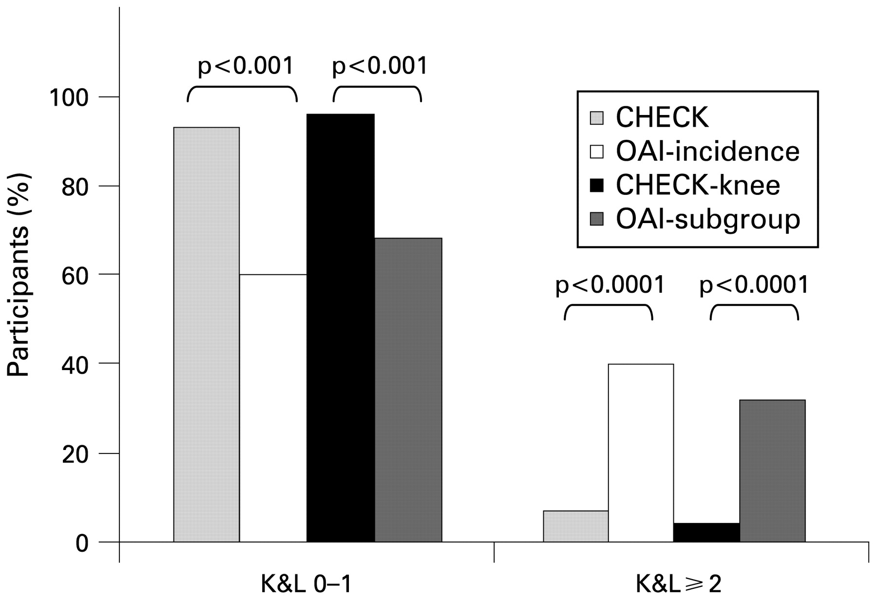
Comparison of radiographic joint damage between Cohort Hip and Cohort Knee (CHECK) and Osteoarthritis Initiative (OAI) and their subpopulations at baseline. The bars depict the percentage of knees with a Kellgren and Lawrence (K&L) score of 0–1 and a K&L score ⩾2. p Values for statistical comparison are given.
Despite the limited number of participants with radiographic knee osteoarthritis in CHECK, 76% of the patients with knee symptoms could be diagnosed as osteoarthritis according to the clinical American College of Rheumatology criteria for the classification of osteoarthritis.1,2 Only a minority of CHECK participants with hip symptoms (24%) fulfilled the clinical classification criteria of hip osteoarthritis.
The evident difference in radiographic joint damage between both cohorts was not accompanied by a similar difference in pain and physical function. On the contrary, as shown in fig 2, on each of the WOMAC subscales participants of CHECK presented with more pain, stiffness and problems in function than patients in the OAI. This was observed for the whole cohorts as well as the subgroups of both cohorts (all p<0.001). Women reported more pain and functional disability than men, which was almost identical with the OAI (all p<0.05; data not shown)

Comparison of the three Western Ontario and McMaster Universities osteoarthritis index (WOMAC) subscales for pain, stiffness and functional disability between Cohort Hip and Cohort Knee (CHECK) and Osteoarthritis Initiative (OAI) and their subpopulations at baseline. Box whisker plot (median and 25th–75th percentiles) and p values are given. A higher score indicates more pain, stiffness and problems in physical functioning.
The specific difference in physical function, contrasting the radiographic difference between both cohorts, was underscored by the difference in PCS in contrast to MCS of the SF-36/12 scale (fig 3); CHECK participants scoring less than patients from the OAI (p>0.001) for the PCS but not for the MCS (not significant). Also for these scales, in both cohorts women scored worse compared with men (all p<0.05; data not shown).

Comparison of the physical component summary (PCS) scale and mental component summary (MCS) scale of the Short Form 36-item health status survey questionnaire (SF-36/12) between Cohort Hip and Cohort Knee (CHECK) and Osteoarthritis Initiative (OAI) and their subpopulations at baseline. Box whisker plot (median and 25th–75th percentiles) and p values are given. A higher score indicates a better health-related quality of life.
Discussion
The CHECK study is the first prospective 10-year follow-up study of osteoarthritis in an early phase of the disease that combines biological, psychological and social aspects of osteoarthritis. Radiographic knee osteoarthritis was present only in a small number of the CHECK participants when compared with the OAI. In contrast, the participants in CHECK had more pain, more stiffness, more limitations in activities and a worse health status. The worse clinical health status is supported by the use of pain medication: At baseline, only 9% of the participants in OAI had taken any pain medication, whereas this was 46% for CHECK (data not shown). Other characteristics such as body mass index or gender appeared not to be explanatory for this observed characteristic difference between both cohorts (data not shown). It could be that the difference in radiographic joint damage between both cohorts is due to differences in implementation of the K&L grading method. However, grading of a random sample of the OAI knee radiographs by those who performed the grading for CHECK excluded this possibility (data not shown). Although not expected to be explanatory, it should be kept in mind that radiographs were taken, although according to standard protocols, in 10 different centres, whereas in the OAI only four centres are involved. It can not be ruled out that the social, cultural and healthcare system differences between the USA and Europe, in particular The Netherlands, account for (part of) the difference in reported health status. Also differences in inclusion between both cohorts can not be ruled out.
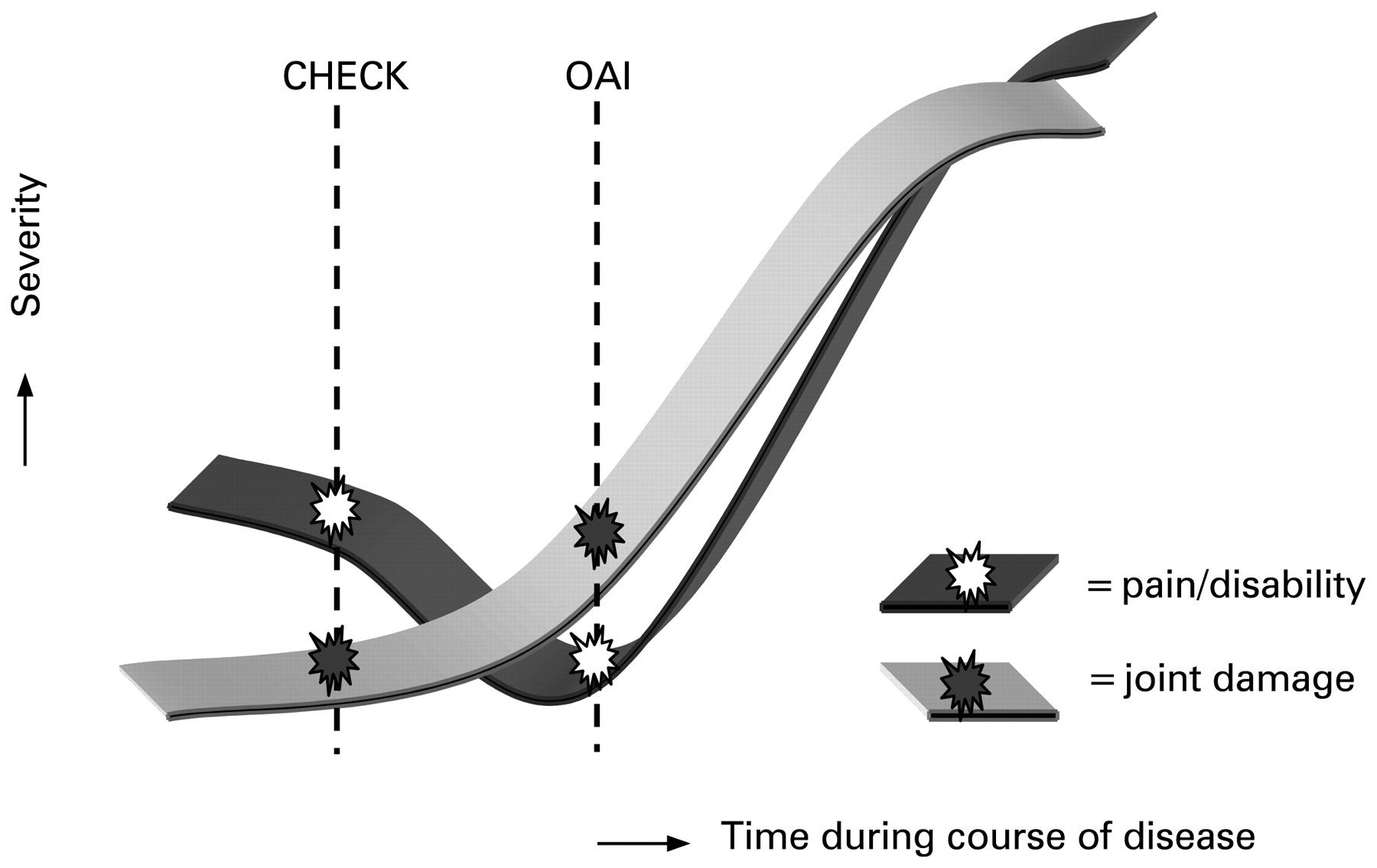
The OAI incidence cohort is recognised as an early osteoarthritis cohort. Taking the radiological findings into account, we conclude that CHECK was started in an even earlier phase of the disease compared with the OAI. Although this is apparently in discordance with the more severe clinical symptoms, the relation between radiographic damage and clinical symptoms has never been clear27 and is the subject of study in both cohorts. Therefore, it is hypothesised that in the early phase of osteoarthritis pain, stiffness and disability (of still unknown origin) are prominent and not yet accompanied by radiographic findings of osteoarthritis (CHECK). In the subsequent phase (OAI) patients are coping with the pain and physical disability, leading to a decrease in the report of these characteristics, while independently (or maybe as a consequence) structural changes, visible on radiographs, develop. In other words, earlier recruitment of patients may carry more perceived symptoms of osteoarthritis (as also seen in rheumatoid arthritis), while in a later stage coping with a new disease may ameliorate the symptoms (fig 4). In the final course of the disease the structural (radiographic) changes progress and lead to further pain and disability. It should be taken into account that, in addition, several other factors, as described in the ICF model, may add to the apparent discrepancy observed between pain and structural joint damage over time.28 Our hypothesis can be tested in the future follow-up of patients in both cohorts, in particular those with the more severe complaints (still) without radiographic joint damage. If it appears that the CHECK population with respect to pain and joint damage, independent of factors such as social background, healthcare system differences, cultural difference, variance in methodology etc, follows the OAI population, then our hypothesis may hold true. Of course other factors, independent of symptoms and joint damage, need to be evaluated regarding observed differences between both cohorts, as such giving both cohorts their surplus value.

Schematic presentation of the hypothesis as put forward in the discussion explaining the (apparent) discrepancy between both cohorts with respect to pain and joint damage. CHECK, Cohort Hip and Cohort Knee; OAI, Osteoarthritis Initiative.
Acknowledgments
The authors would like to thank all participants of the CHECK cohort and all collaborators of the different sites for their initial and still continuing efforts.
REFERENCES
Footnotes
Funding CHECK is funded by the Dutch Arthritis Association on the lead of a steering committee comprising 16 members with expertise in different fields of osteoarthritis chaired by JWJB and coordinated by JW. Involved are: Academic Hospital Maastricht; Erasmus Medical Center Rotterdam; Jan van Breemen Institute/VU Medical Center Amsterdam; Kennemer Gasthuis Haarlem; Martini Hospital Groningen/Allied Health Care Center for Rheumatology and Rehabilitation Groningen; Medical Spectrum Twente Enschede/Twenteborg Hospital Almelo; St Maartenskliniek Nijmegen; Leiden University Medical Center; University Medical Center Utrecht and Wilhelmina Hospital Assen.
Competing interests None.
Ethics approval The study was approved by the medical ethics committees of all participating centres.
Patient consent Obtained.
The OAI is a public–private partnership comprised of five contracts (N01-AR-2-2258; N01-AR-2-2259; N01-AR-2-2260; N01-AR-2-2261; N01-AR-2-2262) funded by the National Institutes of Health (NIH) and conducted by the OAI study investigators. This paper was prepared using an OAI public use dataset and does not necessarily reflect the opinions or views of the OAI investigators, the NIH, or the private funding partners.