Article Text

Abstract
Background: Rheumatoid arthritis is characterised by inflammation and an increased cardiovascular risk. It was recently shown that active early rheumatoid arthritis is associated with dyslipidaemia, which may partially explain the enhanced cardiovascular risk. However, it is unknown when this dyslipidaemia starts.
Objective: To investigate the progression of the lipid profile over time and the influence of inflammatory parameters on this lipid profile, in people who later developed rheumatoid arthritis.
Methods: Levels of total cholesterol, high-density lipoprotein cholesterol (HDLc), triglycerides, apolipoprotein AI (apo AI), apolipoprotein B (apo B) and lipoprotein(a) (Lp(a)) were determined in 1078 stored, deep-frozen, serial blood bank samples, collected between 1984 and 1999, of 79 blood donors who later developed rheumatoid arthritis. These samples were compared with 1071 control samples of unselected blood donors, matched for age, sex and storage time.
Results: Samples of patients who later developed rheumatoid arthritis showed, on average, 4% higher total cholesterol, 9% lower HDLc, 17% higher triglyceride and 6% higher apo B levels than matched controls (p⩽0.05).
The magnitude of the differences in lipid levels between groups, explained by C reactive protein (CRP), was limited. For example, only 3.6% of the difference in HDLc levels between the groups was explained by the CRP concentrations.
Conclusion: Patients who later develop rheumatoid arthritis have a considerably more atherogenic lipid profile than matched blood donors at least 10 years before onset of symptoms. As inflammation only marginally explains the differences between the two groups, a modulating effect of lipids on inflammatory processes is hypothesised.
- anti-CCP, anti-cyclic citrullinated peptides
- apo AI, apolipoprotein AI
- apo B, apolipoprotein B
- CRP, C reactive protein
- HDLc, high-density lipoprotein cholesterol
- IgM-RF, immunoglobulin M rheumatoid factor
- Lp(a), lipoprotein(a)
- sPL-A2, secretory phospholipase A2
Statistics from Altmetric.com
- anti-CCP, anti-cyclic citrullinated peptides
- apo AI, apolipoprotein AI
- apo B, apolipoprotein B
- CRP, C reactive protein
- HDLc, high-density lipoprotein cholesterol
- IgM-RF, immunoglobulin M rheumatoid factor
- Lp(a), lipoprotein(a)
- sPL-A2, secretory phospholipase A2
Rheumatoid arthritis, an inflammatory joint disease, is associated with increased cardiovascular morbidity and mortality, as shown by several inception cohort studies.1–,6 The cause of this enhanced cardiovascular risk is unknown, but inflammation is thought to play an important part,7 which is in line with the accumulating evidence that inflammation has a prominent role in the development of atherosclerosis.8,9 The relevance of inflammation in the development of cardiovascular disease is shown by associations between future cardiovascular events and raised C reactive protein (CRP) levels.10–,13 The underlying pathophysiological mechanism has not yet been completely elucidated and several possibilities have been suggested. Acute-phase proteins might: (1) deteriorate “fatty streaks” into (instable) plaques,9 (2) destabilise plaques and cause plaque ruptures,12 (3) give complement activation13 or (4) facilitate deterioration of the lipid profile.14
Dyslipidaemia may be responsible for the increased cardiovascular risk in patients with rheumatoid arthritis. Several investigators have shown that active rheumatoid arthritis is associated with an unfavourable lipid profile—that is, a decreased total cholesterol and relatively lower high-density lipoprotein cholesterol (HDLc) levels.14–,16 The result is a less favourable atherogenic index, suggesting a relationship between inflammation and dyslipidaemia.
Other lipoproteins have been suggested to play an important part in the development of atherosclerosis—that is, lipoprotein(a) (Lp(a)), apolipoprotein AI (apo AI) and apolipoprotein B (apo B). A few studies have investigated Lp(a) levels in patients with rheumatoid arthritis, and the results are contradictory. Several earlier studies showed considerably lower Lp(a) levels in patients with rheumatoid arthritis and a marked association with acute-phase response.17 A later study found neither considerably raised levels of Lp(a) in patients with rheumatoid arthritis nor a notable relationship between Lp(a) and the acute-phase response.18 Evidence is growing that both apo AI and apo B are useful predictors for future cardiovascular risk. Apo AI may protect against cardiovascular disease, whereas apo B may increase cardiovascular risk.19–,21 Apo AI is the major apolipoprotein in HDLc and has various structural and functional roles in HDLc metabolism. Apo B is the main apolipoprotein of chylomicrons, very low-density, low-density and intermediate-density lipoproteins, and seems to be a better predictor for cardiovascular disease than low-density lipoprotein cholesterol.22 So far, data regarding apolipoproteins in rheumatoid arthritis are sparse and contradictory.
Recently, we showed that in a large group of blood donors, the presence of a preclinical phase preceding the actual clinical phase of rheumatoid arthritis is characterised by serological changes—that is, raised levels of immunoglobulin M rheumatoid factor (IgM-RF) and anti-cyclic citrullinated peptides (anti-CCP).23 In addition, we also found raised CRP levels in the samples of the blood donors who later developed rheumatoid arthritis compared with samples with random control donors.24 Furthermore, the presence of dyslipidaemia in early active rheumatoid arthritis raises the question of whether or not this phenomenon starts in the preclinical phase of rheumatoid arthritis.
This study was undertaken to investigate the lipid profile over time and its relationship with inflammation and serological markers, in patients who later developed rheumatoid arthritis.
METHODS
Participants
The Sanquin Blood Bank Northwest Region, formerly the Red Cross Blood Bank, in Amsterdam, The Netherlands, serves an area with about 2 million inhabitants. The blood bank stores 1 ml aliquots of serum at −30°C, from each donation since 1984. On average, donors donate blood 2–4 times a year over a long-term period. The maximum age of donors is 70 years. Within the area in which the blood bank collects blood from donors, most of the patients with rheumatoid arthritis are registered in the Jan van Breemen Institute, a regional network of rheumatology outpatient clinics. After receiving approval from the local institutional review board, all registered patients with rheumatoid arthritis were sent a letter asking them if they had been a blood donor before the symptoms of rheumatoid arthritis started. When the reply was positive, patients were asked to sign informed consent, permitting the use of stored blood samples for the present study. Ultimately, we identified 79 non-related patients with rheumatoid arthritis, satisfying the 1987 American College of Rheumatology criteria for rheumatoid arthritis.25 Chart review yielded the following data: time of the start of symptoms, time of diagnosis of rheumatoid arthritis, IgM-RF at the time of diagnosis and during follow-up, and the presence of bony erosions on radiographs.
Procedures
From 1984 to 1999, patient samples were collected longitudinally from each consecutive blood donation. This was done during the entire period in which the patients were blood donors. For each sample of every patient with rheumatoid arthritis, one control sample from a random blood donor matched for sex, age (SD 3 years) and time of blood donation was selected as described earlier.23,24 This method of control selection was chosen to ensure identical storage conditions for the materials from patients and controls.
Laboratory measurements
Serum total cholesterol (<5.0 mmol/l) and triglycerides (<2.2 mmol/l) were analysed by an enzymatic method using the appropriate assays supplied by Roche Diagnostics (Almere, The Netherlands), on a Hitachi 911 analyzer (Roche Diagnostics), according to the instructions of the manufacturer. Polyethylene glycol modified enzymes were used to assess HDLc levels (in men, >0.9 mmol/l; in women, >1.1 mmol/l). Apo AI (in men, 1.04–2.02 g/l; in women, 1.08–2.25 g/;), apo B (in men, 0.66–1.33 g/l; in women, 0.60–1.17 g/l) and Lp(a) (<30 mg/dl) were analysed by an immunoturbidimetric method, using appropriate assays (Roche Diagnostics). CRP (<10 mg/l) was measured using a latex-enhanced highly sensitive assay (Roche Diagnostics). Secretory phospholipase A2 (sPLA2; <5 ng/ml) was measured by means of a highly sensitive ELISA (Sanquin Research at CLB, Amsterdam, The Netherlands).
IgM-RF and anti-CCP antibodies were measured using in-house ELISAs on an ES 300 analyzer (Roche Diagnostics).23,24 IgM-RF was calibrated with a national reference serum containing 200 IU/ml. No reference serum is available for anti-CCP. The applied criteria for positive tests were ⩾30 IU/ml for IgM-RF and ⩾50 AU/ml for anti-CCP.
Statistics
We divided the data into 1-year periods before the start of the symptoms and we used the samples from 10 to 0 years before the start of the symptoms to estimate the time course of the different lipids. Samples from 15–10 years before the start of the symptoms were omitted because of the small number of available samples. The time course was estimated using a random coefficient multilevel regression analysis—that is, a longitudinal regression technique that allows both starting levels and progression over time to differ between subjects.26 Multilevel regression analysis can be seen as a longitudinal linear regression analysis, which combines many cross-sectional linear regression models into one model of one variable over time. In our study, we looked at the lipid levels over time and investigated the influence of being a future patient or a control and the influence of inflammation parameters on the progression of the various lipid levels over time. Multilevel regression analysis quantifies these influences, or rather associations, between the lipid levels and these two sets of variables, and tests within for significance.
In addition to this increase of statistical power by combining the cross-sectional data of various time points into one association, the multilevel method enables the user to correct for confounding caused by multiple testing within subjects.
The differences in triglyceride and Lp(a) levels between patients and controls were reported as the differences in geometric means, as these variables were non-normally distributed. Subsequently, the natural logarithm of these levels was used for the analyses.
Preceding the time course analysis, it was investigated whether the various lipid levels were constant over time or if there were increases or decreases in certain 1-year periods.
The first step of modelling the data was carried out to determine differences between patients and controls in the lipid levels over time. These differences were reported as absolute values and as percentages of the mean lipid levels as measured in the control groups. The possibility of interaction between time and group was also investigated in this model. In the second step of this analysis, CRP, IgM-RF and anti-CCP were added to the model to investigate whether the difference between patients and controls could be explained by CRP, IgM-RF or anti-CCP. The differences in the various lipid concentrations between patients and controls, which could be explained by CRP, IgM-RF or anti-CCP, were reported as percentages of the observed total differences between the two groups. We also added another acute-phase reactant to the model—that is, sPLA2—to investigate whether this would have additional value.
In another analysis, the longitudinal relationships between the lipids and CRP, IgM-RF and anti-CCP were analysed for patients and controls combined into one group. Firstly, a model investigating variables measured at the same time was used. Secondly, a “time-lag” –model, in which the relationship between the lipid concentrations of a particular 1-year period with inflammation or serological markers of a previous 1-year period, was investigated.26
The random coefficient analyses were carried out using the statistical program MLwiN.27 A p value <0.05 was considered significant.
RESULTS
Patient characteristics
We included 79 patients with rheumatoid arthritis, 61% were women, with a mean age of 51 years at the onset of symptoms. The range of available serum samples was 1–51, with a median of 13 samples per patient. The first sample was taken at a median of 7.5 (range 0.1–14.5) years before the onset of symptoms. For serum samples from seven patients, no matched control sera were available, resulting in a total of 1078 serum samples of patients and 1071 from controls.
Lipid levels
Figure 1⇓ shows the development of lipid levels over time as measured in blood samples of the patients and controls. An investigation of the total cholesterol, HDLc, triglyceride, apo AI, apo B and Lp(a) levels, using the 1-year periods separately, yielded no trend breach in the progression of lipid levels over time; in other words, the increase or decrease in lipid levels was constant over time. This justified time course estimations in which the 1-year periods were investigated as one whole period.
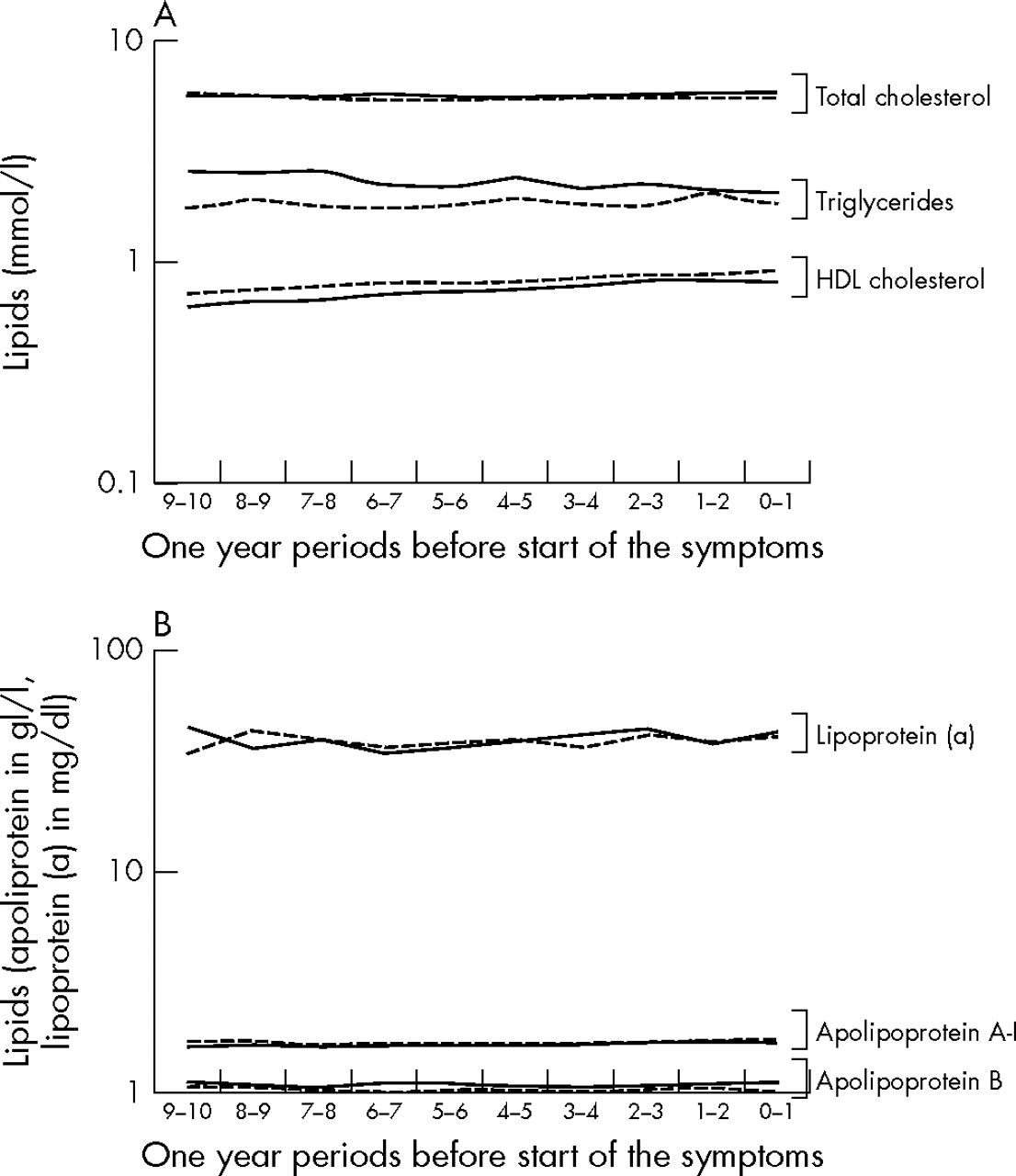
Development of lipid levels over time in patients and controls. Values in patients who later developed rheumatoid arthritis are represented by solid lines and the values in controls are dashed lines.
The first step modelling the data yielded significant differences in lipid levels between patients and controls. Mean total cholesterol levels were higher in the patients than in the controls, on average 0.21 mmol/l (p = 0.05). Triglyceride and Apo B levels were also higher in patients than in controls, on average 0.26 mmol/l (p<0.001) and 0.06 mg/l (p = 0.02), respectively. The HDLc levels were lower in patients, on average 0.08 mmol/l (p<0.001). Expressed as percentages of the calculated mean of the controls, we found that the total cholesterol levels in patients were on average 3.8% higher, triglyceride levels were 16.8% higher, apo B levels were 5.8% higher and HDLc levels were 9.0% lower. We found no effect modification between time and division into groups, indicating that the variance of the lipid levels over time was similar in both groups.
The magnitude of the observed difference in lipids between patients and controls, as explained by CRP, was 4.0% for total cholesterol, 0.4% for triglyceride, 3.6% for HDLc and 2.0% for apo B levels. The influence of IgM-RF and anti-CCP on the differences in total cholesterol, triglyceride, HDLc and apo B levels was also minor, ranging from <1% to a few per cent. In addition, the other inflammation marker sPL-A2 had a similar effect on the differences in lipids between the two groups and it had no additional value above CRP (data not shown). Table 1⇓ shows the differences in lipid levels between patients and controls, after adjustment for the influence of CRP, IgM-RF and anti-CCP.
Multilevel regression analyses of TC, HDLc, apo AI, apo B and Lp(a) levels in patients who later developed rheumatoid arthritisand controls
To investigate the longitudinal relationship of inflammation and serological markers with the various lipid levels, patients and controls were combined into one group. It was shown that CRP had a significant influence on total cholesterol, HDLc and apo AI levels. An increase of 10 mg/l in CRP level was associated with decreases of 0.1 mmol/l in total cholesterol (p = 0.01), 0.04 mmol/l in HDLc (p<0.001) and 0.05 mg/l in apo AI levels (p<0.001). We found no significant associations for the other lipids. Looking at the serological markers, we only found a small significant influence of IgM-RF on apo AI (p = 0.02)—that is, an increase of 10 AU/ml in IgM-RF was associated with a decrease of 0.005 g/l in apo AI. The final analysis using this time course model yielded no evidence for a time lag between the lipids and CRP, sPL-A2 or the serological markers. Although the associations reported here were found in the pooled population, it did not matter whether the person was a patient who later developed rheumatoid arthritis or a control, because the same significant relationship between inflammation and serological markers with the lipid levels was found in the separate groups.
DISCUSSION
Our study shows that the lipid profile of blood donors who later developed rheumatoid arthritis is more atherogenic than that of matched controls. This lipid profile is characterised by higher total cholesterol, triglyceride and apo B levels and lower HDLc levels. Even after adjusting for IgM-RF, anti-CCP and CRP levels, the lipid profile remained more atherogenic in patients. This phenomenon starts more than 10 years before the clinical onset of rheumatoid arthritis.
The differences in the various lipid values are small but may have clinical relevance, in the light of results from other studies.28–,30 For instance, in a placebo-controlled study with fibrates, the differences in lipid values between groups receiving active treatment and placebo were similar to those found in our study and the patients treated with fibrates had >20% risk reduction for cardiovascular disease.31
Contrary to the expectations, CRP had only a marginal influence on the differences in lipid levels between patients and controls. Another acute-phase reactant, sPL-A2, showed a comparable, marginal influence on the differences in lipids between patients and controls. The influence of CRP on lipid levels was equal for both groups—that is, increase in of CRP level was associated with a decrease in total cholesterol, HDLc, apo AI and Lp(a) levels, and with an increase in triglycerides and apo B, resulting in a higher atherogenic index (ie, ratio of total cholesterol to HDLc).
Growing evidence indicates that inflammation has an important role in the pathogenesis of cardiovascular disease, particularly in atherosclerosis.10 In addition to a postulated direct effect of inflammation on endothelial cells, there is mounting evidence that inflammation can also increase the cardiovascular risk by deterioration of the lipid profile. This is supported by the demonstration of a decrease in HDLc and apo AI levels and an increase in triglycerides and apo B levels during an acute-phase response.32 Other investigators found an association between increase in lipids as oxidised low-density lipoprotein cholesterol and proinflammatory cytokines as CRP, interleukin 6 and tumour necrosis factor α.33 Our findings confirm these effects of inflammation on the various lipid concentrations. As the changes in CRP only explained a small part of the observed differences in lipid levels that remained after adjusting for CRP, an alternative explanation is required.
Two possible explanations are plausible. Firstly, in view of the association between the development of rheumatoid arthritis and a less favourable lipid profile, a (marginally) deteriorated lipid profile may render a person more susceptible to inflammation or inflammatory diseases. In other words, one or more of the examined lipids could have a regulatory effect on inflammation. Several reports show anti-inflammatory effects of HDLc and particularly apo AI. It is suggested that apo AI is able to inhibit interactions between T lymphocytes and monocytes, thereby modulating the inflammatory response.34 Moreover, another study showed the ability of apo AI to inhibit interleukin 1 and tumour necrosis factor α, which further supports the theory of an active modulating role of lipids in inflammation.35
Secondly, a less favourable lipid profile is related to the development of rheumatoid arthritis by a common or linked background. This could be a socioeconomic (including dietary) or genetic background. A few studies have indicated a genetic predisposition for dyslipidaemia in patients with rheumatoid arthritis.36 Further unravelling of the human genome will probably elucidate this predisposition. Further investigations into genetic markers that could single out the population at risk should be undertaken.
In summary, our study supports the observation that patients with rheumatoid arthritis have a more atherogenic lipid profile even in the preclinical phase of rheumatoid arthritis, which ultimately could explain the increased cardiovascular risk in patients with rheumatoid arthritis. Furthermore, we show that inflammation is associated with a (further) deterioration of the lipid profile. However, contrary to expectations, inflammation can explain only a small part of the observed differences in lipids between people who later develop rheumatoid arthritis and controls. Whether lipids modulate the susceptibility to the development of inflammatory diseases such as rheumatoid arthritis remains to be elucidated.
Acknowledgments
We thank the Dutch Arthritis Foundation for financial support. We also thank Mrs N Schram for her technical support in the collection of serum samples and Professor JP Vandenbroucke for his advice and critical reading of the manuscript.
REFERENCES
Footnotes
Published Online First 7 June 2006
Funding: This research was financially supported by The Dutch Arthritis Foundation.
Competing interests: None.
Ethical approval: The local ethics committee approved the study.