Article Text

Abstract
STUDY DESIGN Cross sectional.
RESEARCH QUESTIONS (a) Is any clinical variable of ankylosing spondylitis (AS) associated with the presence of ossification of the posterior longitudinal ligament (OPLL)? and (b) Is OPLL present in patients with AS from different geographical or genetic backgrounds?
METHODS Three groups were assembled: (1) a prospective group of 103 consecutive AS patients from two community based rheumatology clinics from Guadalajara, who were evaluated using: a questionnaire with disease characteristic variables; clinical assessment by a neurologist; lateral radiographic views of the cervical spine and somatosensory evoked potentials (SSEP). (2) Fifty one spondyloarthropathies (SpA) patients from Mexico city whose cervical spine films were retrospectively reviewed. (3) Thirty nine AS patients from Edmonton, Canada whose cervical spine films were retrospectively reviewed and compared with 72 controls.
RESULTS Group 1: 74% of the 103 patients were men and 86% were HLA-B27 positive. The mean age was 35 years, and mean (SD) disease duration 10 (8) years. OPLL was reported in 16 patients (15.5%; 95%C I 9, 22). OPLL was statistically associated with older age (p=0.001), longer disease duration (p=0.001), clinical myelopathy (p=0.03), worst functional index (p=0.042), restricted axial movement measurements (all p<0.001), radiological sacroiliitis (p<0.001 for linear association), osteitis pubis (p=0.009), hip involvement (p=0.006 for linear association), and abnormal SSEP (p=0.008). Group 2: 92% of 51 patients were men; the mean age was 30 years and the mean (SD) disease duration 11 (7) years. OPLL was reported in 15 (29%, 95%CI 17, 41) patients (nine AS, two psoriatic arthritis, three juvenile AS, and one Reiter’s syndrome). Group 3: 95% of the 39 patients were men; the mean of age was 46 years and disease duration of 18 (10) years. OPLL was reported in nine (23%; 95%CI 10, 36) patients, including one with psoriatic arthritis, and two with Crohn’s disease. OPLL was observed in two of the control group.
CONCLUSIONS The prevalence of OPLL in AS and SpA is higher than previously recognised and seems to be associated with variables identifying more severe axial disease.
- ankylosing spondylitis
- ossification of the posterior longitudinal ligament
- psoriatic arthritis
- Reiter’s syndrome
Statistics from Altmetric.com
- ankylosing spondylitis
- ossification of the posterior longitudinal ligament
- psoriatic arthritis
- Reiter’s syndrome
The posterior longitudinal spinal ligament extends within the spinal canal from the body of the axis vertebra to the sacrum. Ossification of the posterior longitudinal ligament (OPLL) occurs predominantly in the cervical spine, most often between the C2 and C4 vertebral levels.1 ,2 Depending on the study design and diagnostic approach used, the frequency of OPLL has varied from 0.8 to 3.2%3-5 in non-Japanese Asians,6 and up to 20% in a necropsy study of Japanese over 60 years of age.2 OPLL may contribute to significant cord compression resulting in marked myelopathy and even incontinence.2 ,4 ,7-14 Neurological deterioration over time reflects progression in OPLL thickness, and calcification, both horizontally and longitudinally, over decades.14-16 Lack of familiarity with OPLL may lead to an inappropriate delay, not only in diagnosis, but in operative managements as well.2 ,8 ,14 ,17 ,18
OPLL has been associated with several diseases, including diffuse idiopathic skeletal hyperostosis,10 ,18-20acromegaly,21 cervical spondylosis,4 the use of retinoids,22 ,23 and in ankylosing spondylitis (AS).24-31 Two per cent of a Japanese series of OPLL, also had AS,2 and a 15.5% prevalence of OPLL has recently been reported by our group in Mexican AS patients.32However, whether this feature is limited to that population or is present in other settings is not known.
The research questions we aimed to answer in this study were: (a) Is there any clinical variable of AS associated with the presence of OPLL?; and (b) Is OPLL present in patients with AS from other geographical areas and different genetic backgrounds?
Methods
Four groups were analysed to answer the research questions posed in this study relating to the association of OPLL and AS: (A) A prospective group of AS patients from Guadalajara; (B) a group of patients with spondyloarthropathies (SpA) from Mexico city; and (C) a group of SpA patients from Edmonton, Canada (the last two were studied retrospectively); (D) a control group of 72 consecutive cervical films from non-AS patients taken from the radiology department.
ASSOCIATION BETWEEN OPLL AND AS
Group A: AS patients from Guadalajara
Consecutive patients with a diagnosis of primary AS according to the New York criteria who attended one of two secondary care outpatient rheumatology clinics in Guadalajara, Mexico, between July and December 1993 were included in the study.
All patients had a prospective rheumatological and neurological assessment conducted by a single rheumatologist and neurologist. Patient characteristics, disease characteristics, comorbid conditions, Dougados’ activity index,33 and drug history were recorded using a structured questionnaire and chart review (see table1). The physical examination included axial mobility measurements (modified Schober and lateral Schober)32 and neurological examination. A second neurologist performed somatosensory evoked potentials (SSEP) of the median nerve. All specialists were blinded to the data obtained by others when performing their assessments. Molecular typing for HLA-B27 was performed.32
Selected patient and clinical data for the different patient settings
Lateral views were reviewed for this study. Initially, films were evaluated by one radiologist and two rheumatologists blinded to the patients’ identity and clinical status. To confirm the presence of OPLL, a third radiologist reviewed radiographs of patients with OPLL and the same number of films of patients without OPLL, blinded to the clinical status and previous assessments. Interobserver or intraobserver variability assessments were not done.
OPLL was diagnosed following the previously reported descriptions.2 ,4 ,25 ,27-31 34-36 Agreement was by consensus at final review (table 2).
Selected clinical and patient data of the patients with OPLL from group A
Presence of OPLL in AS patients from different settings
The retrospective groups of SpA from Mexico City and Edmonton, Canada (groups B and C) were studied. Radiological files of patients with SpA from Mexico and from Edmonton were searched for those having cervical films. Relevant clinical data were obtained from patient charts, including age, diagnosis and disease duration at the time of radiography. All films from the three groups showing OPLL were coded and mixed with 72 cervical films of the control group (group D), and were further reviewed again by two rheumatologists (CRR and ASR) blinded to clinical data. Agreement was by consensus at final review. Radiological sacroiliitis and osteitis pubis were also noted in the study groups.
STATISTICAL ANALYSIS
Statistical analyses were performed only in the prospective group (A). The other groups (B and C) were not analysed for associations because of the inherent selection bias in these samples. Differences between two continuous variables were compared using two tailed t tests. Differences between three or more continuous variables were compared using one way analysis of variance, using Scheffe’s method for multiple comparisons. Differences between proportions were compared using χ2 tests and Fisher’s exact tests. Logistic regression analysis was performed to evaluate the association of various independent variables on OPLL. Statistical significance was set at p⩽0.05.
Results
DESCRIPTION OF GROUPS
There were 193 available cervical radiographic files for this study. Table 1 describes some patient characteristics and clinical characteristics of the studied groups, by setting. The prospective group (A) consisted of 103 patients. The patient characteristics and clinical characteristics have been previously reported.32
For the group from Mexico City (B) there were 250 radiographic files from approximately 400 patients seen since 1975. Of these files, 51 contained cervical films available for the study. The majority of the patients had AS, but other SpA were also included (table 1).
In group C from Edmonton, Canada, of the 99 available files from approximately 180 patients seen since 1994, 39 radiographic files contained cervical films. All but three patients were white (one from Lebanon, one from China, and one native) and 37 (95%) were men. All of the patients had AS, but some were associated with psoriasis and Crohn’s disease.
Lateral cervical radiographs from 72 non-AS patients were used as controls; the mean (SD) age was 34 (8) years.
IS ANY CLINICAL ASPECT OF AS ASSOCIATED WITH OPLL?
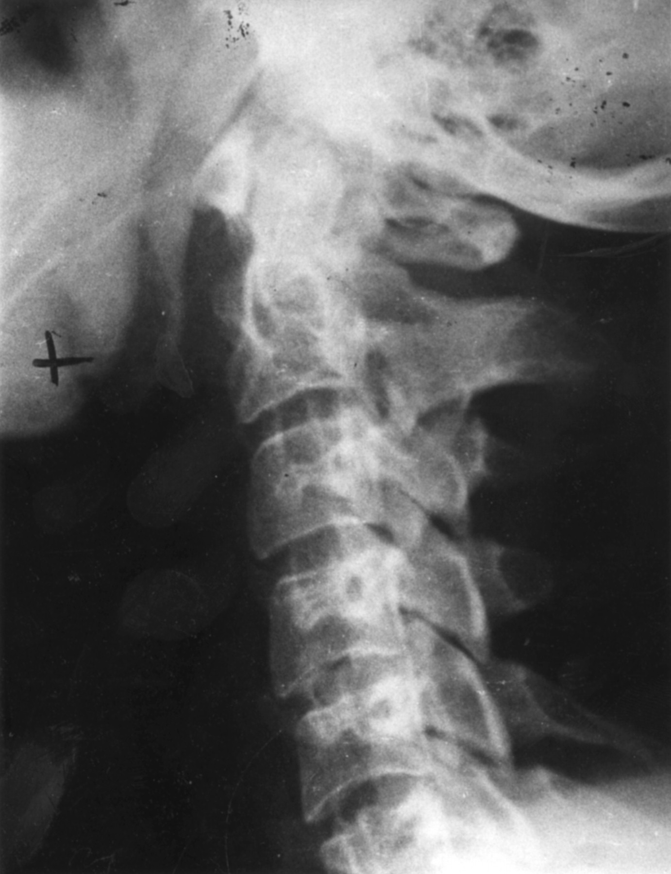
This issue was considered only in group A. OPLL was found in 16 (15.5%; 95%CI 9, 22) of these 103 patients from Guadalajara (fig 1). Table 2 shows the differences in clinical features between patients with and without OPLL. OPLL was statistically associated with older age (p=0.001), longer AS duration (p=0.01), the worst functional index (p=0.042), restricted axial movement measurements (all with p<0.001), clinical myelopathy (p=0.03). OPLL was associated with radiological features of sacroiliitis grade (p<0.001 for linear association), osteitis pubis (p=0.009), ossification of the anterior longitudinal ligament (OALL) (p=0.001), hip joint involvement (p=0.006 for linear association), and with anterior atlantoaxial subluxation (p=0.04). A borderline association with sex and peripheral arthritis was found.

A 44 year old man from the Guadalajara group, with ankylosing spondylitis of 19 years duration. OPLL is present from C3 to C5 (white arrow).
Logistic regression was performed to evaluate the effect of diverse characteristics of the patients on the presence of OPLL; OPLL was significantly associated with occiput wall distance and radiological sacroiliitis, even after adjusting for the other selected independent variables. Other variables such as neck complaints, functional class or HLA-B27 were not statistically associated with OPLL.
PRESENCE OF OPLL IN THE GROUPS (B AND C)
Table 3 shows the differences in clinical features between patients with and without OPLL from these groups. In the patients from Mexico City (B), OPLL was found in 15 (29%; 95%CI 17, 41) patients, including two with psoriatic arthritis and one with Reiter’s syndrome (figs 2 and 3). Patients with OPLL were on average, six years older, had six years longer disease duration, and had a higher proportion of grade IV radiological sacroiliitis and osteitis pubis than those without OPLL.
Selected demographic and clinical data of the patients with OPLL from groups B and C

A 36 year old man from the Mexico city group, with juvenile ankylosing spondylitis of 25 years duration. OPLL is present from C2 to C5.

A 28 year old man with juvenile ankylosing spondylitis of 17 years duration. OPLL is present at C4–5 level.
In the patients from Edmonton (C), OPLL was found in nine (23%, 95%CI 10, 36) patients, including one with psoriatic arthritis, and two with Crohn’s disease. The mean age of patients was similar between those with and without OPLL, as was the mean of the disease duration and proportions with radiological sacroiliitis and osteitis pubis. OPLL was present in two of 72 controls (D); one film from a 72 year old man with DISH and another in an otherwise healthy 21 year old woman.
Discussion
The prevalence of OPLL in patients with AS has not been studied, but the association of both diseases has been occasionally reported.1 ,2 ,24-31 Tsuyama et al stated that 2% of the patients with OPLL in Japan were simultaneously affected by AS1 ,2; however, no detailed information of these patients was given. There are eight reported cases of OPLL in AS patients.24-31 Seven of these were men and the age of the patients ranged from 26 to 73 years. Three patients were orientals. The AS duration, when noted, ranged from two years to 36 years. The presenting complaints of OPLL were tetraparesis after a mild hyperextension injury to the neck in one patient25 and spinal cord compression with gait problems in another24; in the remaining five the OPLL was an unexpected radiological finding in AS patients not having clinically evident neurological symptoms. There is a report of concordant twins, both with AS, but only one had OPLL.28 In all cases the diagnosis of OPLL was made on plain lateral radiographs and in all cases the OPLL was documented in the cervical spine.
In a study designed to assess the prevalence and neurological implications of atlantoaxial subluxation in AS, we found, unexpectedly, a frequency of 15.5% of OPLL.32 With this figure, two main questions arose: what is the clinical importance of OPLL in patients with AS?; and is the high frequency of OPLL restricted to this population or is it present in other geographically and genetically distinct groups?
In group A, OPLL was statistically associated with several variables indicating longer disease duration and more severe axial disease, such as worse Dougados’ functional index33 and restricted axial movement measurements, as well as with radiological features indicating more structural damage, including radiological sacroiliitis, osteitis pubis, hip involvement, OALL, and anterior atlantoaxial subluxation.
Several patients presented some neurological alterations, including clinical data suggesting myelopathy and abnormal SSEP. Although all these were statistically associated with OPLL, in no case was a typical picture of spinal cord compression reported. In the absence of appropiate data from normal controls, the clinical importance of these statistical associations is difficult to determine. Further studies on this aspect should take into account a number of factors that can be concurrent in patients with AS and produce neurological alterations, such as specific type of myelopathy,37myopathy38 and alterations in SSEP,39 all of them ascribed to AS. However, evoked spinal cord potentials recorded with an electrode placed in the cervical epidural space have been described as useful in evaluating myelopathy secondary to OPLL.40 ,41
There are some limitations in this study that merit further comments. The groups from Mexico City and Edmonton were selected from databases because of the availability of cervical radiographs from radiology records. This presupposes a retrospective assessment with selection bias. Consequently, the figures on the frequency of OPLL and its possible associations must be viewed with caution and only as indicating that OPLL is present in some patients with SpA and favouring the notion that OPLL may be associated with variables indicating more severe disease. A further problem is the relative subjectivity in the diagnosis of OPLL and the requirement for a precisely projected lateralx ray beam.8 ,14 ,36 ,42 ,43This was why we felt it important to include some non-AS controls in our study.
With this study it is not possible to ascertain if the OPLL seen in our patients represents an association of OPLL and SpA, or whether OPLL is another manifestation of enthesopathy of the SpA. However, based on the frequency of OPLL in our three groups and its association with numerous clinical variables, we might speculate that the latter possibility is more probable.
In summary, the point prevalence of OPLL was assessed in one prospective and two retrospective groups of SpA from three geographically and genetically different populations. OPLL was found in the three groups with a frequency between 15.5% and 29%, including patients with related SpA, such as Reiter’s syndrome, psoriatic arthritis, and Crohn’s disease. Two of 72 (3%) of the control films (D) were positive. In group A, OPLL was found to be associated with more severe axial disease. Whether OPLL may progress in these patients will require further study.