Article Text

Abstract
Objectives: To confirm and define the genetic association of STAT4 and systemic lupus erythematosus (SLE), investigate the possibility of correlations with differential splicing and/or expression levels, and genetic interaction with IRF5.
Methods: 30 tag SNPs were genotyped in an independent set of Spanish cases and controls. SNPs surviving correction for multiple tests were genotyped in five new sets of cases and controls for replication. STAT4 cDNA was analysed by 5′-RACE PCR and sequencing. Expression levels were measured by quantitative PCR.
Results: In the fine mapping, four SNPs were significant after correction for multiple testing, with rs3821236 and rs3024866 as the strongest signals, followed by the previously associated rs7574865, and by rs1467199. Association was replicated in all cohorts. After conditional regression analyses, two major independent signals, represented by SNPs rs3821236 and rs7574865, remained significant across the sets. These SNPs belong to separate haplotype blocks. High levels of STAT4 expression correlated with SNPs rs3821236, rs3024866 (both in the same haplotype block) and rs7574865 but not with other SNPs. Transcription of alternative tissue-specific exons 1, indicating the presence of tissue-specific promoters of potential importance in the expression of STAT4, was also detected. No interaction with associated SNPs of IRF5 was observed using regression analysis.
Conclusions: These data confirm STAT4 as a susceptibility gene for SLE and suggest the presence of at least two functional variants affecting levels of STAT4. The results also indicate that the genes STAT4 and IRF5 act additively to increase the risk for SLE.
Statistics from Altmetric.com
Systemic lupus erythematosus (SLE) has a strong genetic component supported by high familial aggregation and twin and family studies.1 Like most autoimmune diseases, the HLA has an important contribution2 3 4 and we recently showed that the HLA is the strongest genetic factor in people of European ancestry followed by IRF5 and ITGAM.2 3 4 5 Other somewhat weaker but well-established associations have been found with FCGRIIA,6 PTPN22,7 PDCD1,8 TNFSF4,9 BLK2 3 and most recently, BANK1.10 A genetic association with the signal transducer and activator of transcription 4 (STAT4) was identified in rheumatoid arthritis (RA) with SNP rs7574865 and this association was also found in SLE.11 From the RA studies, the genetic association was defined to the third intron of STAT4. As has been shown for IRF5, several polymorphisms may contribute to the risk, and the risk might also differ among populations.5 Thus, our aim with this study was to revise the STAT4 genetic association using independent sets of Europeans and Latin American populations with a dense set of tag single nucleotide polymorphisms (SNPs) to define if rs7574865 and thus, the third intron signal is the sole genetic contributor to susceptibility in STAT4.
STAT4 is a critical regulator of immune responses, primarily induced by the dendritic cell-produced interleukin 12 (IL12), leading to the development of Th1 cells, which have the ability to secrete high levels of interferon γ (IFNγ). STAT4 is activated after ligation of IL12 to its receptor, which associates with the tyrosine kinases Tyk2 and Jak2.12 13 These are expressed in activated T and B cells and, particularly, NK cells. Activation of STAT4 leads to the formation of homodimers of STAT4 that translocate into the nucleus and induce transcription of IFNγ. In addition, STAT4 activation is also induced by IFNα/β stimulation. This stimulation does not appear to lead to Th1 development, but only to an acute IFNγ secretion by CD4+ T and NK cells where IL18 is also required. IFNα/β induces STAT4 phosphorylation through direct interaction of STAT4 with the IFNαR2 subunit.
Here, we fine mapped a Spanish set of cases and controls. We found evidence for another peak of association beyond the intron 3 SNP rs7574865. This association was replicated in four independent sets of cases and controls. We also found evidence for a correlation between the associated SNPs and expression levels of STAT4 in peripheral blood mononuclear cells (PBMCs). When we analysed the possibility of genetic interaction between STAT4 and IRF5, we found no interaction, but rather an additive increase in the risk for SLE.
Patients and methods
Patients and controls
In total, 1581 cases and 1851 controls were used in this study, all with complete data for the SNPs analysed: 390 cases and 620 controls from Spain, 247 cases and 220 controls from Germany, 221 patients and 207 controls from Italy, 171 patients and 171 controls from Argentina, and 231 cases and 250 controls from Mexico (adults) along with a set of 321 paediatric patients and 383 adult controls. The patient and control sets studied here have been described previously.10 14 All patients fulfilled the 1982 American College of Rheumatology criteria for the classification of SLE.
Selection of tag SNPs
Twenty-nine tag SNPs covering the STAT1 and STAT4 genes, and the intergenic region, were selected using Haploview version 3.32 from the HapMap-CEU population genotype data. Aggressive tagging mode was used to select tag SNPs with a minor allele frequency ⩾5%, with an r2 threshold ⩾0.8. rs7574865 was added to the tag list after being reported as associated with RA.11 SNPs associated in the Spanish fine mapping, after quality control and correction for multiple testing, were typed in the German, Italian, Argentinian, and both Mexican sets.
Genotyping
Spanish samples were genotyped in Granada using TaqMan 5′ exonuclease assay (ABI, Foster City, California, USA). German, Italian and Latin American samples were genotyped at Uppsala University. Mexican paediatric samples were genotyped at Instituto Nacional de Medicina Genómica using the same method. Genotyping consistency between the centres was established to be near to 100%.15
Statistical analyses
The Spanish genotype data was processed using Haploview version 4.0,16 PLINK version 1.0217 and R. Quality control filters were applied to remove SNPs with >10% missing data in cases or controls (one SNP excluded), deviations from Hardy–Weinberg equilibrium (p<0.001, two SNPs excluded), or minor allele frequency <5% in controls (two SNPs excluded). Twenty-five SNPs remained. Subjects with an individual missing genotyping rate >10% were also removed (n = 42). Genotyping rate in the remaining subjects was 97.4%. Pairwise linkage disequilibrium (LD) measures (D′ and r2) between SNPs and maximum-likelihood haplotype frequencies were estimated with the EM algorithm. Multiple testing was corrected by Bonferroni and false discovery rate methods.18
Statistical analysis of the replication sets included only subjects with 100% individual genotyping calls. DerSimonian–Laird and Cochran–Mantel–Haenszel methods implemented in StatsDirect and PLINK were used to estimate the meta-analysis odds ratio for all populations assuming random and fixed effects models on the allelic association, respectively. The heterogeneity test based on partitioning the χ2 statistic implemented in PLINK, was used to test between-population differences. Univariate genotypic odds ratios were estimated by logistic regression.20 These were adjusted by adding the stratification variable “population” to the logistic regression model containing the genotype as the exposure variable. This test is identical to the one degree of freedom Mantel–Haenszel test of the hypothesis that the stratum-specific odds ratios are 1.19 Conditional logistic regression was used to determine independence of the SNPs from rs7574865. All logistic regression analyses were done with R.
Multiple logistic regression was used to evaluate if additive or interaction effects were present between SNPs within STAT4 and IRF5. To measure the ability to discriminate between SLE cases and controls, the area under the receiver operating characteristic curve (C statistic) was calculated. To statistically compare the C statistics we applied the method of DeLong et al.21
5′-RACE and 3′-RACE PCR
Marathon-ready cDNA from different tissues (Clontech, Mountain View, California, USA) was used as template for amplification of tissue-specific 5′- and 3′-UTRs. The following pairs of nested gene-specific primers were used for 5′-RACE PCR: 5′-GAAATTCTACTGAGAGACTCCCATTG-3′ and 5′-GAATCGTTGCCATGGTTTCATTGTTAG-3′ and for 3′-RACE PCR: 5′-CTAAACTATCAGGTAAAGGTTAAGGCATC-3′ and 5′-GGTAAACACTACAGCTCTCAGCCTTG-3′. Adapter primers Ap1 and Ap2 were provided with cDNA. Nested PCR was carried out using 1/30 of the first-round PCR products. Thirty-five cycles of PCR (95°C for 20 s, 60°C for 15 s and 72°C for 3 min) were performed after initial denaturation at 95°C for 5 min in buffer containing 1.5 mM MgCl2, 200 μM of each of dNTPs, 0.4 μM of each of the corresponding primers and 0.5 U of Platinum Taq high fidelity enzyme (Invitrogen, Carlsbad, California, USA). PCR products were analysed by sequencing.
RNA purification and STAT4 expression analysis
Total RNA was purified as described elsewhere10 from PBMCs obtained with agreed consent from 73 healthy volunteers. RNA (2 μg ) was reverse transcribed using oligo-dT primers according to the manufacturer’s protocol (Applied Biosystems, Foster City, California, USA).
STAT4 expression was determined by real-time PCR using SYBR Green detection. Cycling conditions were as follows: 95°C for 5 min, 45 cycles of PCR (95°C for 15 s, 60°C for 10 s and 72°C for 20 s). α Isoform was detected with the following primers: forward 5′-CATCTCAACAATCCGAAGTGATTCA-3′ and common reverse primer 5′-GTCAGAGTTTATCCTGTCATTCAGCAG-3′. β-Isoform-specific forward primer was 5′-TGACCTTGTTATCTCTTTAAGCCGA-3′. Expression levels were normalised against TATA-binding protein gene expression amplified with commercial reagents (Applied Biosystems). All experiments were run in triplicate.
Statistical analysis of gene expression
Analysis of variance and F test were used to determine the difference in the mRNA expression level in relation to each of the SNPs, taking the three possible genotypes as factor levels. Significance of the gene expression was also tested with linear regression.22
Results
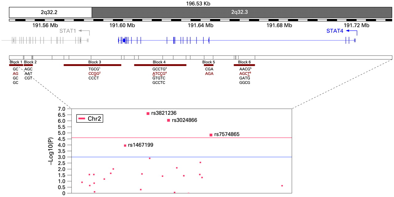
Association was detected for several SNPs across STAT4 and the STAT1–STAT4 intergenic region, with the strongest association with rs3821236 (p = 7.07×10⁻⁸), followed by rs3024866 (p = 3.83×10⁻⁷), rs7574865 (p = 9.37×10⁻⁶) and rs1467199 (p = 7×10⁻⁵), all of which remained significant after correction for multiple tests (table 1). The region LD structure defined six haplotype blocks, two located in STAT1 (blocks 1 and 2), one in the intergenic region (block 3) and three in STAT4 (blocks 4–6) (fig 1 and online supplementary fig 1). Our results indicate association of two additional haplotype blocks than that containing rs7574865 (block 6): block 3, which contains rs1467199, and block 4 harbouring rs3821236 and rs3024866 (fig 1).

Fine mapping of the STAT1–STAT4 region. The physical position (top panel) of the SNPs typed in 390 patients and 480 controls from Spain covering the ∼200 kb STAT1– STAT4 region is shown. The region linkage disequilibrium (LD) structure defined six haplotype blocks (middle panel) of which three were associated with disease susceptibility (see block structure and R2 values in supplementary fig 1). Risk haplotypes are shown in red and main single-marker hits replicated are underlined (see table 2 and supplementary table 1). The bottom panel shows the significance of the association data presented as the –log10 (p value) for 25 tag SNPs passing genotyping quality control. The blocks have been defined using the solid spine of the LD method in Haploview version 4.0. The p values of the risk and protective haplotypes at each block in the Spanish population are as follows: 1block 3 risk haplotype p value = 0.0037; 2block 3 protective haplotype p value = 7×10⁻⁴; 3block 4 risk haplotype p value = 2.64×10⁻⁶; 4block 4 protective haplotype p value = 2.24×10⁻⁵; 5block 6 risk haplotype p value = 9.52×10⁻⁶; 6block 6 protective haplotype p value = 0.0331.
Results from the fine mapping conducted in Spanish patients with systemic lupus erythematosus and matched controls
To replicate the genetic associations and increase statistical power, we genotyped the associated SNPs in five independent sets from Italy, Germany, Argentina, and Mexico (one adult and one paediatric set). Homogeneity test showed that the odds ratios for the SNPs could be combined, except rs3024866 and rs7574865, which had some heterogeneity across the strata. None of the SNPs provided association with the German population except for a borderline association with rs7574865 (table 2 and supplementary table 1). The Mexican paediatric set showed association only with rs1467199 (p = 0.008, supplementary table 2). Despite the low heterogeneity introduced by the German set (homogeneity p = 0.01), it was included in the meta-analysis. The Mexican paediatric set displayed the highest heterogeneity and it could not be combined (compare supplementary table 2 with table 2). In the meta-analysis, SNPs rs3821236 (p = 5.96×10⁻²⁰) and rs7574865 (p = 4.44×10⁻²³) showed the strongest association across all strata in the allelic and genotypic tests, but rs3024866 was also associated (p = 2.31×10⁻¹²) (table 2). Although rs1467199 reached significant association in the meta-analysis, at the individual population level it was only replicated in the Argentine set (rs1467199-CG p = 1.63×10⁻², rs1467199-GG p = 0.053) (supplementary table 1).
Population-specific replication and general stratified allelic association analysis of the main associated single nucleotide polymorphisms (SNPs) in the fine mapping
We tested whether the associated SNPs constituted independent effects. rs7574865 is not in strong LD with rs3024866 (R2 = 0.29) or rs3821236 (R2 = 0.42), which are located 62 kb and 42 kb from rs7574865, respectively, and therefore they are not proxies. rs1467199 has R2 = 0.30 with rs3821236, R2 = 0.18 with rs3024866 and R2 = 0.13 with rs7574865 (fig 1). The low pairwise correlation coefficients (R2) suggest that the individual SNP associations reflect independent effects, except for rs3024866, which has a relatively high R2 with rs3821236 (R2 = 0.64) (fig 1). This was confirmed by conditional logistic regression analysis: rs3821236 remained significant when conditioning on rs7574865, and vice versa (supplementary table 3). Thus, rs3821236 and rs7574865 represent two independent genetic effects within STAT4. rs3821236 is located in the 16th intron, ∼60 kb downstream from the third intron where the association has been confined in previous studies.11 SNPs rs3821236 and rs3024866 are tagging a 26 kb haplotype block (block 4) covering part of the gene between intron 8 and 16 (fig 1 and supplementary fig 1) that contain three of the six markers associated with SLE in the Spanish fine mapping after multiple test correction (table 1). After a conditional haplotype-based association test the haplotype block 4 (SNPs rs3821236 and rs932169) remain significant (p = 0.002277) after controlling for rs7574865, confirming its independency.
Usage and splicing of 5′- and 3′-UTRs in STAT4
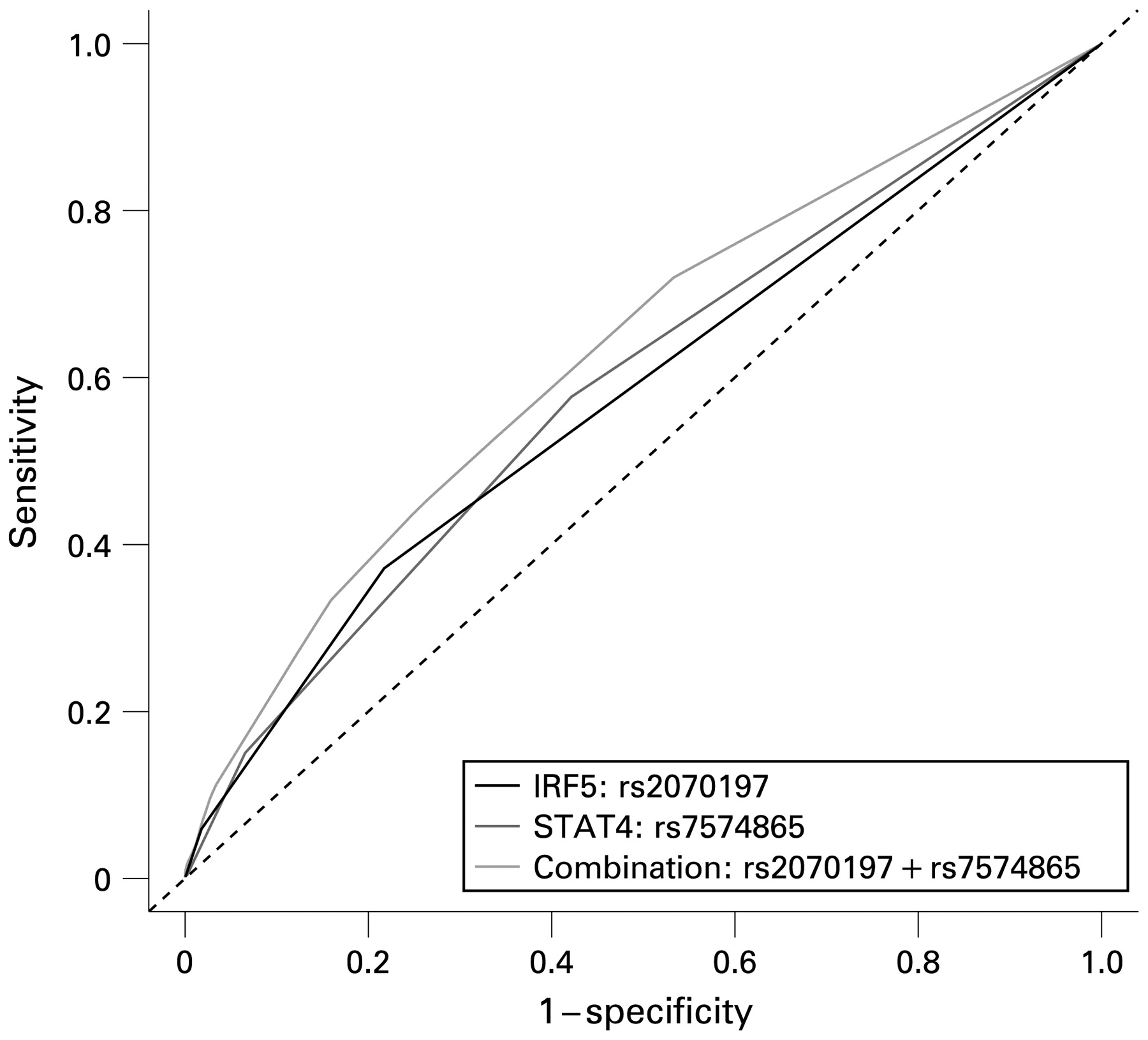
Using the receiver operating characteristic curve and C statistic, we observed that rs7574865 was a somewhat better risk predictor (C = 0.590) than rs3821236 (C = 0.577), but the difference did not reach statistical significance (supplementary table 4). Thus, each SNP alone provides the same level of prediction of the risk for SLE, independently.
Statistical analysis of the interaction with IRF5
IRF5 was found as a well-established non-major histocompatibility complex association with SLE. To study if the effect of STAT4 was independent of IRF5, or if there were epistatic effects between SNPs in these genes we performed multiple logistic regression analysis and C statistic including the genotype data for three SNPs of IRF5 (rs2004640, rs2070197 and rs10954213) from our previous studies,5 14 The analyses revealed no significant interaction effects between STAT4 and IRF5 SNPs, but close to complete independent effects on SLE risk as shown by a slight change in estimates when combined in a multivariate model (data not shown).
The three SNPs of IRF5 had C statistics of 0.587 0.585 and 0.565, respectively. (supplementary table 5). rs2070197 tags the major risk haplotype and has the highest predictive ability.14 Addition of rs7574865 to the IRF5 SNPs increased the predictive value of the models significantly (rs7574865 with rs2004640: C statistic = 0.632, p = 1.66×10⁻⁵; rs7574865 with rs2070197: C statistic = 0.636, p = 3.28×10⁻¹¹ and rs7574865 with rs10954213: C statistic = 0.624, p = 9.62×10⁻⁷). The results support an additive effect of SNPs of both genes, particularly rs7574865 and rs2070197, to increase risk for SLE (fig 2).

Predictive ability of STAT4 and IRF5 single nucleotide polymorphisms (SNPs) for systemic lupus erythematosus (SLE). The predictive ability of the two genes for SLE was investigated using the C statistic. The test to compare two dependent receiver operating characteristic curves is used. Within STAT4, the SNP rs7574865 is the strongest predictor for SLE and adds a significant fraction to the predictive ability of the IRF5 SNP rs2070197. Between genes the best combination is rs7574865+rs2070197 having an overall C statistic of 0.636.
Tissue-specific alternative transcripts of STAT4
In order to investigate if differential splicing could be related to the genetic association of STAT4, functional annotation of gene transcripts expressed in different tissues was performed. Until now, two isoforms have been described for STAT4, α and β.23 Only the known α and β isoforms were detected when we tested spleen testis, kidney, lung, pancreas, intestine and uterus for new isoforms.
Since 5′- and 3′-UTRs may substantially affect gene expression,14 we performed detailed analysis of non-coding 5′-exons and 3′-UTRs. The pattern of 5′-UTRs was diverse in different tissues (table 3), but all of them led to the known isoforms of STAT4.
Analysis of STAT4 expression levels
Levels of STAT4 gene expression in human mononuclear cells were assessed by quantitative real-time PCR. Since expression of the β transcript was much lower than of the α transcript and its expression followed the trend of the α transcript in all samples, it was excluded from the analysis. By using analysis of variance to test if differences in gene expression correlated with genotypes, we found a modest upregulation of STAT4 mRNA with the CC risk genotype of rs3024866 (p = 0.0281). By regression analysis, risk alleles of SNPs rs3821236 and rs7574865 correlated with higher STAT4 expression. Importantly, SNP rs1467199 did not correlate with STAT4 expression (fig 3).

Association between STAT4 polymorphism and expression levels. Analysis of variance and F test were used to determine difference in the mRNA relative expression levels between subjects carrying different genotypes, taking the three possible genotypes as factor levels and the major allele homozygous as reference. Multiple comparisons of means revealed that subjects with rs3024866-CC genotype have higher STAT4 expression levels than those with rs3024866-TT (p value = 0.0281)
Discussion
We confirmed the genetic association between STAT4 and SLE and through a fine mapping effort we identified a second effect independent of rs7574865 located in the vicinity of intron 16.
The publication of Remmers, et al,11 identifying STAT4 as a susceptibility gene for RA and SLE was published while our fine mapping effort was continuing, and after having identified a strong signal for this gene in a 100k scan in Argentine subjects (data not shown). The data from the Spanish mapping suggested the presence of several peaks independent of rs7574865. Analyses using a larger set of joined samples confirmed the independent effect of rs3821236 in intron 16. Conditional SNP and haplotype regression analysis supported this result.
We observed differences between north and south European samples: the German set contributed, albeit weakly, to the association only at rs7574865, whereas the association of rs3821236 was contributed particularly by the Spanish, Italian and Latin American sets. In our view it is highly plausible that several independent risk haplotypes are involved in disease susceptibility, with some having stronger effects in some populations than others.
The weak result of the German set could not be due to a lack of power, as the size of the other sets is comparable.
We are approaching a phase in complex disease genetics where identification of the genes involved in disease susceptibility is becoming a reality. Therefore, it is of interest that we understand the relationship between the various genetic effects. Here we examined the possibility of genetic interaction between IRF5 and STAT4. We observed no genetic interaction but we observed a significant increase in the predictive ability for SLE when STAT4 and IRF5 SNPs were added. These results suggest that the IRF5 and STAT4 SNPs act additively to increase the risk for SLE. While this work was under revision, an independent study corroborated this additive effect.24
Identification of the functional variants for STAT4 might prove relatively difficult considering the large size of this gene and the location of the associated SNPs. Complete resequencing of the gene and genotyping may be needed to reveal the true functional variants. We observed no differences in splicing of the gene in PBMCs. Instead we found a correlation between expression levels of STAT4 in PBMCs and the STAT4 associated SNPs, rs7574865, rs3024866 and rs3821236 but not rs1467199. It should be noted that PBMC samples are expected to have varying numbers of T and NK cells, high variation in gene expression and a weak correlation. Further, in complex diseases we also expect effects caused by functional variants to be modest and difficult to define.14
How does a modest increase in STAT4 expression contribute to the risk for SLE? STAT4 is a transcription factor through which the functional effects of IL12 are conducted, leading to IFNγ production. The STAT4 pathway has been studied in mice and patients with SLE, with contradictory results. In two studies, mice deficient for STAT4 show increased development of glomerulonephritis,25 26 whereas a third study reported opposite findings, in line with our observations.27
We detected only two isoforms of STAT4, one of which was expressed at extremely low levels (β), and tissue-specific promoters as observed by the presence of alternative, tissue-specific 5′-UTRs (table 3). Given that such promoters could adjust transcription of the gene in a particular tissue, this poses an additional obstacle for defining the role of genetic variation. Other authors showed that the SLE risk haplotype of STAT4 is overexpressed in mesenchymal cells,24 supporting a tissue-specific component involved in the regulation of STAT4 expression. Using purified cell populations (eg, NK cells, which constitute only 5% of the blood leukocytes but have high basal level of STAT4, kidney mesangial cells, etc) may be critical for correct assessment of the altered levels of STAT4 gene expression as well as definition of the splicing isoforms that could be involved.
Major effects of the expression of human STAT4 may be important in the kidney. Activation of STAT4 leads to increased expression of IFNγ. A study has shown that increased expression of IL12 and IFNγ in the kidneys of MRL-lpr/lpr mice precedes the development of glomerulonephritis.28 29. Thus, the localised action of the various genes in specific tissues may be of importance. This is also important to consider in view of the recent results showing a strong correlation between rs7574865 and end-organ disease, in particular, kidney disease.30
Our results do support an important role of STAT4 in SLE susceptibility, a role that appears to vary between different populations and derives from two different and independent risk variants, whose functional nature needs to be examined.
Acknowledgments
We express our sincere gratitude to Susanna Lewén for help with purification of PBMCs and total RNA, and Hong Yin for help with the preparation of DNA samples. We also thank Adriana I Scollo, Armando M Perichon and Mariano CR Tenaglia, CEDIM, Diagnóstico Molecular y Forense SRL, Rosario, Argentina, for their help in DNA preparation of the Argentinian samples. We thank particularly the Lupus Patient Association of Asturias for their help in the collection of samples and all the patients for their contribution.
The Argentine collaborative group participants are: Hugo R Scherbarth MD, Pilar C Marino MD, Estela L Motta MD Servicio de Reumatología, Hospital Interzonal General de Agudos “Dr Oscar Alende”, Mar del Plata, Argentina; Susana Gamron MD, Cristina Drenkard MD, Emilia Menso MD Servicio de Reumatología de la UHMI 1, Hospital Nacional de Clínicas, Universidad Nacional de Córdoba, Córdoba, Argentina; Alberto Allievi MD, Guillermo A Tate MD Organización Médica de Investigación, Buenos Aires, Argentina; Jose L Presas MD Hospital General de Agudos Dr Juán A Fernandez, Buenos Aires, Argentina; Simon A Palatnik MD, Marcelo Abdala MD, Mariela Bearzotti PhD Facultad de Ciencias Medicas, Universidad Nacional de Rosario y Hospital Provincial del Centenario, Rosario, Argentina; Alejandro Alvarellos MD, Francisco Caeiro MD, Ana Bertoli MD Servicio de Reumatología, Hospital Privado, Centro Medico de Córdoba, Córdoba, Argentina; Sergio Paira MD, Susana Roverano MD, Hospital José M Cullen, Santa Fe, Argentina; Cesar E Graf MD, Estela Bertero PhD Hospital San Martín, Paraná; Cesar Caprarulo MD, Griselda Buchanan PhD Hospital Felipe Heras, Concordia, Entre Ríos, Argentina; Carolina Guillerón MD, Sebastian Grimaudo PhD, Jorge Manni MD Departamento de Inmunología, Instituto de Investigaciones Médicas “Alfredo Lanari”, Buenos Aires, Argentina; Luis J Catoggio MD, Enrique R Soriano MD, Carlos D Santos MD Sección Reumatología, Servicio de Clínica Medica, Hospital Italiano de Buenos Aires y Fundación Dr Pedro M Catoggio para el Progreso de la Reumatología, Buenos Aires, Argentina; Cristina Prigione MD, Fernando A Ramos MD, Sandra M Navarro MD Servicio de Reumatología, Hospital Provincial de Rosario, Rosario, Argentina; Guillermo A Berbotto MD, Marisa Jorfen MD, Elisa J Romero PhD Servicio de Reumatología Hospital Escuela Eva Perón Granadero Baigorria, Rosario, Argentina; Mercedes A Garcia MD, Juan C Marcos MD, Ana I Marcos MD Servicio de Reumatología, Hospital Interzonal General de Agudos General San Martín, La Plata; Carlos E Perandones MD, Alicia Eimon MD Centro de Educación Médica e Investigaciones Clínicas (CEMIC), Buenos Aires, Argentina; Cristina G Battagliotti MD Hospital de Niños Dr Orlando Alassia, Santa Fe, Argentina.
Bernardo Pons-Estel is the coordinator of the Argentine collaborative group.
The German collaborative group participants are: K Armadi-Simab, MD, Wolfgang L Gross, MD, Abteilung Rheumatologie, University Hospital of Schleswig-Holstein, Campus Luebeck, Rheumaklinik Bad Bramstedt, Luebeck, Germany, Erika Gromnica-Ihle, MD, Rheumaklinik Berlin-Buch, Berlin, Germany, Hans-Hartmut Peter, MD, Medizinische Universitaetsklinik, Abteilung Rheumatologie und Klinische Immunologie, Freiburg, Germany, Karin Manger, MD, Medizinische Klinik III derFAU Erlangen-Nuernberg, Erlangen, Germany, Sebastian Schnarr, MD, Henning Zeidler, MD, Abteilung Rheumatologie, Medizinische Hochschule Hannover, Hannover, Germany, Reinhold E Schmidt, MD, Abteilung Klinische Immunologie, Medizinische Hochschule Hannover, Hannover, Germany.
The Italian collaborative participants are: Gian Domenico Sebastiani (UOC di Reumatologia Ospedale San Camillo, Roma – Italy), Enrica Bozzolo (IRCCS San Raffaele Hospital, Milan, Italy), Mauro Galeazzi, (Department of Clinical Medicine and Immunology Sciences, Section of Rheumatology, Siena University, Siena, Italy), Nadia Barizzone (Department of Medical Sciences, University of Eastern Piedmont, Novara, Italy) and Maria Giovanna Danieli and Professor Armando Gabrielli (Dipartimento di Scienze Mediche e Chirurgiche, Università Politecnica delle Marche, Ancona, Italy).
The AADEA (Andalusian Association of Autoimmune Diseases) group participants are: José L Callejas-Rubio, Servicio de Medicina Interna, Hospital Clínico San Cecilio, Granada; Juan Jiménez-Alonso and Mario Sabio, Servicio de Medicina Interna, Hospital Virgen de las Nieves, Granada; Julio Sánchez-Román and Francisco J García-Hernández, Servicio de Medicina Interna, Hospital Virgen del Rocio, Sevilla; Enrique De-Ramon and Mayte Camps, Servicio Medicina Interna, Hospital Carlos Haya, Málaga; Rosa García-Portales, Servicio Reumatología, Hospital Virgen de la Victoria, Málaga; Miguel A López-Nevot, Servicio de Inmunología, Hospital Virgen de las Nieves, Granada.
REFERENCES
Supplementary materials
web only data 68/11/1746
Files in this Data Supplement:
Footnotes
Additional figures and tables are published online only at http://ard.bmj.com/content/vol68/issue11
A-KA, AMD-V, SVK, ES, JM and MEA-R, at their respective positions, contributed equally to this work
Funding This work has been supported in part by grants from the European CVDIMMUNE project from the European Commission LSHM-CT-2006-037227, the Swedish Research Council (12673), the Torsten and Ragnar Söderbersstiftelse, the Swedish Association Against Rheumatism, the King Gustaf the Vth 80th-Jubilee Foundation and the Knut and Alice Wallenberg Foundation for supporting MEAR through the Royal Swedish Academy of Sciences. This study was also supported by grant SAF2006-00398 from the Spanish Ministerio de Educacion y Ciencia, grant PI052409 from the Fondo de Investigación Sanitaria (Spain), C2.12 from BMBF Kompetenznetz Rheuma in Germany and FISM, Regione Piemonte (CIPE) and the Consejo Nacional de Ciencia y Tecnología (CONACYT: SALUD-2004-01-153). MEAR is a Greenberg Scholar at the OMRF.
Competing interests JW and HA are employees of Merck Serono Inc and produced the Argentine 100k data on which our investigation and search for STAT4 variants was first based.
Ethics approval Ethics committee approval from each of the participating institutions.