Article Text

Abstract
Background Adhesion molecule CD44 enables T lymphocytes' adhesion to endothelium. During inflammation, increased expression of CD44 contributes to T cell migration into target organs. Infiltration of peripheral tissues is crucial in the development of SLE organ damage and the different isoforms of CD44 seem to be involved in this process. Both CD44v3 and CD44v6 isoforms have been found in kidney biopsies of SLE patients, and CD44v3 in the skin only1,2. A higher expression of CD44v3 and v6 has been identified on T cells from SLE patients compared to healthy subjects (HS) and the expression levels seem to correlate with disease activity3.
Objectives The aim of this study was to investigate the expression of the CD44v3/v6 isoforms on T cells of SLE patients and their correlation with disease activity and clinical phenotype.
Methods We enrolled 23 patients (23F, mean age±SD 45.7±13 years, mean disease duration±SD 13±8years) affected by SLE according to the 1997 ACR criteria, and 14 HS (14F, mean age±SD 34.28±12.7 years). Disease activity was measured by SLEDAI-2K. 10 patients were in remission (SLEDAI-2K=0) and 13 patients had an active disease (SLEDAI-2K≥4). Expression of CD44v3 and v6 on T cells was determined by flow cytometry analysis.
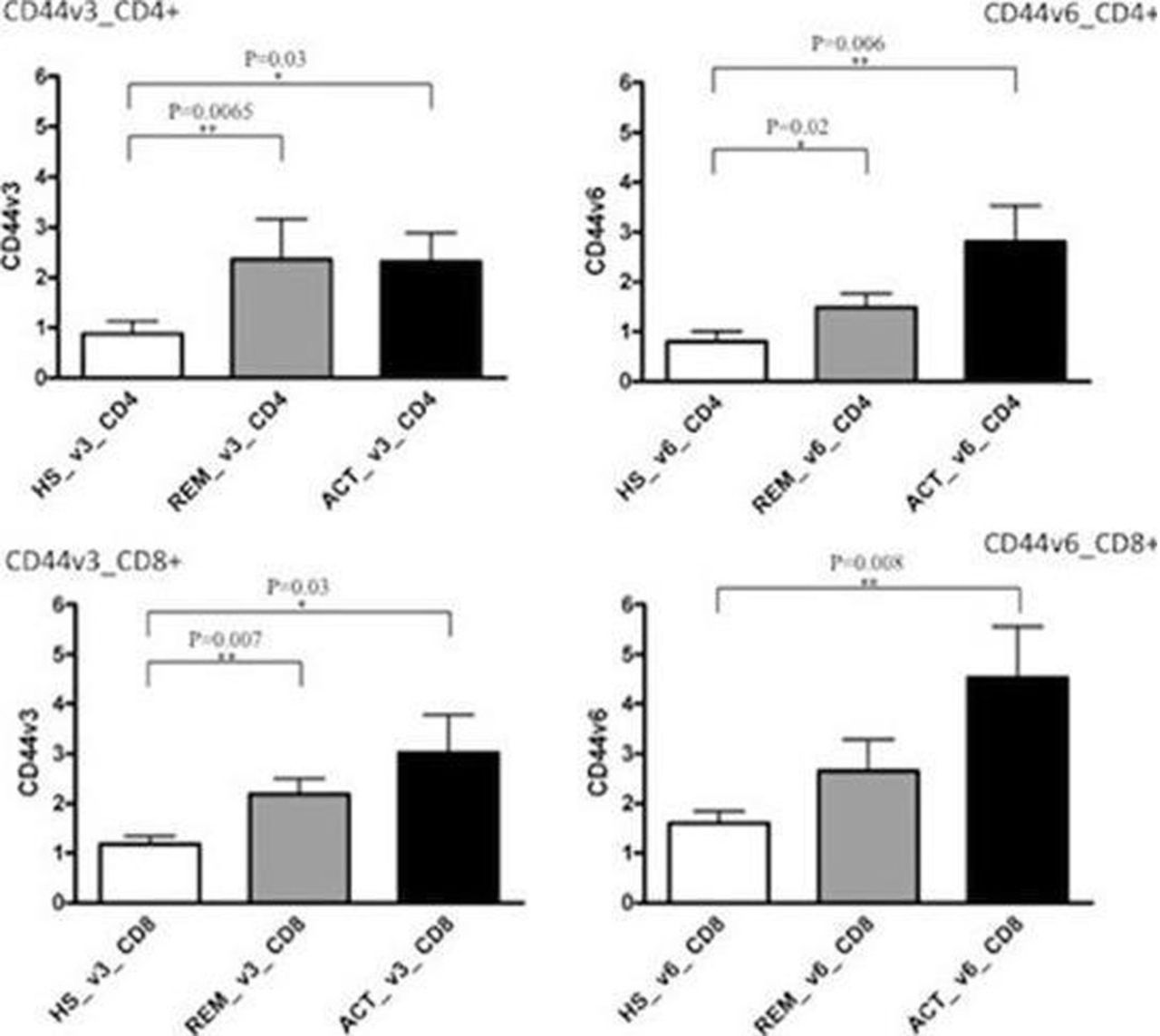
Results Expression of CD44v3 and v6 was significantly higher in active and remission patients compared to HS on CD4+ and CD8+ T cells. SLE patients with active disease showed a trend of major expression of CD44v3 and v6 on CD4+ and CD8+ cells compared to patients in remission (Fig.1). CD44v3/CD44v6 expression ratio on CD4+ and CD8+ T cells was shifted towards isoform v3 on CD4+ cells and towards isoform v6 on CD8+ cells in SLE patients in remission and HS. In active disease this ratio was shifted towards isoform v6 on both T cells populations (Table 1). By using a ROC curve analysis, CD44v6 on CD4+ T cells resulted the most sensitive and specific one (sensitivity 82.6%, specificity 78.6%). Finally, we observed a significant correlation between CD44v3 on CD4+ cells and skin involvement (P=0.027, r=0.632).

Conclusions Our study confirms previous evidences suggesting a higher expression of CD44v3 and v6 on T cells from SLE patients compared to HS. Higher expression of CD44v3 and v6 on patients with active disease suggests their possible use as biomarkers of disease activity. The good specificity and sensitivity of CD44v6 on CD4+ T cells, and the shift of the ratio towards this isoform in active patients, suggest a stronger value of this isoform as a biomarker of disease activity.
References
Cohen RA, Bayliss G, Crispín JC et al. T cells and in situ cryoglobulin deposition in the pathogenesis of lupus nephritis. Clin Immunol 2008;128:1–7.
Seiter S, Schadendorf D, Tilgen et al. CD44 variant isoform expression in a variety of skin-associated autoimmune diseases. Clin Immunol Immunopathol 1998;89:79–93.
Crispín JC, Keenan BT, Finnell MD et al. Expression of CD44 variant isoforms CD44v3 and CD44v6 is increased on T cells from patients with systemic lupus erythematosus and is correlated with disease activity. Arthritis Rheum 2010;62:1431–7.
References
Disclosure of Interest None declared