Article Text

Abstract
Background Aromatase Inhibitors (AIs) block physiological estrogen production in peripheral tissues and significantly improve overall survival rates of post-menopausal, hormone receptor-positive breast cancer patients by reducing tumor recurrences. However, half of patients taking these drugs develop aromatase inhibitor induced arthralgia (AIIA), which is characterized by severe pain and inflammation in various joints. Since AIIA leads to suspension of therapy in 20% of patients, reducing incidence may provide sustained AI treatment and enhanced long-term survival.
Objectives In order to establish a better understanding of the inflammatory mechanism and to create a platform that can be used to explore interventional strategies, our objective in this study was to design a novel animal model of AIIA.
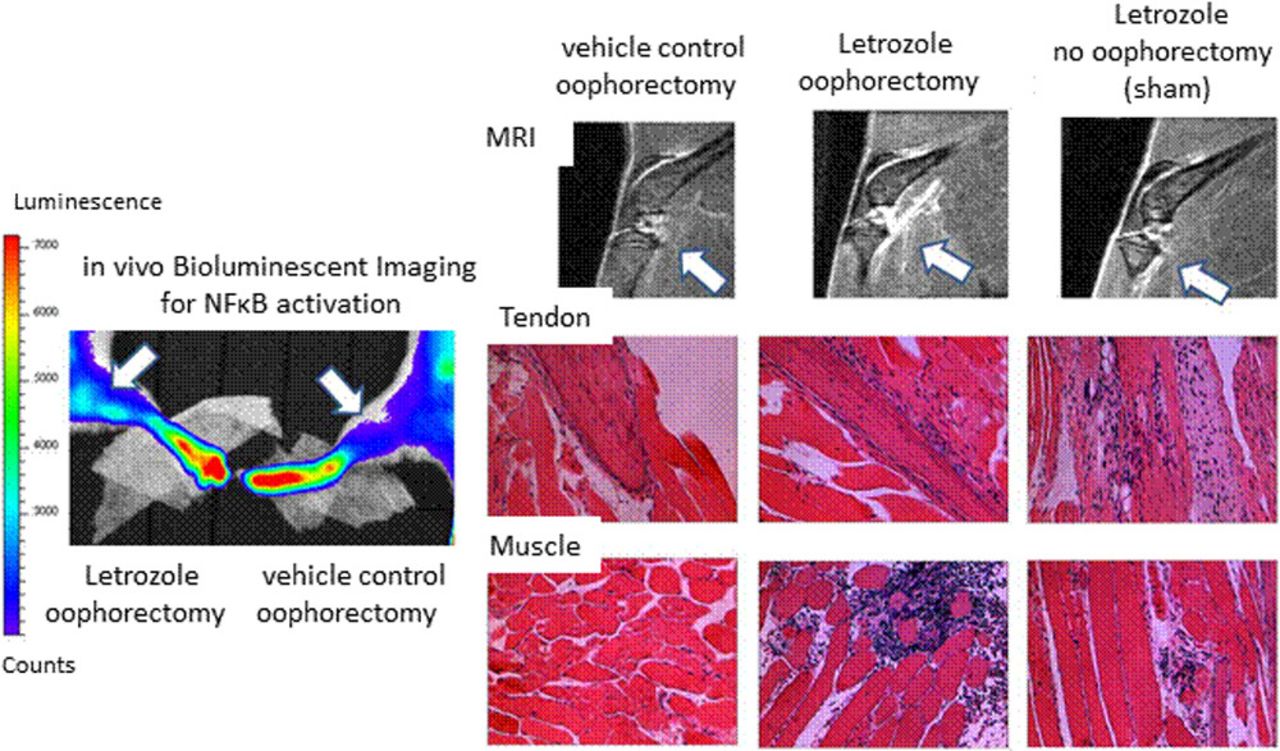
Methods Female BALB/C-Tg (NFκB-RE-luc)-Xen mice, which have a firefly luciferase cDNA reporter gene under the regulation of 3 κB responsive binding sites, were oophorectomized and treated with AI (letrozole) by daily subcutaneous injections. Control groups included oophorectomized mice receiving vehicle control injections and non-oophorectomized mice treated with AI. Bioluminescent imaging of hind limbs was performed after 3 weeks on the in vivo imaging system (IVIS) to measure NFκB activation. At 5 weeks, knee joints and surrounding tissue were imaged on the BioSpec 94/30 micro-MRI. Legs were collected for histopathological analysis and serum cytokine levels were measured at experimental endpoint.
Results Bioluminescent imaging showed significantly enhanced NFκB activation in the hind limbs compared to oophorectomized controls receiving vehicle treatment. Analysis of knee joints and legs by MRI imaging showed enhanced signal detection in the joint space and surrounding tissue following AI treatment. Surprisingly, enhanced MRI detection was also demonstrated in non-oophorectomized mice that were treated with AI. Histopathological analysis further demonstrated mild inflammation in the synovial tissue and joint damage in mice treated receiving AI both with and without oophorectomy. Moreover, tenosynovitis and inflammatory muscle tissue infiltrates were detected in AI-treated mice and serum cytokine levels of IL-2, IL-4, IL-6, and CXCL1 were significantly elevated.

Conclusions Collectively, these data establish a novel mouse model of AIIA and suggest that the pathogenesis of AI-induced inflammation is estrogen-independent. Future studies will be directed into the characterization of this inflammatory mechanism to provide insight into potential therapeutic strategies directed at mitigating this adverse inflammatory burden.
References
Henry NL, Giles JT, Ang D, et al. Prospective characterization of musculoskeletal symptoms in early stage breast cancer patients treated with aromatase inhibitors. Breast cancer research and treatment. 2008;111(2):365–372.
Mao JJ, Stricker C, Bruner D, et al. Patterns and risk factors associated with aromatase inhibitor-related arthralgia among breast cancer survivors. Cancer. 2009;115(16):3631–3639.
References
Acknowledgements Stefanie Spielman Fund for Breast Cancer Research and The Wexner Medical Center.
Disclosure of Interest None declared