Article Text

Abstract
Background Musculoskeletal ultrasonography (US) has become an important diagnostic tool for the diagnosis and follow-up of inflammatory joint diseases in pediatric rheumatology. However, there is lack of clinical studies in pediatric joint ultrasonography concerning normal findings of healthy joints and periarticular structures in children.
Objectives The aim of the study was to evaluate normal values for the thickness of healthy joint cartilage, capsule and tendons of the hand (finger/wrist), elbow, hip, knee, ankle and forefoot in children by musculoskeletal ultrasound (US). Furthermore, power Doppler activity was measured.
Methods The joints were assessed by four physicians experienced in musculoskeletal US with equally preset Esaote Mylab Twice with high frequency (13 to 18 MHz) probes. N=75 healthy children (41 boys, 34 girls) between 4.9–11.1 years (girls) and 4.3–13.1 years (boys) were included in the study.
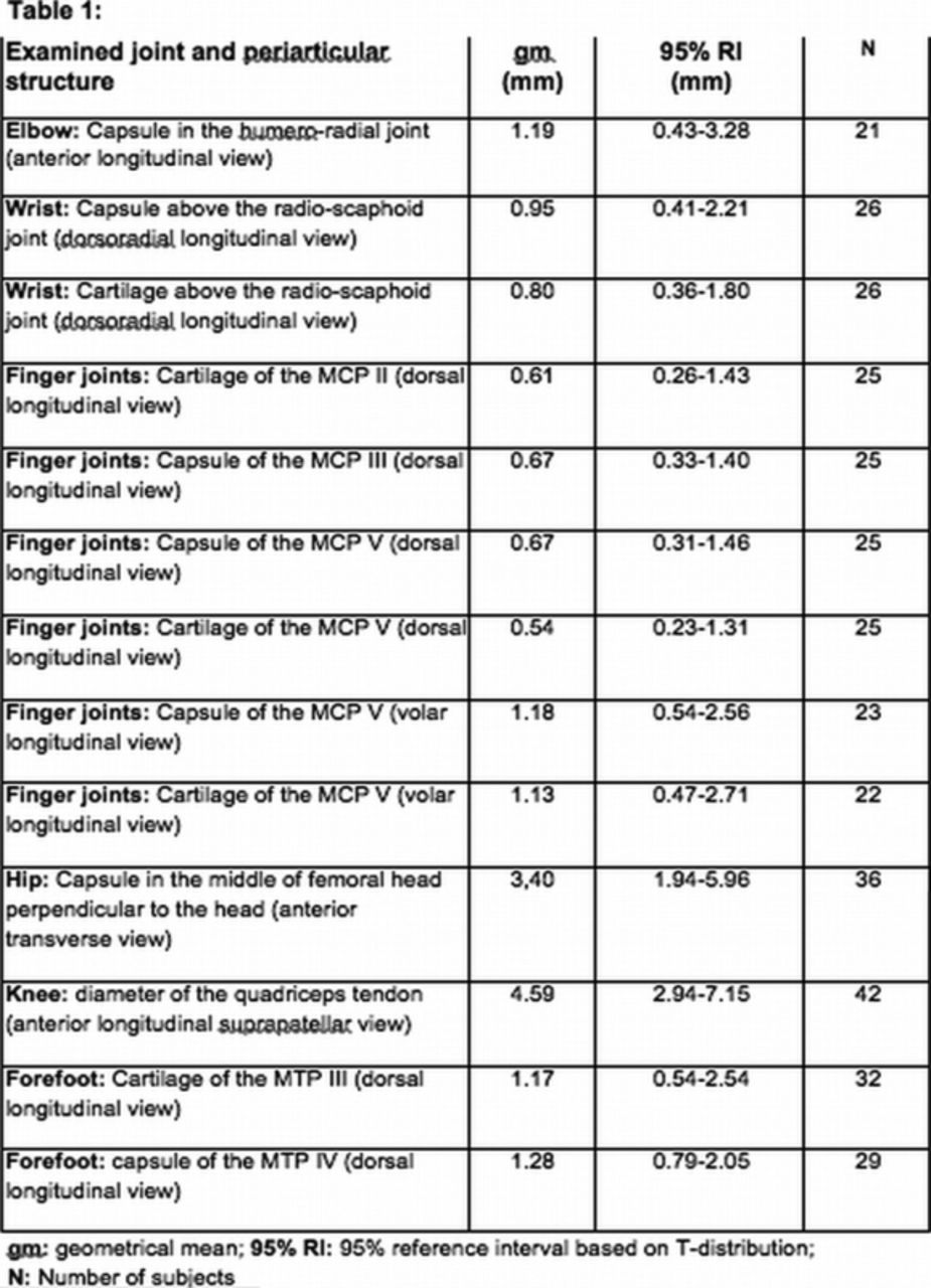
Results If there was no significant difference between girls and boys, common reference intervals were calculated (see attached Table 1). Otherwise, 95% separate reference intervals were defined for boys and girls each (not presented in abstract). Power Doppler US (PDUS) signals were measured in all joints. PDUS signals were found regularly, notably in the wrist and finger joints.
Conclusions The reference intervals for healthy children are helpful for the interpretation of pathological findings in juvenile idiopathic arthritis (JIA). The intervals show a wide range; therefore pathology should be examined carefully. Especially in younger children, joint measurements might have different values due to the growing structures, which need further evaluation. Furthermore, PDUS signals in children can be quite common, depending on the joint region and the selected standard scan.
Disclosure of Interest None declared