Article Text

Abstract
Objective Accelerated atherosclerosis is a major source of morbidity in systemic lupus erythematosus (SLE). However, the cause of SLE-accelerated atherosclerosis remains unclear.
Methods CD4+ T cells from C57/Bl/6 (B6) or SLE-susceptible B6.Sle1.2.3 (B6.SLE) mice were transferred into LDLr−/−, Rag−/− mice. T cells were examined for cytokine production and expression of interleukin-10 receptor (IL-10R) and functional markers. T cells were isolated based on FoxP3GFP expression and transferred to LDLr−/−, Rag−/− mice to establish a role for B6.SLE effector T cells (Teff) in atherosclerosis.
Results Mice receiving whole B6.SLE CD4+ T cells displayed no other SLE phenotype; however, atherosclerosis was increased nearly 40%. We noted dysregulated IL-17 production and reduced frequency of IL-10R expression by B6.SLE regulatory T cells (Treg). Functional assays indicated resistance of B6.SLE Teff to suppression by both B6.SLE and B6 Treg. Transfer experiments with CD4+FoxP3− Teff and CD4+FoxP3+ Treg from B6.SLE and B6 mice, respectively, resulted in increased atherosclerosis compared with B6 Teff and Treg recipients. Treg isolated from mice receiving B6.SLE Teff with B6 Treg had increased production of IL-17 and fewer expressed IL-10R compared with B6 Teff and Treg transfer.
Conclusions Transfer of B6.SLE Teff to LDLr−/−, Rag−/− mice results in accelerated atherosclerosis independent of the source of Treg. In addition, the presence of B6.SLE Teff resulted in more IL-17-producing Treg and fewer expressing IL-10R, suggesting that B6.SLE Teff may mediate phenotypic changes in Treg. To our knowledge, this is the first study to provide direct evidence of the role of B6.SLE Teff in accelerating atherosclerosis through resistance to Treg suppression.
This is an Open Access article distributed in accordance with the Creative Commons Attribution Non Commercial (CC BY-NC 3.0) license, which permits others to distribute, remix, adapt, build upon this work non-commercially, and license their derivative works on different terms, provided the original work is properly cited and the use is non-commercial. See: http://creativecommons.org/licenses/by-nc/3.0/
Statistics from Altmetric.com
Introduction
Systemic lupus erythematosus (SLE) is an autoimmune disorder characterised by T and B cell dysfunction, autoantibody production and end organ damage, with 30% of deaths due to premature cardiovascular disease (CVD). Multiple studies demonstrate accelerated atherosclerosis in SLE patients, with risk of developing atherosclerosis increased, on average, 2-to 10-fold compared with healthy controls.1–3 Although aetiology of SLE-accelerated atherosclerosis remains unknown, studies in patients and animal models implicate some likely suspects including the CD40–CD40L signalling pathway,2 ,4–7 decreased TGF-β,8 decreased regulatory T cells (Treg),9–12 increased proportions and activation of Th1 and Th17 cells10 ,13–15 and production of autoantibodies against β2GPI–oxLDL complexes.16
T cell populations including Th1 and Th17 cells can be pro-atherogenic,9 ,13 ,17–19 while Treg have been shown to be atheroprotective, likely through production of anti-inflammatory cytokines including interleukin (IL)-10.11 ,20–23 It is well established that, in both mice and humans, SLE T cells are hyperactivated and produce increased amounts of IFNγ and IL-17, inflammatory cytokines associated with Th1 and Th17 cells, respectively.10 ,24–26 The B6.Sle1.2.3 (B6.SLE) mouse model contains three genetic loci (Sle1, Sle2 and Sle3) which confer SLE susceptibility.27 When all three loci are present, mice display a fully penetrant lupus phenotype that develops with age and is similar to the human disease.27 ,28 One aspect of this phenotype is T cell activation. Using a bone marrow transplant model, our laboratory has demonstrated that LDLr−/− mice receiving SLE bone marrow (LDLr.SLE) have increased atherosclerosis compared with mice receiving B6 bone marrow (LDLr.B6 controls).24 ,29 This increased atherosclerosis was accompanied by an increase in the proportion of T cells within the atherosclerotic lesions, raising the question of whether SLE T cells are important for acceleration of lesion progression. Although, due to their role in autoantibody production, B cells are the focus of the majority of clinical trials of new SLE therapies, T cells also play a significant role in disease progression. Given the role of T cells in atherosclerosis, they may be an important target to consider when developing therapies to treat SLE and CVD. In the current study, we focus on the role of SLE CD4+ T cells in accelerated atherosclerosis.
Methods
Mice
C57BL/6J mice (B6), B6.129S7-Rag1tm1Mom/J (Rag−/−), B6;129S7-Ldlrtm1Her/J (LDLr−/−) and B6.Cg-Foxp3tm2(EGFP)Tch/J mice were originally obtained from The Jackson Laboratory. Rag−/− and LDLr−/− mice were crossed to obtain LDLr−/−, Rag−/− mice. The congenic strain B6.Sle1.2.3 (B6.SLE), originally obtained from Edward K Wakeland UTSW, Dallas, Texas, USA, has been described.24 ,27 ,28 B6.Cg-Foxp3tm2(EGFP)Tch/J mice were crossed with the B6.SLE strain to obtain B6.SLE mice with FoxP3-driven GFP expression (B6.SLEFoxP3−GFP). All mice are on the C57BL/6 background and maintained in our colony. Female mice were used for all studies. All procedures were approved by the Vanderbilt Institutional Animal Care and Use Committee.
Statistical analyses
Statistical significance between two groups was calculated using a Student t test. Differences among three or more variables were calculated using a one-way analysis of variance (ANOVA) with a Bonferroni post-test. A p value of <0.05 was considered statistically significant. All calculations were performed and graphs generated using GraphPad Prism Software (Graph Pad Software, Inc, La Jolla, California, USA). More detailed methods can be found in the online supplementary material.
Results
B6.SLE CD4+ T cells are sufficient to induce SLE-accelerated atherosclerosis
An increased prevalence of atherosclerosis has been associated with SLE in both humans and mouse models.1–3 ,24 ,29–32 To date, it is not clear whether one specific cell type is responsible for accelerated atherosclerosis in SLE. Results from our previous studies indicated that, along with larger atherosclerotic lesions, more CD4+ T cells were present in the lesions of LDLr.SLE mice.24 ,29 ,33 This suggests a role for CD4+ T cells in SLE-accelerated atherosclerosis. Therefore, we hypothesised that B6.SLE CD4+ T cells were sufficient to accelerate atherosclerosis. To test this hypothesis, we isolated CD4+ T cells (>90% purity) from 6-month-old B6 and B6.SLE mice and adoptively transferred them to LDLr−/−, Rag−/− mice, an atherosclerotic mouse model lacking functional B and T cells. Two weeks following transfer, all animals were placed on a Western diet for 10 additional weeks (see study design, figure 1A). B6.SLE donors had active lupus, confirmed by the presence of dsDNA autoantibody titres (figure 1B). At sacrifice, however, there were no differences between recipient mice for dsDNA autoantibody titres indicating that there was no significant transfer of B cells or autoantibody production in the recipient animals (figure 1B). Additionally, there was no difference in total spleen cell number, body weight, spleen to body weight ratio or urine protein grade (figure 1C and see online supplementary figure S1A, B and E). Numbers of CD4+ T cells recovered from spleens were similar between B6 and B6.SLE recipients (figure 1D). Excitingly, atherosclerotic lesion area in the aortic root was increased 36% in mice receiving B6.SLE CD4+ T cells (figure 1E). This increase in atherosclerosis was independent of serum cholesterol and triglyceride levels (see online supplementary figure S1C,D), supporting a specific role of CD4+ T cells in lupus-accelerated atherosclerosis. Collectively, these data demonstrate that the CD4+ T cells from B6.SLE mice are sufficient to accelerate atherosclerosis when transferred to a non-lupus, atherosclerosis-susceptible mouse.
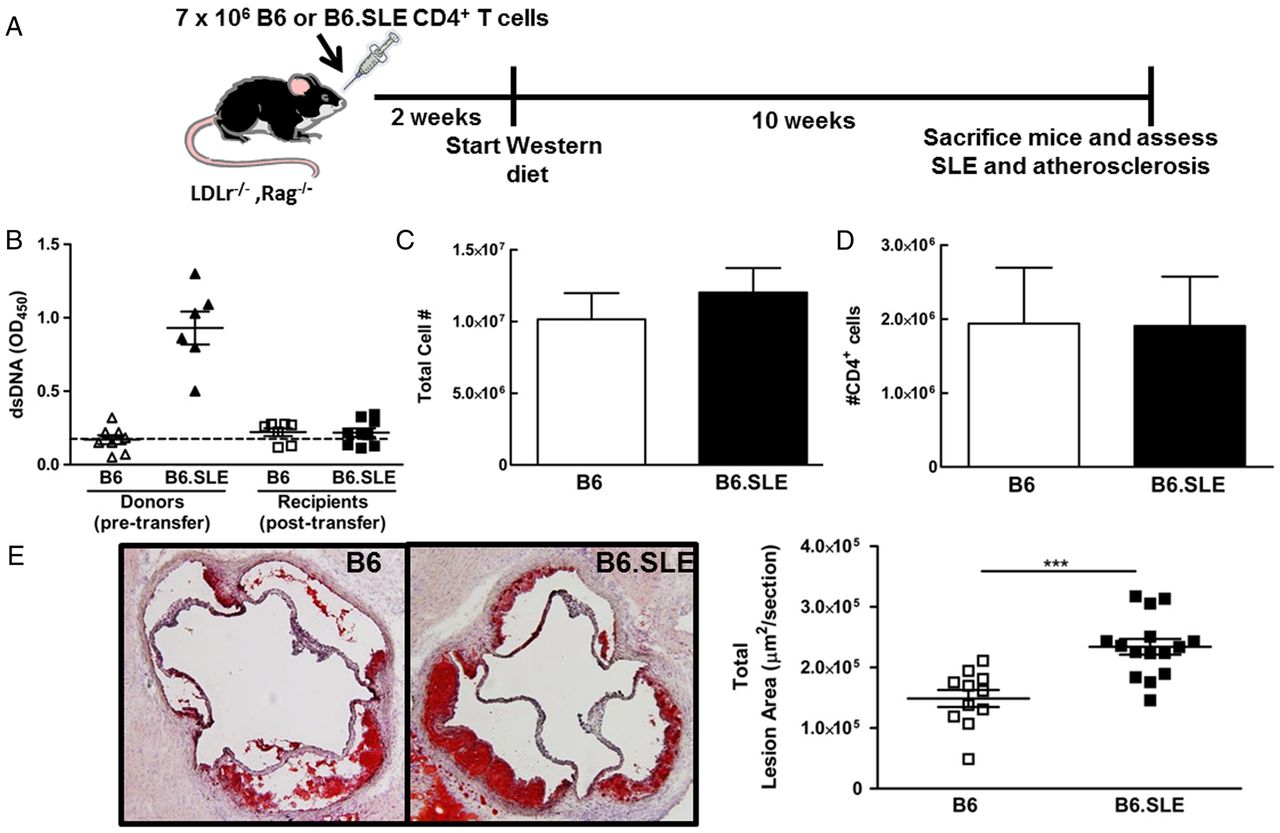
B6.SLE CD4+ T cells are sufficient to accelerate atherosclerosis. (A) Study design. (B) dsDNA titres from donor mice (pretransfer) and recipient mice at the time of sacrifice. (C) Total number of cells in the spleens of recipient mice. (D) The total number of CD4 T cells recovered from the spleens of recipient mice. (E) Representative images of Oil Red O stained sections of the aortic root (left) and quantification of the total lesion area based on the Oil Red O staining (right). Data are representative of n=7–15 mice per group. ***Significant at p<0.001. SLE, systemic lupus erythematosus.
B6.SLE Treg have altered function and phenotype
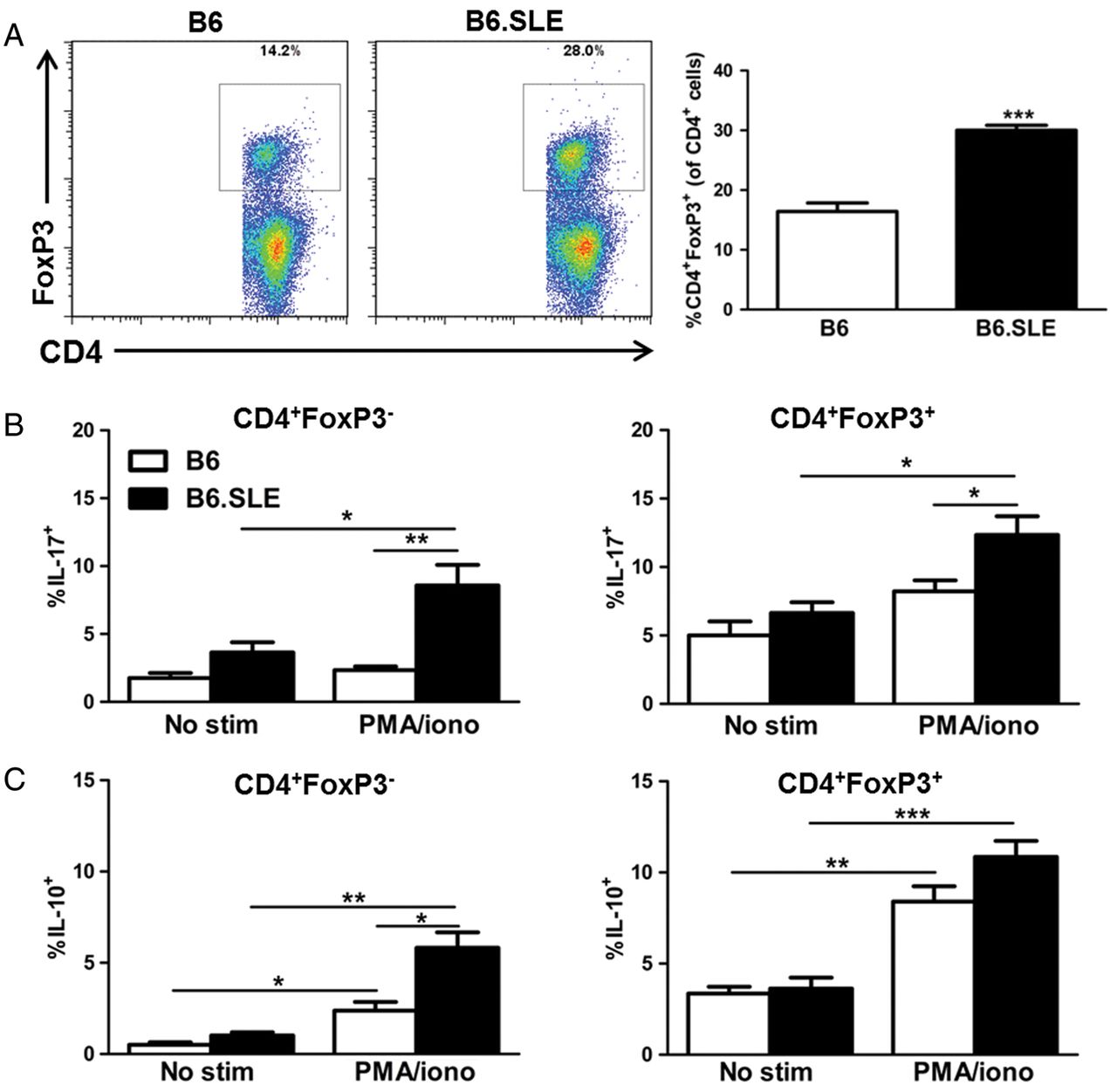
Treg dysfunction or reduced numbers of Treg may contribute to the inflammatory capabilities of B6.SLE CD4+ T cells (see online supplementary figure S2 and ref 28). Data from human and mouse studies are unclear as to whether Treg are proportionally or functionally different in SLE patients versus healthy subjects.34–37 Although there are a number of effector T cells (Teff) and Treg markers, here we define Teff as CD4+FoxP3− and Treg as positive for both CD4 and FoxP3. Examination of donor Treg showed a twofold increase in the proportion of CD4+FoxP3+ Treg in B6.SLE compared with B6 mice (figure 2A).

An increased proportion of B6.SLE Teff and Treg are IL-17+. (A) Dot plots of FoxP3 versus CD4 (gated on CD4+ cells), left panel and the resulting quantification of CD4+FoxP3+ cells, right panel. Whole splenocytes were cultured with or without anti-CD3/CD28 stimulation for 5 h and then subjected to intracellular staining for IL-17 (B) and IL-10 (C). The percentage of cytokine positive cells was measured by flow cytometry. (B) The percent of IL-17+ cells in the CD4+FoxP3− (left) and CD4+FoxP3+ (right) populations. (C) The percent of IL-10+ cells in the CD4+FoxP3− (left) and CD4+FoxP3+ (right) populations. Data are representative of n=4–6, 6-month-old mice per group. *, ** and *** indicate significance at p<0.05, 0.01 and 0.001, respectively. SLE, systemic lupus erythematosus.
First, given the dysregulation of cytokine production by CD4+ T cells in the B6.SLE model (see online supplementary figure S2D–F), and to determine whether SLE Treg possessed qualitative differences in cytokine production compared with their B6 counterparts, we examined cytokine secretion by CD4+FoxP3− Teff and CD4+FoxP3+ Treg after phorbol 12-myristate 13-acetate (PMA) and ionomycin stimulation of whole splenocytes. Not unexpectedly, B6.SLE CD4+FoxP3− cells exhibited a 3.6-fold increase in the proportion of IL-17-producing cells compared with B6 mice (figure 2B, left panel). Surprisingly, however, B6.SLE CD4+FoxP3+ Treg also exhibited a large increase in the percentage of IL-17+ cells (figure 2B, right panel). Greater than 6% of Treg produced IL-17; a twofold increase over unstimulated B6.SLE CD4+FoxP3− Teff. This percentage was further increased upon stimulation, with greater than 12% of Treg secreting IL-17. These data suggest dysregulation of IL-17 production by CD4+FoxP3+ Treg in B6.SLE mice.
Treg exert their anti-inflammatory effects in part through IL-10.38 IL-10 is classically anti-inflammatory but is associated with SLE disease activity.39 CD4+FoxP3− T cells in B6 and B6.SLE mice did not differ in terms of the proportion of IL-10+ cells at baseline. However, after stimulation, B6.SLE CD4+FoxP3− cells had a twofold increase in IL-10+ cells compared with B6 (figure 2C, left panel), as would be expected in a model of SLE. Although both B6 and B6.SLE Treg responded to stimulation by increasing the proportion of cells expressing IL-10, there was no significant difference between strains of mice (figure 2C, right panel).
B6.SLE Treg have reduced levels of IL-10R expression
A recent report indicated that Treg IL-10 receptor (IL-10R) expression facilitates suppression of Th17 responses.40 Because we observed increased IL-17 production by Treg (figure 2B) but also increased production of IL-10 in B6.SLE mice (figure 2C and see online supplementary figure S2F), we hypothesised that IL-10R expression might be reduced in B6.SLE. The proportion of CD4+FoxP3−IL-10R+ cells was similar between B6 and B6.SLE (figure 3A,B). However, there was a 1.5-fold reduction of IL-10R+ cells in the B6.SLE Treg compartment compared with B6. Expression levels as measured by mean fluorescence intensity (MFI) were not different between B6 and B6.SLE (figure 3C).

Reduced IL-10 receptor expression in B6.SLE Treg and Teff populations. Whole splenocytes were obtained from 6-month-old mice and examined for surface expression of the IL-10R by flow cytometry. Samples were gated on CD4+ cells, and then the cell populations of interest were examined. (A) Contour plots showing IL-10R expression on CD4+FoxP3− (left) and CD4+FoxP3+ (right) cells from B6 (top) and B6.SLE (bottom) mice. (B) Quantification showing the proportion of each cell population expressing IL-10R. (C) Quantification of the mean fluorescence intensity of IL-10R staining on Treg and Teff populations. Data are representative of at least six mice per group. * Indicates significance at p<0.05. SLE, systemic lupus erythematosus.
B6.SLE Teff are resistant to Treg-mediated suppression
Despite the increased proportion of Treg, Teff activation persists (see online supplementary figure S2). This, in addition to increased IL-17 production and reduced IL-10R expression by B6.SLE Treg, led us to hypothesise that these cells are dysfunctional in their ability to suppress Teff. To test this hypothesis, we isolated Treg and Teff and performed in vitro functional assays. Because FoxP3 is an intracellular marker, we isolated these cells based on CD25 expression. CD4+CD25+ cells served as Treg in this assay while Teff were defined as CD4+CD25−. Suppressive function was determined as the ability of Treg to suppress proliferation of Teff. When challenged with strain-matched Teff, B6.SLE Treg were less suppressive compared with B6 Treg (figure 4A,B). Surprisingly, when challenged with B6 Teff, B6.SLE Treg were as suppressive as B6 Treg, suggesting that B6.SLE Teff are resistant to suppression. To further confirm this, we incubated both B6 and B6.SLE Treg with B6.SLE Teff. The ability of Treg from both strains to suppress B6.SLE Teff proliferation was reduced (figure 4A,B) compared with their inhibition of B6 Teff. These data suggest that, similar to results seen in some SLE patients,37 ,41 ,42 B6.SLE Teff are resistant to Treg-mediated suppression.

B6.SLE Teff are resistant to Treg-mediated suppression. Suppression assays were performed from purified CD4+CD25+ (Treg) and CD4+CD25 − (Teff) cells. (A) Percent inhibition of proliferation by B6 and B6.SLE Treg challenged with B6 or B6.SLE Teff. (B) Comparison of inhibition at the 1:4 ratio of Treg to Teff. (C) All samples were gated based on CD4+ T cells, and then the individual cell populations of interest were examined. Representative histograms of GITR expression (top panel) on CD4+ cell populations in B6 (red line) and B6.SLE (blue line) mice; quantification of GITR expression based on mean fluorescence intensity (top right panel); representative histograms of inducible costimulatory molecule (ICOS) expression (bottom panel) on CD4+ cell populations in B6 (red line) and B6.SLE (blue line) mice; Quantification of ICOS expression based on mean fluorescence intensity (bottom right panel). Data reflect n=6–8 mice per group from one of two representative experiments. *, ** and *** indicate significance at p<0.05, 0.01 and 0.001, respectively. GITR, glucocorticoid-induced TNFR-related protein; SLE, systemic lupus erythematosus.
We next measured surface expression of glucocorticoid-induced TNFR-related protein (GITR), a surface receptor upregulated on newly activated T cells. Surface expression of GITR on Teff is thought to extend survival and confer resistance to suppression by Treg.43 Supporting the functional assay, B6.SLE CD4+ T cell populations exhibited a twofold increase in GITR expression compared with B6 (figure 4C, top panel). Surface expression of inducible costimulatory molecule (ICOS), typically upregulated on the surface of activated T cells and important in Th2 responses,44 was increased twofold on B6.SLE CD4+ T cells compared with B6 (figure 4C, bottom panel). There were no differences between Teff or Treg cell populations. These data support results from figure 4A and online supplementary figure S1 indicating T cell activation and resistance of Teff to Treg-mediated suppression in the B6.SLE model.
B6 Treg are not sufficient to prevent B6.SLE Teff-mediated acceleration of atherosclerosis
Data thus far support the hypothesis that Teff resistance to Treg-mediated suppression in B6.SLE mice may accelerate atherosclerosis. To test this hypothesis, we performed a T cell transfer study in which CD4+FoxP3− Teff were isolated from 6-month-old B6FoxP3−GFP and B6.SLEFoxP3−GFP mice while CD4+FoxP3+ Treg were isolated from B6FoxP3−GFP mice. A control group of LDLr−/−, Rag−/− mice received both CD4+FoxP3+ Treg and CD4+FoxP3− Teff from B6FoxP3−GFP mice (B6 TE:B6 TR) while the experimental group received B6.SLEFoxP3−GFP Teff with B6FoxP3−GFP Treg (B6.SLE TE:B6 TR). The study followed the timeline as shown in figure 1A. As in our previous transfer experiment, we noted no differences in body weight, spleen to body weight ratio, total number of splenocytes or in the total number of CD4+ T cells in the spleens at sacrifice (see online supplementary figure S3A–D). Furthermore, there were no differences in urine protein grade, as all recipients had little to no urine protein (see online supplementary figure S3E). Upon examination of the aortic root, the total lesion area was increased 44% in the B6.SLE TE:B6 TR experimental group compared with the B6 TE:B6 TR mice (figure 5A). In addition, the percentage of CD4+ T cells in the lesions was slightly, but significantly, higher in the B6.SLE Teff recipients compared with control (figure 5B). When examining the T cell populations within the spleens of recipients, we discovered that, despite the presence of B6 Treg, there was a greater than twofold increase in the percentage of Teff in the SLE TE:B6 TR group which were producing IL-17 (figure 5C, left panel). Interestingly, in recipients of SLE TE:B6 TR, there was a 2.8-fold increase in the percentage of Treg which were producing IL-17 (figure 5C, right panel). Accompanying this increase in the proportion of IL-17+ cells was a significant reduction in the percentage of IL-10R+ cells in the SLE TE:B6 TR group in both Treg and Teff (figure 5D). These data implicate B6.SLE Teff as the primary player in SLE-accelerated atherosclerosis, and suggest Teff-mediated dysregulation of Treg IL-17 production and IL-10R expression as a potential mechanism by which Teff cause this acceleration.

B6.SLE Teff accelerate atherosclerosis independent of the source of Treg. (A) Representative images of Oil Red O stained sections of the aortic root (top) and quantification of the total lesion area based on the Oil Red O staining (bottom). (B) Representative images of immunofluorescent staining for CD4 + cells (red), with 4′, 6-diamidino-2-phenylindole (DAPI) staining (blue) for nuclei (top) and quantification of the proportion of total cells that were shown to express CD4 (bottom). (C) Whole splenocytes from recipients were cultured with or without anti-CD3/CD28 stimulation for 5 h and then subjected to intracellular staining for IL-17. The percentage of cytokine positive cells was measured by flow cytometry. The percent of IL-17+ cells in the CD4+FoxP3− (left) and CD4+FoxP3+ (right) populations. (D) IL-10R expression was measured on Teff and Treg from recipients. Cells were gated based on CD4+ expression, then separated into Teff and Treg populations. IL-10R staining was measured by flow cytometry. Data reflect n=6 mice per group. *, ** and *** indicate significance at p<0.05, 0.01 and 0.001, respectively. SLE, systemic lupus erythematosus.
Discussion
SLE and atherosclerosis are complex chronic inflammatory diseases characterised by immune dysfunction. Evidence supports interplay between the two diseases, with studies showing accelerated atherosclerosis in SLE. Increased activation of T cells has been demonstrated in both SLE patients and in mouse models of SLE including (NZB×NZW)F1,45 MRL/MpJ-Fas(lpr/lpr)/J46 and B6.SLE mice.24 ,27 ,28 Given the important effector and regulatory functions of T cells in atherosclerosis and that accelerated CVD is a major cause of death for SLE patients, it has been suggested that T cells may be important to consider when developing therapies for SLE and SLE-accelerated atherosclerosis (reviewed in47). However, most clinical studies to date have focused on B cells due to their role in producing the autoantibodies that lead to immune complex formation and the resultant end organ damage in SLE. The current study was undertaken to determine the effects of B6.SLE CD4+ T cells on atherosclerosis. While mice are not optimal for study of plaque vulnerability and rupture, common properties of atherosclerotic lesions in SLE patients, they provide a platform to study the complicated mechanisms by which the immune system contributes to plaque development. We chose the LDLr−/−, Rag−/− model, allowing us to determine the direct effects of CD4+ T cells on atherosclerosis, independent of other SLE immune cell populations including the autoantibody-producing B cells. Importantly, at the time of sacrifice, T cell recipients did not have an SLE phenotype, attributing any effects on atherosclerosis to the T cells. Here, we demonstrate a direct role for B6.SLE CD4+ T cells in accelerated atherosclerosis.
While Teff including Th1 cells are generally thought to be atherogenic48 ,49 (and reviewed in50), Treg have been shown to have antiatherogenic properties. Ait-Oufella et al11 showed a negative correlation between Treg number and atherosclerosis development (reviewed in51), and it has been directly demonstrated that depletion of Treg results in increased atherosclerosis.52 In our study, B6.SLE mice had an increased proportion of CD4+FoxP3+ Treg, yet atherosclerosis was increased with CD4+ T cell transfer from these mice. Treg and Th17 dysregulation is a common feature in SLE. We made the unexpected observation that an increased proportion of B6.SLE Treg was secreting IL-17, a cytokine typically attributed to Th17 cells. IL-17 contributes to tissue damage in SLE and is correlated with disease activity.53 ,54 Interestingly, the proportion of these cells making IL-17 was higher than that of Teff, suggesting that Treg are a part of the IL-17 dysregulation observed in SLE and have the potential to contribute to tissue damage. IL-17 is also believed to play a role in atherosclerosis, although whether it contributes to or reduces atherosclerosis is unclear due to the existence of studies supporting both possibilities.13 ,19 ,55 Since IL-17-producing cells likely play a role in atherosclerosis, the fact that significant proportions of B6.SLE Treg secrete IL-17 identifies them as potential contributors to SLE-accelerated atherosclerosis. A report by Chaudhry et al40 concluded that Treg IL-10R expression is required to suppress inflammation mediated by Th17 cells. This led us to ask the question as to whether IL-10R expression was reduced in B6.SLE T cell populations. Reduced IL-10R expression could explain the persistence of inflammation despite increased IL-10 in the context of SLE and why an increase of this antiatherogenic cytokine does not lead to reduced atherosclerosis. Indeed, the proportion of IL-10R+ cells in the Treg populations from B6.SLE mice was reduced. These data led us to hypothesise that B6.SLE Treg have reduced suppressive capacity. On the contrary, results from Treg functional assays demonstrated intact suppressive function of B6.SLE CD4+CD25+ Treg but that B6.SLE Teff are resistant to Treg-mediated suppression. These data are comparable with some studies in SLE patients which point to Teff resistance rather than Treg dysfunction.37 ,56
Given the damaging effects of IL-17 in SLE, uncovering the underlying mechanisms behind the dysregulation in IL-17 and IL-10R may be important for our understanding of the interplay between SLE and atherosclerosis. Indeed, a recent study in SLE patients with atherosclerosis has shed light on a potential atherosclerotic role for IL-17 in SLE by demonstrating that these patients have increased serum levels of IL-17 compared with SLE patients and healthy controls without atherosclerosis.57 Importantly, results from our study implicate B6.SLE Teff as being responsible for this dysregulation of Treg IL-17 production and IL-10R expression. With transfer of B6.SLE Teff and B6 Treg, atherosclerosis was increased. This increase, despite the presence of wild-type Treg, indicates that Teff resistance to Treg-mediated suppression may be a strong contributing factor in this mechanism. Not only did B6 Treg fail to suppress B6.SLE Teff-accelerated atherosclerosis, their phenotype appeared to be altered by the presence of B6.SLE Teff, resulting in an increased proportion of B6 Treg producing IL-17 and a reduced percentage of cells expressing IL-10R. This suggests that B6.SLE Teff resistance may be a direct result of their ability to alter IL-10R expression and, subsequently, IL-17 production by Treg. One limitation of this study, however, is our ability to definitively know whether the altered Treg phenotype in SLE TE:B6 TR recipients was due to changes in the original B6 Treg or conversion of B6.SLE Teff to Treg with a dysfunctional phenotype. We are currently developing the reporter mice which would allow us to map Treg that become ex-Treg58 and these studies are the focus of future experiments.
With evidence mounting for the antiatherogenic role of Treg, increased focus has been placed on these cells as a therapeutic target for atherosclerosis. Recent studies have identified a number of potential mechanisms involved in Treg suppression that may prove useful for enhancement of suppression and/or Treg numbers including MYD88 signalling in dendritic cells,20 the IL-33–ST2 interaction,59 CD39 and CD73 signalling, CTLA-4 and LAG-3.60 However, data from the current study favour Teff resistance over Treg-mediated suppression as a major culprit in SLE and suggest that, at least in the context of SLE and atherosclerosis, targeting mechanisms of Teff resistance may prove most effective. Given our data, we postulate that therapies targeting IL-17 production, and perhaps IL-10R expression, would prove effective in treatment of SLE-accelerated atherosclerosis.
Multiple aspects of T cell dysregulation in B6.SLE mice suggest these cells have the potential to accelerate atherosclerosis. To date, speculation regarding their role in SLE-accelerated atherosclerosis has far outweighed any direct evidence supporting this hypothesis. In the current study, we demonstrate (1) B6.SLE CD4+ T cells are sufficient to accelerate atherosclerosis, (2) B6.SLE Teff are resistant to Treg-mediated suppression, (3) dysregulation of IL-17 production by Treg, with a possible role for the IL-10R in this process and (4) B6.SLE Teff are likely the primary T cell type involved in the observed accelerated atherosclerosis, potentially through their ability to alter Treg IL-17 production and IL-10R expression. These data provide strong evidence for a causal role of CD4+ T cells and, specifically, for B6.SLE Teff, in SLE-accelerated atherosclerosis. To our knowledge, this is the first study to provide such evidence. These results emphasise the importance of considering T cell-targeted therapies when developing strategies to treat SLE and SLE-accelerated atherosclerosis and suggest that, in SLE, Teff may be a more desirable target for therapy than Treg.
Acknowledgments
The authors thank Mr Roman Covarrubias and Drs Curtis L Gabriel and Yanice Mendez-Fernandez for thoughtful discussion regarding the content of this manuscript.
References
Supplementary materials
Supplementary Data
This web only file has been produced by the BMJ Publishing Group from an electronic file supplied by the author(s) and has not been edited for content.
Files in this Data Supplement:
- Data supplement 1 - Online supplement
Footnotes
-
Handling editor Tore K Kvien
-
Contributors AJW designed, performed and interpreted experiments presented in this manuscript. She also wrote and edited most of the manuscript content. JPR performed the experiments and edited the text. NSW performed experiments presented in this manuscript. ASM designed, performed, interpreted the experiments and edited the text. She is also the principal investigator of the laboratory.
-
Funding The work presented in this study was supported by National Institutes of Health (NIH) grants R01HL088364 and R01HL089310 (to ASM), a predoctoral NRSA F32HL105024 from the NIH (to NSW) and postdoctoral fellowships from the AHA (12POST11940044) and NIH (NRSA 1F32HL114244) (to AJW).
-
Competing interests None.
-
Provenance and peer review Not commissioned; externally peer reviewed.